Mesoderm

From Embryology
Revision as of 11:01, 16 July 2019 by Z8600021 (talk | contribs)
(diff) ← Older revision | Latest revision (diff) | Newer revision → (diff)
Embryology - 19 Mar 2024    Facebook link Pinterest link Twitter link  Expand to Translate  
Google Translate - select your language from the list shown below (this will open a new external page)

العربية | català | 中文 | 中國傳統的 | français | Deutsche | עִברִית | हिंदी | bahasa Indonesia | italiano | 日本語 | 한국어 | မြန်မာ | Pilipino | Polskie | português | ਪੰਜਾਬੀ ਦੇ | Română | русский | Español | Swahili | Svensk | ไทย | Türkçe | اردو | ייִדיש | Tiếng Việt    These external translations are automated and may not be accurate. (More? About Translations)

Introduction

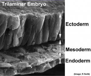
The trilaminar embryo

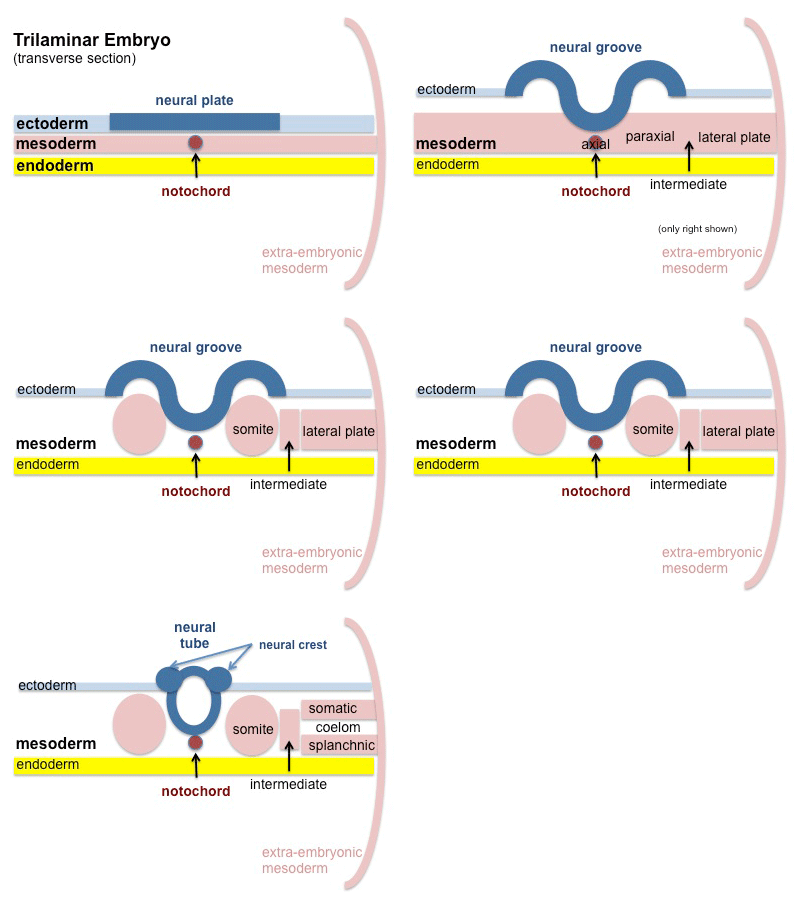
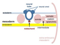
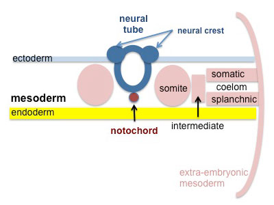
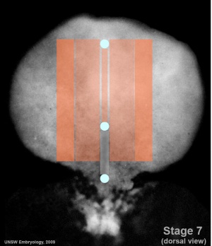
The mesoderm forms the middle layer of the early trilaminar embryo germ layers (ectoderm, mesoderm and endoderm) formed by gastrulation. The segmentation of the initial mesoderm into somites, and their regular addition, is often used to stage embryonic development (23 somite embryo).


This middle germ layer forms connective tissues and muscle throughout the body, with the exception of in the head region where some of these structures have a neural crest (ectoderm) origin.

  • connective tissues - cartilage, bone, blood, blood vessel endothelium, dermis, etc.
  • muscle - cardiac, skeletal, smooth.

Students often mix-up the terms mesoderm (middle layer) with mesenchyme (embryonic connective tissue). It is true that mesoderm initially does have a mesenchymal cellular organisation, but can also form a range of epithelial structures (surrounding somites, mesothelium lining of body cavities).

Mesoderm Links: endoderm | mesoderm | ectoderm | Lecture - Mesoderm | Lecture - Musculoskeletal | 2016 Lecture | notochord | somitogenesis | somite | splanchnic mesoderm | skeletal muscle | smooth muscle | heart | Notochord Movie | musculoskeletal | cartilage | bone | sonic hedgehog | Category:Mesoderm
Historic Embryology  
Historic Disclaimer - information about historic embryology pages 
Mark Hill.jpg
Pages where the terms "Historic" (textbooks, papers, people, recommendations) appear on this site, and sections within pages where this disclaimer appears, indicate that the content and scientific understanding are specific to the time of publication. This means that while some scientific descriptions are still accurate, the terminology and interpretation of the developmental mechanisms reflect the understanding at the time of original publication and those of the preceding periods, these terms, interpretations and recommendations may not reflect our current scientific understanding.     (More? Embryology History | Historic Embryology Papers)

Historic Papers: 1883 Mesoderm | 1910 Chick Somites | 1933 | 1935 Rabbit Somites

Historic Textbooks: 1892 Primitive Segments | 1907 Somites | 1910 Skeleton | 1914 Somite | 1920 Chick Mesoderm | 1921 Connective Tissue | 1951 Frog Mesoderm

Some Recent Findings

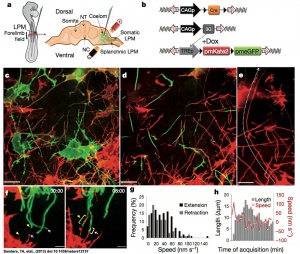
Mesenchymal cells of the developing limb bud possess long and highly dynamic cytoplasmic extensions.[1]
  • BMP and FGF signaling interact to pattern mesoderm by controlling basic helix-loop-helix transcription factor activity[2] "The mesodermal germ layer is patterned into mediolateral subtypes by signaling factors including BMP and FGF. How these pathways are integrated to induce specific mediolateral cell fates is not well understood. We used mesoderm derived from post-gastrulation neuromesodermal progenitors (NMPs), which undergo a binary mediolateral patterning decision, as a simplified model to understand how FGF acts together with BMP to impart mediolateral fate. Using zebrafish and mouse NMPs, we identify an evolutionarily conserved mechanism of BMP and FGF mediated mediolateral mesodermal patterning that occurs through modulation of basic helix-loop-helix (bHLH) transcription factor activity. BMP imparts lateral fate through induction of Id helix loop helix (HLH) proteins, which antagonize bHLH transcription factors, induced by FGF signaling, that specify medial fate. We extend our analysis of zebrafish development to show that bHLH activity is responsible for the mediolateral patterning of the entire mesodermal germ layer."
  • BRACHYURY directs histone acetylation to target loci during mesoderm development[3] "T-box transcription factors play essential roles in multiple aspects of vertebrate development. Here, we show that cooperative function of BRACHYURY (T) with histone-modifying enzymes is essential for mouse embryogenesis. A single point mutation (TY88A) results in decreased histone 3 lysine 27 acetylation (H3K27ac) at T target sites, including the T locus, suggesting that T autoregulates the maintenance of its expression and functions by recruiting permissive chromatin modifications to putative enhancers during mesoderm specification. Our data indicate that T mediates H3K27ac recruitment through a physical interaction with p300. In addition, we determine that T plays a prominent role in the specification of hematopoietic and endothelial cell types. Hematopoietic and endothelial gene expression programs are disrupted in TY88A mutant embryos, leading to a defect in the differentiation of hematopoietic progenitors. We show that this role of T is mediated, at least in part, through activation of a distal Lmo2 enhancer." blood
More recent papers  
Mark Hill.jpg
PubMed logo.gif

This table allows an automated computer search of the external PubMed database using the listed "Search term" text link.

  • This search now requires a manual link as the original PubMed extension has been disabled.
  • The displayed list of references do not reflect any editorial selection of material based on content or relevance.
  • References also appear on this list based upon the date of the actual page viewing.


References listed on the rest of the content page and the associated discussion page (listed under the publication year sub-headings) do include some editorial selection based upon both relevance and availability.

More? References | Discussion Page | Journal Searches | 2019 References | 2020 References

Search term: Mesoderm Development | Extra-embryonic Mesoderm Development | Axial Mesoderm | Paraxial Mesoderm | Intermediate Mesoderm | Splanchnic Mesoderm | Somatic Mesoderm |Mesoderm Images

Older papers  
These papers originally appeared in the Some Recent Findings table, but as that list grew in length have now been shuffled down to this collapsible table.

See also the Discussion Page for other references listed by year and References on this current page.

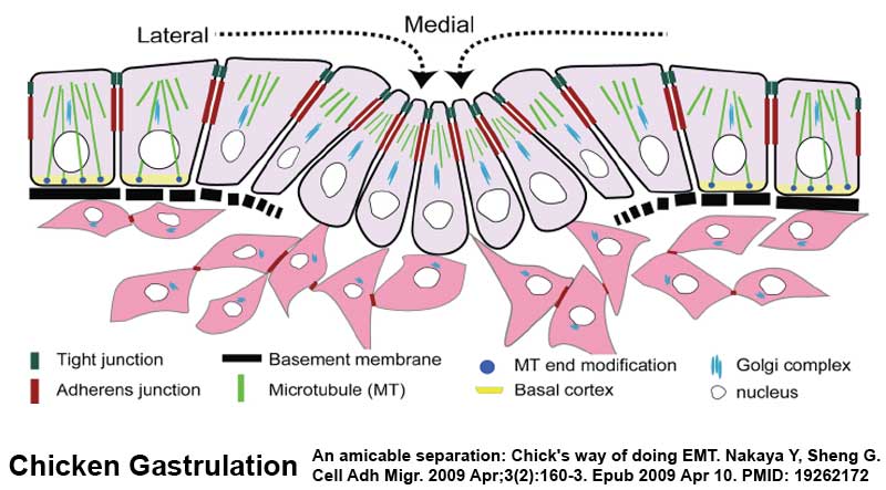
  • STRIP1, a core component of STRIPAK complexes, is essential for normal mesoderm migration in the mouse embryo[4] "Regulated mesoderm migration is necessary for the proper morphogenesis and organ formation during embryonic development. Cell migration and its dependence on the cytoskeleton and signaling machines have been studied extensively in cultured cells; in contrast, remarkably little is known about the mechanisms that regulate mesoderm cell migration in vivo. Here, we report the identification and characterization of a mouse mutation in striatin-interacting protein 1 (Strip1) that disrupts migration of the mesoderm after the gastrulation epithelial-to-mesenchymal transition (EMT). STRIP1 is a core component of the biochemically defined mammalian striatin-interacting phosphatases and kinase (STRIPAK) complexes that appear to act through regulation of protein phosphatase 2A (PP2A), but their functions in mammals in vivo have not been examined. Strip1-null mutants arrest development at midgestation with profound disruptions in the organization of the mesoderm and its derivatives, including a complete failure of the anterior extension of axial mesoderm. Analysis of cultured mesoderm explants and mouse embryonic fibroblasts from null mutants shows that the mesoderm migration defect is correlated with decreased cell spreading, abnormal focal adhesions, changes in the organization of the actin cytoskeleton, and decreased velocity of cell migration. The results show that STRIPAK complexes are essential for cell migration and tissue morphogenesis in vivo." (More? cell migration | NCBI Gene - STRIP1)
  • An atlas of transcriptional, chromatin accessibility, and surface marker changes in human mesoderm development[5] "Mesoderm is the developmental precursor to myriad human tissues including bone, heart, and skeletal muscle. Unravelling the molecular events through which these lineages become diversified from one another is integral to developmental biology and understanding changes in cellular fate. To this end, we developed an in vitro system to differentiate human pluripotent stem cells through primitive streak intermediates into paraxial mesoderm and its derivatives (somites, sclerotome, dermomyotome) and separately, into lateral mesoderm and its derivatives (cardiac mesoderm)."
  • A role for Vg1/Nodal signaling in specification of the intermediate mesoderm[6] "The intermediate mesoderm (IM) is the embryonic source of all kidney tissue in vertebrates. The factors that regulate the formation of the IM are not yet well understood. Through investigations in the chick embryo, the current study identifies and characterizes Vg1/Nodal signaling (henceforth referred to as 'Nodal-like signaling') as a novel regulator of IM formation. ... We postulate that Nodal-like signaling regulates IM formation by modulating the IM-inducing effects of BMP signaling." Renal System Development
  • Signaling gradients during paraxial mesoderm development[7] "These studies indicate that high levels of Wnt and FGF signaling are required for the segmentation clock activity. Furthermore, we discuss how these signaling gradients act in a dose-dependent manner in the progenitors of the paraxial mesoderm, partly by regulating cell movements during gastrulation. Finally, links between the process of axial specification of vertebral segments and Hox gene expression are discussed."
  • Transcriptional profiling of the nucleus pulposus: say yes to notochord[8] "This editorial addresses the debate concerning the origin of adult nucleus pulposus cells in the light of profiling studies by Minogue and colleagues. In their report of several marker genes that distinguish nucleus pulposus cells from other related cell types, the authors provide novel insights into the notochordal nature of the former. Together with recently published work, their work lends support to the view that all cells present within the nucleus pulposus are derived from the notochord. Hence, the choice of an animal model for disc research should be based on considerations other than the cell loss and replacement by non-notochordal cells."

Overview

mesoderm
The nested tables below show an overview of the different mesoderm-derived tissues (links go to topic pages).
embryonic
axial  
notochord
intervertebral disc nucleus pulposis
paraxial  
unsegmented paraxial  
head mesoderm
segmented paraxial  
somite
sclerotome  
axial skeleton
syndetome  
tendon
vertebral body intervertebral disc
dermomyotome  
dermotome  
myotome  
intermediate  
renal genital
lateral plate
somatic  
body wall connective tissue
intra-embryonic coelom  
pericardial cavity pleural cavity peritoneal cavity
splanchnic 
splanchnic mesoderm
heart blood vessels gastrointestinal tract connective tissue gastrointestinal tract smooth muscle respiratory connective tissue
extra-embryonic
placental membranes
placental villi
amnion chorion yolk sac villi connective tissue villi blood vessels
Overview: Ectoderm | Mesoderm | Endoderm Layers: ectoderm | mesoderm | endoderm


Hierarchical View  
mesoderm - (hierarchical view)
  1. embryonic mesoderm
    1. axial
      1. notochord
        1. axial skeleton - nucleus pulpous
    2. paraxial
      1. unsegmented paraxial
      2. segmented paraxial somites
        1. sclerotome
          1. syndetome - tendon
          2. axial skeleton - vertebral body, intervertebral disc
        2. dermomyotome
          1. dermis - integumentary dermis, hypodermis
          2. myotome skeletal muscle
    3. intermediate
      1. renal
      2. genital
    4. lateral plate
      1. somatic
      2. intraembryonic coelom
        1. pericardial
        2. pleural
        3. peritoneal
      3. splanchnic mesoderm
  2. extra-embryonic mesoderm
    1. placental membranes
      1. amnion, chorion, yolk sac
    2. placental villi
      1. villi connective tissue, villi blood vessels
Germ Layers: ectoderm | mesoderm | endoderm


Mesoderm Structures  
Mesoderm Origin
Alphabetical list of anatomical structures derived from mesoderm.
A
  • accessory foramen
  • acetabular notch
  • acetabular part of hip bone
  • acetabular rim
  • acropodial skeleton
  • adductor blade
  • adductor crest
  • adductor mandibulae complex
  • adrenal cortex
  • adrenal gland capsule
  • adrenal gland cortex zone
  • adrenal medulla (stroma)
  • adrenal tissue
  • adrenal/interrenal gland
  • adult mammalian kidney
  • adventitia of epididymis
  • adventitia of seminal vesicle
  • adventitia of ureter
  • agger limitans anterior of ilium
  • agger limitans anterior of ischium
  • alisphenoid bone
  • amnion
  • ampulla of uterine tube
  • anterior dentation of pectoral fin spine
  • anterior distal condyle of femur
  • anterior distal serration of pectoral fin spine
  • anterior humeral ridge
  • anterior lateral mesoderm
  • anterior lateral plate mesoderm
  • anterior subdivision of masseter
  • antitrochanter
  • antotic pillar
  • aorta collagen fibril
  • aortic body
  • apical region of heart ventricle
  • apophysis distalis of tibiale fibulare
  • apophysis distalis of tibiofibula
  • apophysis proximalis
  • appendix epididymis
  • appendix testis
  • arachnoid barrier layer
  • ascending process of the astragalus
  • associated mesenchyme of midgut
  • astragalus head
  • astragalus-calcaneum unit
  • atrial foramen intermedium
  • atrioventricular region
  • atrium auricular region
  • atrium myocardial trabecula
  • autopodial skeleton
  • axochord
B
  • basal zone of heart
  • base of glans penis
  • basipterygium element
  • basisphenoid bone
  • bicipital crest
  • bicipital tuberosity
  • blood vessel
  • blood vessel elastic tissue
  • blood vessel layer
  • body of ilium
  • bone marrow
  • bone of free limb or fin
  • branch of ilio-marsupialis muscle
  • branchiomeric muscle
  • brevis shelf
  • broad ligament of uterus
  • bulb of aorta
  • bulb of penis
  • bulbus arteriosus inner layer
  • bulbus arteriosus middle layer
  • bulbus arteriosus outer layer
  • bursal follicle
  • bursal plica
C
  • calcaneal tuber
  • canal of Nuck
  • capitulum of humerus
  • capitulum of radio-ulna
  • capitulum of radius
  • capitulum ulnae
  • caput epididymis
  • caput ossis cruris
  • cardiac chamber
  • cardiac jelly
  • cardiac mesenchyme
  • cardiac septum
  • cardiac skeleton
  • cardiac valve leaflet
  • cardial valve
  • cardiogenic splanchnic mesoderm
  • cardiopharyngeal field
  • carotid body
  • cauda epididymis
  • celiac trunk
  • chorioallantoic membrane
  • chorion
  • chorionic plate
  • circulatory system dorsal vessel
  • clitoris
  • cnemial crest
  • collum antibrachii
  • collum ilei
  • columnar area
  • condyle of femur
  • condyle of humerus
  • condyle of tibia
  • corona of glans penis
  • coronary vessel
  • coronoid process of ulna
  • corpus cavernosum clitoridis
  • corpus cavernosum penis
  • corpus epididymis
  • corpus luteum
  • corpus spongiosum of penis
  • corpuscles of Stannius
  • cortex of humerus
  • cotyloid notch
  • cranial muscle
  • crista dorsalis humeri
  • crista hypertrophica ischium
  • crista ischii
  • crista lateralis humeri
  • crista medialis humeri
  • crista radii
  • crista terminalis
  • crista tibiofibularis
  • crus of clitoris
  • crus of penis
  • cumulus oophorus
  • cusp of cardiac valve
D
  • dartos muscle of labia majora
  • dartos muscle of scrotum
  • deep part of masseter muscle
  • deep part of temporalis
  • deltoid
  • deltoid pre-muscle mass
  • deltoid process
  • deltopectoral crest
  • descending thoracic aorta
  • descending trunk of arch of aorta
  • diaphysis of femur
  • diaphysis of fibula
  • diaphysis of metacarpal bone
  • diaphysis of metatarsal bone
  • diaphysis of radius
  • diaphysis of tibia
  • diaphysis of ulna
  • dilated medial process of metacarpal IV
  • distal cartilage of external anterior process of basipterygium
  • distal cartilage of internal anterior process of basipterygium
  • distal cartilage of middle anterior process of basipterygium
  • distal cartilage of posterior process of basipterygium
  • distal keel of metacarpal III
  • dorsal fin actinotrichium
  • dorsal iliac process
  • dorsal iliac ridge
  • dorsal lateral plate region
  • dorsal patch of Leydig's organ
  • dorsal ridge
  • dorsal vessel heart
  • duct of bulbourethral gland
  • duct of epididymis
  • duct of lesser vestibular gland
  • duct of major vestibular gland
  • duct of seminal vesicle
E
  • ectepicondylar flange
  • ectepicondylar foramen
  • ectocondylar tubercle
  • elastica externa of notochord
  • element Y of fore mesopodium
  • embryonic/larval lymph gland
  • eminentia arcuata
  • endocardial ring
  • endometrial gland
  • endomysium
  • entepicondylar foramen
  • epaxial musculature
  • epicardial fat
  • epicardium
  • epicondyle of humerus
  • epididymal fat pad
  • epididymis
  • epileon
  • epimysium
  • epiphysis of femur
  • epiphysis of fibula
  • epiphysis of humerus
  • epiphysis of metacarpal bone
  • epiphysis of metatarsal bone
  • epiphysis of phalanx
  • epiphysis of radius
  • epiphysis of tibia
  • epiphysis of ulna
  • epipubis
  • epoophoron
  • esophagus smooth muscle circular layer
  • esophagus smooth muscle longitudinal layer
  • excurrent foramen of ectepicondylar foramen
  • external anterior process of basipterygium
  • extra-ocular muscle
  • extraembryonic membrane mesenchyme
  • extra-embryonic mesoderm
  • extraglomerular mesangium
  • eyelid subcutaneous connective tissue
F
  • fallopian tube
  • fascia of Camper
  • femoral ridge
  • fibrous ring of heart
  • fibular crest
  • flexural organ
  • floor plate
  • floor plate of neural tube
  • foramen nutritium exterius
  • foramen perforans carpi
  • foramen perforans tarsi
  • foramen primum
  • foramen secundum
  • fossula tuberis superioris
  • fovea capitis of femur
  • fovea capitis of humerus
  • fundus of urinary bladder
G
  • gastro-splenic ligament
  • gastrophrenic ligament
  • genioglossus muscle
  • genital swelling
  • glans
  • glomerular basement membrane
  • glomerular mesangium
  • glomerular tuft
  • gubernacular bulb
  • gubernacular bulb, extra-abdominal part
  • gubernacular bulb, intra-abdominal part
  • gubernacular cord
  • gubernaculum (male or female)
  • Guérin's valve
H
  • head mesenchyme from mesoderm
  • head of femur
  • head of radius
  • heart
  • heart elastic tissue
  • heart layer
  • heart ventricle wall
  • hematopoietic system
  • hematopoietic tissue
  • hemopoietic organ
  • hepatoduodenal ligament
  • hepatogastric ligament
  • humeral diverticulum of clavicular air sac
  • humerus diaphysis
  • hyaloid canal
  • hyoid muscle
  • hyoid pre-muscle mass
  • hypaxial musculature
  • hypodermis
  • hypodermis skeletal muscle layer
I
  • iliac blade
  • iliac crest
  • iliac endochondral element
  • iliac fossa
  • iliac neck
  • iliac peduncle
  • iliac peduncle of the pubis
  • iliac spine
  • ilial protuberance
  • ilial ridge
  • ilial shaft
  • ilio-marsupialis muscle
  • ilioischiadic foramen
  • incisura terminalis
  • incurrent foramen of ectepicondylar foramen
  • individual digit of digitopodial skeleton
  • inflow tract
  • inner medulla vasa recta ascending limb
  • inner medulla vasa recta descending limb
  • interilial region
  • intermediate cell mass of mesoderm
  • intermediate mesenchyme
  • intermedium (fore)
  • internal anterior process of basipterygium
  • internal spermatic fascia
  • internal trochanter
  • internal urethral orifice
  • intertarsal sesamoid
  • interventricular foramen intermedium
  • interventricular foramen of heart
  • interventricular groove
  • intra-ocular muscle
  • intrinsic muscle of tongue
  • intumescentia bilateralis inferior
  • intumescentia bilateralis superior
  • ischial cartilage
  • ischial endochondral element
  • ischial foot
  • ischial peduncle
  • ischial ramus
  • ischial spine
  • ischiopubic ramus
J
  • jugular body
  • jugular bulb
  • juxtaductal region of aortic arch
L
  • labium majora (genital)
  • labium minora (genital)
  • lamina densa of glomerular basement membrane
  • lamina propria of ureter
  • lamina propria of urethra
  • lamina propria of urinary bladder
  • lamina propria of vagina
  • lamina rara externa
  • lamina rara interna
  • lamina terminalis of ischium
  • large intestine smooth muscle circular layer
  • large intestine smooth muscle longitudinal layer
  • late distal segment
  • lateral epicondyle of femur
  • lateral floor plate
  • lateral malleolus of fibula
  • lateral plate mesoderm
  • lateral process of basipterygium
  • lateral tuber of ulna
  • lateral tubercle of astragalus
  • latissimus dorsi muscle
  • latissimus dorsi pre-muscle mass
  • latissimus dorsi process
  • left renal pelvis
  • lesser tubercle of humerus
  • levator scapulae muscle
  • levator scapulae pre-muscle mass
  • lieno-renal ligament
  • limb endochondral element
  • lower part of vagina
M
  • major vestibular gland
  • male preputial gland
  • manual claw
  • manual digit mesenchyme
  • manus connective tissue
  • marginal zone of spleen
  • maxillary process mesenchyme from head mesenchyme
  • medial blade of ilium
  • medial epicondyle of femur
  • medial floor plate
  • medial pelvic process
  • mediale
  • membranous urethra of Male or Female
  • mesangium
  • mesenchyme from somatopleure
  • mesenchyme from splanchnopleure
  • mesenchyme of umbilical cord
  • mesenteric fat pad
  • mesenteric lymph node
  • mesial pelvic ridge
  • meso-epithelium
  • mesonephric capsule
  • mesonephric collecting duct
  • mesonephric early proximal tubule
  • mesonephric late distal segment
  • mesonephric late proximal tubule
  • mesonephric mesenchyme
  • mesonephric nephron
  • mesonephric nephron progenitor
  • mesonephros
  • mesopodial skeleton
  • metanephric capsule
  • metanephric collecting duct
  • metanephric cortex mesenchyme
  • metanephric cortical collecting duct
  • metanephric descending thin limb
  • metanephric long descending thin limb bend
  • metanephric mesenchyme
  • metanephric nephron
  • metanephric prebend segment
  • metanephric pyramid
  • metanephric renal pelvis
  • metanephros
  • metanephros cortex
  • metaphysis of femur
  • metaphysis of fibula
  • metaphysis of humerus
  • metaphysis of radius
  • metaphysis of tibia
  • metapodial skeleton
  • metoptic pillar
  • middle anterior process of basipterygium
  • mixed endoderm/mesoderm-derived structure
  • mucosa of seminal vesicle
  • mucosa of ureter
  • mucosa of urethra
  • mucosa of urinary bladder
  • mucosa of uterine tube
  • mucosa of vagina
  • Mullerian duct
  • muscle layer of epididymis
  • muscle layer of oviduct
  • muscle layer of spongiose part of urethra
  • muscle layer of urinary bladder
  • muscle of digastric group
  • muscle rectus abdominis superficialis
  • muscle tissue
  • muscular coat of seminal vesicle
  • muscular coat of ureter
  • muscular layer of prostatic urethra
  • muscular layer of vagina
  • musculus rectus abdominis profundus
N
  • navicular fossa of spongiose part of urethra
  • neck of femur
  • neck of fibula
  • neck of humerus
  • neck of radius
  • neck of talus
  • neck of urinary bladder
  • nephrostome
  • neural lobe of neurohypophysis
  • Nobelian rod
  • notochord
  • notochord posterior region
  • nucleus pulposus
O
  • obturator foramen
  • obturator process of ischium
  • olecranon
  • olecranon fossa
  • omentum
  • optic foramen
  • orbitosphenoid
  • orbitosphenoid septum
  • os clitoris
  • os penis
  • outer medulla vasa recta ascending limb
  • outer medulla vasa recta descending limb
  • outflow tract
  • outflow tract pericardium
  • ovarian follicle
  • ovarian ligament
P
  • paired fin radial element
  • paired fin radial skeleton
  • paired limb/fin field
  • paired limb/fin skeleton
  • papillary muscle of heart
  • parenchyma of spleen
  • parietal serous membrane
  • pars cylindriformis ilei
  • pars reflexa of masseter
  • part of afferent arteriole forming the juxtaglomerular complex
  • patella cartilage element
  • patella pre-cartilage condensation
  • pearly penile papule
  • pectinate muscle
  • pectoral appendage cartilage tissue
  • pectoral fin actinotrichium
  • pectoral fin hook
  • pectoral muscle
  • pectoral pre-muscle mass
  • pectoral process of humerus
  • pedal claws
  • pedal digit mesenchyme
  • pelvic appendage cartilage tissue
  • pelvic fin actinotrichium
  • pelvic fin hook
  • pelvic girdle bone/zone
  • pelvic girdle skeleton
  • pelvic intercleithral cartilage
  • penile spine
  • penis
  • perichordal ring
  • perichordal tissue
  • perimysium
  • pes connective tissue
  • pharyngeal arch mesenchyme from head mesenchyme
  • placenta
  • placenta junctional zone
  • placenta labyrinth (mouse)
  • placenta metrial gland
  • placental labyrinth villous
  • placental membrane
  • placentome of cotyledonary placenta
  • podocyte slit diaphragm
  • podocyte slit junction
  • popliteal area
  • postacetabular buttress
  • postacetabular zone
  • postaxial process of the femur
  • postaxial process of the fibula
  • postaxial process of the ulnare
  • postductal region of aortic arch
  • posterior dentation of pectoral fin spine
  • posterior distal condyle of femur
  • posterior kidney
  • posterior lateral plate mesoderm
  • posterior process of basipterygium
  • posterior process of ilium
  • posterior pronephric duct
  • posterior subdivision of masseter
  • posterodorsal process of ilium
  • postminimus
  • postpubis
  • preacetabular expansion
  • preacetabular process
  • preductal region of aortic arch
  • prehallux skeleton
  • preoptic pillar
  • prepectoral space
  • prepollex skeleton
  • prepubic process
  • prepuce
  • presumptive atrium primitive heart tube
  • presumptive axochord
  • presumptive cardiac ventricle primitive heart tube
  • primary heart field
  • primitive heart tube
  • pronephric mesoderm
  • pronephric nephron
  • pronephros
  • prostatic urethra
  • prostatic utricle
  • proximal convoluted tubule segment 1
  • proximal convoluted tubule segment 2
  • proximal head of humerus
  • pubic boot
  • pubic endochondral element
  • pubic peduncle
  • pubic ramus
  • pubic symphysis
  • pubis-ischium contact
  • pubo-ischium
  • puboischiadic bar
  • puboischiadic plate
R
  • radial head of humerus
  • raphe of penis
  • raphe of perineum
  • raphe of scrotum
  • recessus coccygealis
  • rectal diverticulum
  • rectouterine fold
  • rectus abdominis muscle
  • remnant of cardiac valve
  • remnant of processus vaginalis
  • remnant of urachus
  • remnnant of ductus deferens
  • renal glomerulus
  • rete ovarii
  • rete testis
  • retroperitoneal fat pad
  • rhomboid
  • rhomboid pre-muscle mass
  • right atrium valve
  • right renal pelvis
  • rostral blood island
  • round ligament of uterus
  • rugal fold of vagina
S
  • sac of scrotum
  • Schweigger-Seidel sheath
  • sciatic notch
  • scrotal sweat gland
  • scrotum
  • scrotum skin
  • secondary heart field
  • section of aorta
  • seminal vesicle
  • seminiferous tubule of testis
  • septum of scrotum
  • serous membrane
  • sinotubular junction
  • sinus of Valsalva
  • skeleton of digitopodium
  • skin of clitoris
  • skin of penis
  • small intestine smooth muscle circular layer
  • small intestine smooth muscle longitudinal layer
  • spina pelvis anterior
  • spina pelvis posterior
  • spiral valve of conus arteriosus
  • spleen B cell corona
  • spleen capsule
  • spleen follicular dendritic cell network
  • spleen germinal center
  • spleen lymphoid follicle
  • spleen perifollicular zone
  • spleen primordium
  • spleen pulp
  • splenic cord (spleen)
  • spongiose part of urethra
  • spongiotrophoblast layer
  • sternocleidomastoid
  • stomach muscularis externa
  • stomach smooth muscle circular layer
  • stomach smooth muscle inner oblique layer
  • stomach smooth muscle outer longitudinal layer
  • stroma of bone marrow
  • styloid process of ulna
  • stylopodial skeleton
  • subcutaneous adipose tissue
  • subcutaneous lymph node
  • submucosa of urinary bladder
  • submucosa of uterine tube
  • subscapularis muscle
  • sulcus distalis ossis cruris
  • sulcus longitudinalis
  • sulcus pro musculo extensori cruris brevis
  • sulcus proximalis ossis cruris
  • superficial cervical fascia
  • superficial part of masseter muscle
  • superficial part of temporalis
  • supinator process
  • supraacetabular buttress
  • supraacetabular crest
  • supraacetabular rim
  • supracondylar tubercle
  • supracondyle tubercle
  • suprazygomatic part of temporalis
  • suspensory ligament of ovary
  • suspensory ligament of testis
T
  • teres major muscle
  • teres major pre-muscle mass
  • theca cell layer
  • theca externa
  • theca interna
  • thoracic duct
  • tibial crest
  • tibial plateaux
  • trabecula carnea
  • trabecula of spleen
  • tracheoesophageal septum
  • transformed artery
  • transformed vein
  • transverse pelvic ridge
  • trapezius muscle
  • trigone of urinary bladder
  • trochanter
  • trochanteric crest
  • trochanteric shelf
  • trochlea of humerus
  • trochlear groove of humerus
  • trochlear notch
  • trunk mesoderm derivative
  • tunica vaginalis testis
U
  • ulnar condyle
  • ulnar metaphysis
  • ulnar tuberosity
  • upper part of cisterna chyli
  • urachus
  • urachus mesenchyme
  • ureter
  • ureteral orifice
  • ureteral valve
  • urethra
  • urethra mesenchymal layer
  • urethra muscle tissue
  • urethral crest
  • urethral gland
  • urethral meatus
  • urinary bladder
V
  • vacuolated notochordal tissue
  • vagina
  • vagina orifice
  • vagina sebaceous gland
  • vaginal hymen (vagina)
  • vaginal sphincter
  • vasa recta ascending limb
  • vasa recta descending limb
  • venous valve
  • ventral humeral ridge
  • ventral patch of Leydig's organ
  • ventral ridge system
  • ventral tubercle of humerus
  • ventral wall of dorsal aorta
  • vesicular appendage of epoophoron
  • visceral serous membrane
W
Y
  • ypsiloid cartilage
Z
  • zeugopodial skeleton
  • zygomaticomandibularis muscle
Data Origin: Bioportal Uberon is an integrated cross-species anatomy ontology representing a variety of entities classified according to traditional anatomical criteria such as structure, function and developmental lineage.   

Links: mesoderm | Mesoderm table | Mesoderm collapse table | Ectoderm table | Endoderm table | Neural crest table

Mesoderm Movies

Mesoderm 001 icon.jpg
 ‎‎Week 3 Mesoderm
Page | Play
Notochord 01 icon.jpg
 ‎‎Week 3 Notochord
Page | Play
Notochord 02 icon.jpg
 ‎‎Week 3 Notochord
Page | Play
Vertebra 003 icon.jpg
 ‎‎Vertebra
Page | Play
Somite 001 icon.jpg
 ‎‎Musculoskeletal
Page | Play
Mesoderm migration movie 1 icon.jpg
 ‎‎Mesoderm Move
Page | Play
Presomitic mesoderm movie 3 icon.jpg
 ‎‎Presomite Mesod
Page | Play
Somitogenesis 01 icon.jpg
 ‎‎Somitogenesis
Page | Play

Mesoderm Formation during Gastrulation
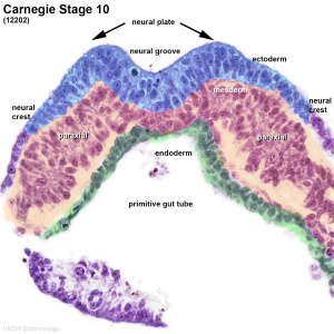
Human embryo (stage 10) mesoderm

Chicken-gastrulation2.jpg


Links: gastrulation

Patterning

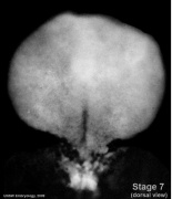
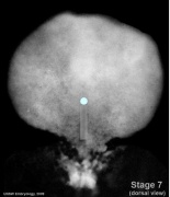
Notochord secreting sonic hedgehog, shown in white

Mesoderm cartoon.gif

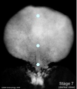
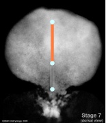
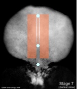
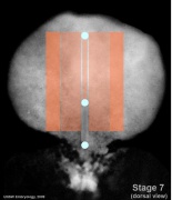
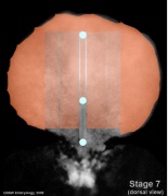
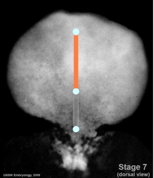
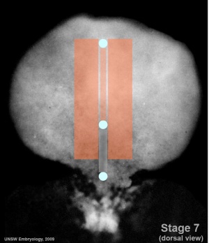
Mesoderm-cartoon1.jpgMesoderm-cartoon2.jpgMesoderm-cartoon3.jpgMesoderm-cartoon4.jpg


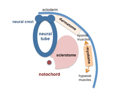
Somite cartoon5.png

Somite patterning

Embryonic Mesoderm

mesoderm - (hierarchical view)
  1. embryonic mesoderm
    1. axial
      1. notochord
        1. axial skeleton - nucleus pulpous
    2. paraxial
      1. unsegmented paraxial
      2. segmented paraxial somites
        1. sclerotome
          1. syndetome - tendon
          2. axial skeleton - vertebral body, intervertebral disc
        2. dermomyotome
          1. dermis - integumentary dermis, hypodermis
          2. myotome skeletal muscle
    3. intermediate
      1. renal
      2. genital
    4. lateral plate
      1. somatic
      2. intraembryonic coelom
        1. pericardial
        2. pleural
        3. peritoneal
      3. splanchnic mesoderm
  2. extra-embryonic mesoderm
    1. placental membranes
      1. amnion, chorion, yolk sac
    2. placental villi
      1. villi connective tissue, villi blood vessels
Germ Layers: ectoderm | mesoderm | endoderm

Mesoderm Regions (week 3)


Mesoderm-cartoon4.jpg

Axial Mesoderm

axial mesoderm
axial mesoderm

(notochord)

Paraxial Mesoderm

paraxial mesoderm
paraxial mesoderm

Intermediate Mesoderm

intermediate mesoderm

renal and genital

Somatic Mesoderm

Splanchnic Mesoderm

Extra-embryonic Mesoderm

The origin of extra-embryonic mesoderm (EEM) has been extensively discussed in the literature. This mesoderm lies outside the embryo, associated with fetal membrane and placenta development. This mesoderm formed at gastrulation along with the embryonic mesoderm from the proximal side of the primitive streak. A recent study in mouse, has shown that the primitive streak absence and excessive epiblast Nodal activity in pre-gastrulation stage, but not in the primitive streak cells during gastrulation, disrupts extraembryonic mesoderm development. [9]


Links: placenta | placental membranes

Molecular Factors

References

  1. Sanders TA, Llagostera E & Barna M. (2013). Specialized filopodia direct long-range transport of SHH during vertebrate tissue patterning. Nature , 497, 628-32. PMID: 23624372 DOI.
  2. Row RH, Pegg A, Kinney B, Farr GH, Maves L, Lowell S, Wilson V & Martin BL. (2018). BMP and FGF signaling interact to pattern mesoderm by controlling basic helix-loop-helix transcription factor activity. Elife , 7, . PMID: 29877796 DOI.
  3. Beisaw A, Tsaytler P, Koch F, Schmitz SU, Melissari MT, Senft AD, Wittler L, Pennimpede T, Macura K, Herrmann BG & Grote P. (2018). BRACHYURY directs histone acetylation to target loci during mesoderm development. EMBO Rep. , 19, 118-134. PMID: 29141987 DOI.
  4. Bazzi H, Soroka E, Alcorn HL & Anderson KV. (2017). STRIP1, a core component of STRIPAK complexes, is essential for normal mesoderm migration in the mouse embryo. Proc. Natl. Acad. Sci. U.S.A. , 114, E10928-E10936. PMID: 29203676 DOI.
  5. Koh PW, Sinha R, Barkal AA, Morganti RM, Chen A, Weissman IL, Ang LT, Kundaje A & Loh KM. (2016). An atlas of transcriptional, chromatin accessibility, and surface marker changes in human mesoderm development. Sci Data , 3, 160109. PMID: 27996962 DOI.
  6. Fleming BM, Yelin R, James RG & Schultheiss TM. (2013). A role for Vg1/Nodal signaling in specification of the intermediate mesoderm. Development , 140, 1819-29. PMID: 23533180 DOI.
  7. Aulehla A & Pourquié O. (2010). Signaling gradients during paraxial mesoderm development. Cold Spring Harb Perspect Biol , 2, a000869. PMID: 20182616 DOI.
  8. Shapiro IM & Risbud MV. (2010). Transcriptional profiling of the nucleus pulposus: say yes to notochord. Arthritis Res. Ther. , 12, 117. PMID: 20497604 DOI.
  9. Jin JZ, Zhu Y, Warner D & Ding J. (2016). Analysis of extraembryonic mesodermal structure formation in the absence of morphological primitive streak. Dev. Growth Differ. , 58, 522-9. PMID: 27273137 DOI.

Reviews

Risbud MV, Schaer TP & Shapiro IM. (2010). Toward an understanding of the role of notochordal cells in the adult intervertebral disc: from discord to accord. Dev. Dyn. , 239, 2141-8. PMID: 20568241 DOI.

Burke AC. (2007). Development and evolution of the vertebrate mesoderm. Dev. Dyn. , 236, 2369-70. PMID: 17705304 DOI.

Articles

Martin BL & Kimelman D. (2010). Brachyury establishes the embryonic mesodermal progenitor niche. Genes Dev. , 24, 2778-83. PMID: 21159819 DOI.

Korecki CL, Taboas JM, Tuan RS & Iatridis JC. (2010). Notochordal cell conditioned medium stimulates mesenchymal stem cell differentiation toward a young nucleus pulposus phenotype. Stem Cell Res Ther , 1, 18. PMID: 20565707 DOI.

Trainor PA, Tan SS & Tam PP. (1994). Cranial paraxial mesoderm: regionalisation of cell fate and impact on craniofacial development in mouse embryos. Development , 120, 2397-408. PMID: 7956820

Historic

Florian J. (1933). The Early Development of Man, with Special Reference to the Development of the Mesoderm and Cloacal Membrane. J. Anat. , 67, 263-76. PMID: 17104422

Search PubMed

Search NLM Online Textbooks: "Mesoderm" : Developmental Biology | The Cell- A molecular Approach | Molecular Biology of the Cell | Endocrinology


Search Pubmed: Mesoderm | Notochord

External Links

External Links Notice - The dynamic nature of the internet may mean that some of these listed links may no longer function. If the link no longer works search the web with the link text or name. Links to any external commercial sites are provided for information purposes only and should never be considered an endorsement. UNSW Embryology is provided as an educational resource with no clinical information or commercial affiliation.

Take the Quiz

1 Mesenchyme refers to the middle layer of the trilaminar embryo

true
false

2 The intraembryonic coelom forms within :

somites
lateral plate
neural tube
intermediate mesoderm

3 All paraxial mesoderm segments into somites.

true
false

4 Somites are developmental structures that contribute the following adult structures :

vertebra, notochord, dermis, skeletal muscle
vertebra, intervertebral discs, dermis, skeletal muscle
kidney, body wall connective tissue, sensory ganglia
kidney, gastrointestinal tract smooth muscle, mesentry


Glossary Links

Glossary: A | B | C | D | E | F | G | H | I | J | K | L | M | N | O | P | Q | R | S | T | U | V | W | X | Y | Z | Numbers | Symbols | Term Link

Cite this page: Hill, M.A. (2024, March 19) Embryology Mesoderm. Retrieved from https://embryology.med.unsw.edu.au/embryology/index.php/Mesoderm

What Links Here?
© Dr Mark Hill 2024, UNSW Embryology ISBN: 978 0 7334 2609 4 - UNSW CRICOS Provider Code No. 00098G