Menstrual Cycle - Histology: Difference between revisions
| Line 94: | Line 94: | ||
** Originally described by Gardner and Dukes (1955).<ref><pubmed>14361525</pubmed></ref> | ** Originally described by Gardner and Dukes (1955).<ref><pubmed>14361525</pubmed></ref> | ||
* clinical features - malodorous, thin homogeneous vaginal discharge with elevated vaginal pH above 4.5. | * clinical features - malodorous, thin homogeneous vaginal discharge with elevated vaginal pH above 4.5. | ||
<gallery> | |||
File:Bacteria - gram-stained vaginal smear 01.jpg|L. crispatus | |||
File:Bacteria - gram-stained vaginal smear 02.jpg|L. crispatus | |||
File:Bacteria - gram-stained vaginal smear 03.jpg|non-L. crispatus with thin lactobacilli | |||
File:Bacteria - gram-stained vaginal smear 04.jpg|non-L. crispatus with thin lactobacilli | |||
File:Bacteria - gram-stained vaginal smear 05.jpg|mixture non-L. crispatus with L. crispatus | |||
File:Bacteria - gram-stained vaginal smear 06.jpg|mixture non-L. crispatus with L. crispatus | |||
File:Bacteria - gram-stained vaginal smear 07.jpg|irregular-shaped Gram positive rod | |||
File:Bacteria - gram-stained vaginal smear 08.jpg|irregular-shaped Gram positive rod | |||
File:Bacteria - gram-stained vaginal smear 09.jpg|mixture Lactobacillus and bacterial vaginosis-associated | |||
File:Bacteria - gram-stained vaginal smear 10.jpg|mixture Lactobacillus and bacterial vaginosis-associated | |||
File:Bacteria - gram-stained vaginal smear 11.jpg|bacterial vaginosis | |||
File:Bacteria - gram-stained vaginal smear 12.jpg|bacterial vaginosis | |||
</gallery> | |||
{{Histology vaginal smear}} | |||
==References== | ==References== | ||
Revision as of 11:05, 19 November 2011
Introduction
This page presents images from vaginal smears and uterine endometrium dilatation and curettage[1] samples during different phases of the human menstrual cycle.
| Histology Links: stains | fixatives | artifacts | menstrual histology | placenta histology | heart histology | liver histology | Pancreas | Gall Bladder | Colon | Renal | Respiratory Histology | Bone | Category:Histology | UNSW Histology |
| Historic Histology Textbooks: 1941 Histology] | 1944 Oral Histology |
| Phase | Days (range) | Smear | Smear Description | Uterine Endometrium (D&C) |
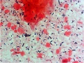
| Menstrual | 1 - 4 | Click on image to see full size. | Both stratum corneum (red) and stratum spinosum (blue) epithelial cells will mostly blood.
Leukocytes and bacteria may also be present. |

|
| Early Proliferative | 5 - 9 | 
|
Mainly large and small basophilic (blue) stratum spinosum cells. | |
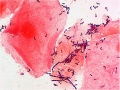
| Mid Proliferative | 9 - 13 | 
|
Stratum spinosum (blue) increase in size.
Dark precipate outside cells are bacteria. |

|
| Late Proliferative, Ovulatory | 13-14 | 
|
mainly stratum corneum (red) which are large and flat.
Appear due to high estrogen levels. |

|
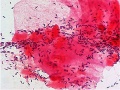
| Secretory | 15 - 22 | 
|
stratum spinosum cells (blue) which are folded or with curled edges.
Appear immediately after ovulation due to increase in progesterone. Leukocytes (small black cells) becoming more numerous. |

|
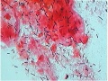
| Late Secretory, (Ischemic) Premenstrual | 23 - 28 | 
|
stratum spinosum cells (blue) mainly with a few stratum corneum cells (red).
Clustering of cells occurs at this stage. Both leukocytes and bacteria are prevelant. |

|
Human vaginal smear histology images in sequence: early proliferative | mid-proliferative | late proliferative | secretory | late secretory
Human Uterus (D and C histology images) in sequence: menstrual | mid-proliferative | late proliferative | secretory | late secretory
See also Uterus Development

Uterine body endometrium and myometrium during the proliferative phase of the menstrual cycle overview

Uterine body endometrium during the proliferative phase of the menstrual cycle

Uterine body endometrium during the secretory phase of the menstrual cycle overview

Uterine body endometrium during the secretory phase of the menstrual cycle


Abnormalities
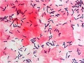
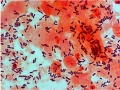
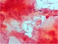
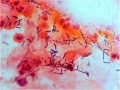
Bacterial Vaginosis
- The normal vaginal flora (lactobacillus morphotypes) is replaced by a mixed microbial flora consisting of Gardnerella vaginalis, Mycoplasma hominis and anaerobes.
- Originally described by Gardner and Dukes (1955).[2]
- clinical features - malodorous, thin homogeneous vaginal discharge with elevated vaginal pH above 4.5.
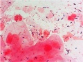
L. crispatus
L. crispatus
non-L. crispatus with thin lactobacilli
non-L. crispatus with thin lactobacilli
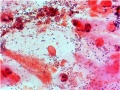
mixture non-L. crispatus with L. crispatus
mixture non-L. crispatus with L. crispatus
irregular-shaped Gram positive rod
irregular-shaped Gram positive rod
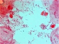
mixture Lactobacillus and bacterial vaginosis-associated
mixture Lactobacillus and bacterial vaginosis-associated
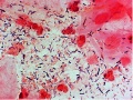
bacterial vaginosis
bacterial vaginosis


- Smear Image Links: L. crispatus | L. crispatus | non-L. crispatus with thin lactobacilli | non-L. crispatus with thin lactobacilli | mixture non-L. crispatus with L. crispatus | mixture non-L. crispatus with L. crispatus | irregular-shaped Gram positive rod | irregular-shaped Gram positive rod | mixture Lactobacillus and bacterial vaginosis-associated | mixture Lactobacillus and bacterial vaginosis-associated | bacterial vaginosis | bacterial vaginosis
- Links: Menstrual Cycle - Histology | Histology - Gram Stain | Bacterial Vaginosis | CDC (USA) Fact Sheet - Bacterial Vaginosis
References
Search Pubmed
Search Pubmed Now: Menstrual Cycle Histology | uterine histology | vaginal smear | pap smear |
External Links
External Links Notice - The dynamic nature of the internet may mean that some of these listed links may no longer function. If the link no longer works search the web with the link text or name. Links to any external commercial sites are provided for information purposes only and should never be considered an endorsement. UNSW Embryology is provided as an educational resource with no clinical information or commercial affiliation.
- Medline Plus - D and C - series | D and C video
Glossary Links
- Glossary: A | B | C | D | E | F | G | H | I | J | K | L | M | N | O | P | Q | R | S | T | U | V | W | X | Y | Z | Numbers | Symbols | Term Link
Cite this page: Hill, M.A. (2024, April 26) Embryology Menstrual Cycle - Histology. Retrieved from https://embryology.med.unsw.edu.au/embryology/index.php/Menstrual_Cycle_-_Histology
- © Dr Mark Hill 2024, UNSW Embryology ISBN: 978 0 7334 2609 4 - UNSW CRICOS Provider Code No. 00098G