Gastrointestinal Tract Development: Difference between revisions
mNo edit summary |
mNo edit summary |
||
| Line 2: | Line 2: | ||
== Introduction == | == Introduction == | ||
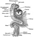
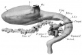
[[File:Gray0982a.jpg|thumb|300px|The early gastrointestinal tract.]] | [[File:Gray0982a.jpg|thumb|300px|The early gastrointestinal tract.]] | ||
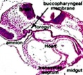
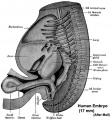
[[File: | [[File:Stage11_sem4.jpg|thumb|300px|Human head ([[Week 4]], [[Carnegie stage 11|Stage 11]]) showing buccopharyngeal membrane breakdown.]] | ||
The gastrointestinal tract (GIT) arises initially during the process of gastrulation from the [[E#endoderm|endoderm]] of the trilaminar embryo (week 3) and extends from the [[B#buccopharyngeal membrane|buccopharyngeal membrane]] to the [[C#cloacal membrane|cloacal membrane]]. The tract and associated organs later have contributions from all the germ cell layers. | The gastrointestinal tract (GIT) arises initially during the process of gastrulation from the [[E#endoderm|endoderm]] of the trilaminar embryo (week 3) and extends from the [[B#buccopharyngeal membrane|buccopharyngeal membrane]] to the [[C#cloacal membrane|cloacal membrane]]. The tract and associated organs later have contributions from all the germ cell layers. | ||
Revision as of 14:08, 22 April 2014
| Embryology - 4 May 2024 |
|---|
| Google Translate - select your language from the list shown below (this will open a new external page) |
|
العربية | català | 中文 | 中國傳統的 | français | Deutsche | עִברִית | हिंदी | bahasa Indonesia | italiano | 日本語 | 한국어 | မြန်မာ | Pilipino | Polskie | português | ਪੰਜਾਬੀ ਦੇ | Română | русский | Español | Swahili | Svensk | ไทย | Türkçe | اردو | ייִדיש | Tiếng Việt These external translations are automated and may not be accurate. (More? About Translations) |
Introduction


The gastrointestinal tract (GIT) arises initially during the process of gastrulation from the endoderm of the trilaminar embryo (week 3) and extends from the buccopharyngeal membrane to the cloacal membrane. The tract and associated organs later have contributions from all the germ cell layers.
During the 4th week three distinct regions (fore-, mid- and hind-gut) extend the length of the embryo and will contribute different components of the GIT. The large mid-gut is generated by lateral embryonic folding which "pinches off" a pocket of the yolk sac, the 2 compartments continue to communicate through the vitelline duct.
The oral cavity (mouth) is formed following breakdown of the buccopharyngeal membrane (oropharyngeal or oral membrane) and contributed to mainly by the pharynx lying within the pharyngeal arches (More? Head Development). Loss of buccopharyngeal membrane opens the tract to amniotic fluid through the remainder of development, and during the fetal period is actively swallowed.
From the oral cavity the next portion of the foregut is initially the pharynx, a single gastrointestinal (oesophagus) and respiratory (trachea) common tube, that lies behind the heart. Note that the respiratory tract will form from a ventral bud arising at this level (More? Respiratory).
This current page provides an introductory overview, use the links below for descriptions of specific components and regions as well as developmental abnormalities.
Note that in historic texts the term entoderm is used to describe endoderm and other terminology may also differ from current descriptions.
Some Recent Findings

|
| More recent papers |
|---|
 This table allows an automated computer search of the external PubMed database using the listed "Search term" text link.
More? References | Discussion Page | Journal Searches | 2019 References | 2020 References Search term: Gastrointestinal Tract Embryology <pubmed limit=5>Gastrointestinal Tract Embryology</pubmed> |
Textbooks
- Human Embryology Larson Chapter 9 p229-260
- The Developing Human: Clinically Oriented Embryology (6th ed.) Moore and Persaud Chapter 12 p271-302
- Before We Are Born (5th ed.) Moore and Persaud Chapter 13 p255-287
- Essentials of Human Embryology Larson Chapter 9 p123-146
- Human Embryology Fitzgerald and Fitzgerald Chapter 19,20 p119-123
More? References | Online Textbooks | Historic Textbooks
| UNSW Students | |
|---|---|
| You have access the following online Embryology textbooks through the UNSW Library. | |

|
Moore, K.L. & Persuad, T.V.N. (2008). The Developing Human: clinically oriented embryology (8th ed.). Philadelphia: Saunders.
|

|
Schoenwolf, G.C., Bleyl, S.B., Brauer, P.R. and Francis-West, P.H. (2009). Larsen’s Human Embryology (4th ed.). New York; Edinburgh: Churchill Livingstone.
|
Objectives
- Understanding of germ layer contributions to the early gastrointestinal tract (GIT)
- Understanding of the folding of the GIT
- Understanding of three main GIT embryonic divisions
- Understanding of associated organ development (liver, pancreas, spleen)
- Brief understanding of mechanical changes (rotations) during GIT development
- Brief understanding of gastrointestinal abnormalities
Germ Layer Contributions
- Endoderm - epithelium and associated glands
- Mesoderm (splanchnic) - mesentry, connective tissues, smooth muscle, blood vessels
- Ectoderm (neural crest) - enteric nervous system (neural tube) - extrinsic innervation
Both endoderm and mesoderm will contribute to associated organs.
Gastrointestinal Tract Movies
| Gastrointestinal Tract Movies | |||||||||||||||||||
|---|---|---|---|---|---|---|---|---|---|---|---|---|---|---|---|---|---|---|---|
|
|
|
|
| |||||||||||||||
|
|
|
|
| |||||||||||||||
|
|
|
|
| |||||||||||||||
| Stage 13 (week 5) | Stage 22 (week 8) | Stage 23 (week 8) | GIT Abnormalities Ultrasound | ||||||||||||||||

3 GIT divisions

During the 4th week the 3 distinct portions (fore-, mid- and hind-gut) extend the length of the embryo and will contribute different components of the GIT. These 3 divisions are also later defined by the vascular (artery) supply to each of theses divisions.
The large mid-gut is generated by lateral embryonic folding which "pinches off" a pocket of the yolk sac, the 2 compartments continue to communicate through the vitelline duct.
The oral cavity (mouth) is formed following breakdown of the buccopharyngeal membrane (oropharyngeal, oral membrane) and contributed to mainly by the pharynx lying within the pharyngeal arches. The opening of the GIT means that it contains amniotic fluid, which is also swallowed later in development.
Foregut

From the oral cavity the next portion of the foregut is initially a single gastrointestinal (oesophagus) and respiratory (trachea) common tube, the pharynx which lies behind the heart. Note that the respiratory tract will form from a ventral bud arising at this level.
- Oral cavity
- Pharynx (esophagus, trachea)
- Respiratory tract
- Stomach
Midgut

From beneath the stomach the initial portion of the small intestine, the duodenum, and the associated pancreas now lie.
Much of the midgut is herniated at the umbilicus external to the abdomen through development. A key step in development is the rotation of this midgut that must occur to place the GIT in the correct abdominal position with its associated mesentry. The GIT itself differentiates to form significantly different structures along its length: oesophagus, stomach, duodenum, jejunum, iliem (small intestine), colon (large intestine).
The mesentries of the GIT are generated from the common dorsal mesentry, with the ventral mesentry contributing to the lesser omentum and falciform ligament.
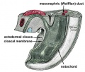
Hindgut
The distral transverse colon, descending colon, sigmoid colon, rectum and cloaca. The cloaca is the common urogenital sinus which will later become partitioned into an anterior urinary and posterior GIT rectal component.
- Links: Intestine Development
Development Overview
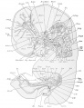
GIT shown in green anchored by dosal and ventral mesogastrium. The space ouside this will be the peritoneal cavity.
Red ring-neural tube with neural crestBlue ring- notocordOrange- somites
Differentiation of associated organs at the level of the forming stomach occurs both dorsally (spleen) and ventrally (liver).
Large blue ring- dorsal aortaDark green ring- Liver
Continued growth of the GIT and the organs leads to organ movements and bending of tract.
Carnegie stage 13 Embryo Overview
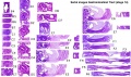
Below is an overview of the sections starting at the level of pharynx compressed dorsoventrally, following the GIT through to the rectum. The most obvious feature is that of a continuous tube initially, attached by dorsoventral mesentry. Outside this tube and mesentry (at the levels below the lung buds) is the intraembryonic coelom that will form the peritoneal cavity. The hepatic diverticulum (liver bud) lies under the septum transversum is the earliest associated GIT organ that has differentiated, and now occupies a substantial region of the abdomen. Clicking on sections below will open the original images.

| |||
| Bifurcation of the pharynx into anterior respiratory and posterior oesophagous. | The stomach forming beneath the lung buds and adjacent to the developing liver. | Below the stomach the GIT has a large dorsal mesogastrium and finer ventral mesogastrium. Associated with the tract is the large portal blood vessel derived from the vitelline circulation. | At the bottom curvature of the embryo the mesentry association with the GIT shows extensive vitelline vessels running out through the umbilicus. The hindgut can then be seen, ending at the common urogenital sinus, the cloaca. |
Innervation

Neural History
- 1857 Meissner was the first to describe a nerve plexus in the submucosa of the bowel wall.
- 1864 Auerbach described the myenteric plexus between the longitudinal and circular muscle layers.
- 1981 LeDouarin describes neural crest contribution to both plexuses.
Myenteric Plexus
- Peristalsis
- Coordinated waves of descending inhibition followed by waves of descending excitation
+ Extrinsic parasympathetic cholinergic nerves (vagal and sacral) excite peristalsis and stimulate
- Sympathetic noradrenergic nerves inhibit the transit of gut contents
Submucosal Plexus
- epithelial movements
- secretion and absorption
Associated Organs

The early tract develops as a simple tube, then a number of endodermal outgrowths from this tube at different levels and contribute to a range of additional organs and tissues. The gastrointestinal associated organs liver, gall bladder and pancreas. Development of these organs is described on separate pages.
There are also a number of additional non-gastrointestinal structures including the respiratory tract and development within the mesentery such as the spleen.
Gastrointestinal Tract Abnormalities
Only a brief description is given on this current page, for more details see Gastrointestinal Tract - Abnormalities.
Lumen Abnormalities
There are several types of abnormalities that impact upon the continuity of the gastrointestinal tract lumen.
- Atresia - interuption of the lumen (esophageal atresia, duodenal atresia, extrahepatic biliary atresia, anorectal atresia)
- Stenosis - narrowing of the lumen (duodenal stenosis, pyloric stenosis).
- Duplication - incomplete recanalization resulting in parallel lumens, this is really a specialized form of stenosis.
Meckel's Diverticulum
This GIT abnormality is a very common and results from improper closure and absorption of the omphalomesenteric duct (vitelline duct) in development. This transient developmental duct connects the yolk to the primitive GIT.
Intestinal Malrotation

- Links: Intestinal Malrotation
Intestinal Aganglionosis
(intestinal aganglionosis, Hirschsprung's disease, aganglionic colon, megacolon, congenital aganglionic megacolon, congenital megacolon) A condition caused by the lack of enteric nervous system (neural ganglia) in the intestinal tract responsible for gastric motility (peristalsis).
Gastroschisis
Gastroschisis (omphalocele, paraomphalocele, laparoschisis, abdominoschisis, abdominal hernia) is a congenital abdominal wall defect which results in herniation of fetal abdominal viscera (intestines and/or organs) into the amniotic cavity. Incidence of gastroschisis has been reported at 1.66/10,000, occuring more frequently in young mothers (less than 20 years old).
By definition, it is a body wall musculoskeletal defect, not a gastrointestinal tract defect, which in turn impacts upon GIT development.
Molecular
The endoderm of the developing gastrointestinal tract is a source for patterning signals for both within the tract and also for the surrounding organs and tissues.
- Sox2 - expressed in the anterior part of the primitive gut[3]
- Cdx2 - expressed in the posterior part of the primitive gut[3]
- GDNF - regulate migration of enteric neural crest cells[4]
- endothelin - regulate migration of enteric neural crest cells[4]
References
Online Textbooks
- Developmental Biology (6th ed) Gilbert, Scott F. Sunderland (MA): Sinauer Associates, Inc.; c2000. The Digestive Tube and Its Derivatives | Endodermal development of a human embryo
- The Gastrointestinal Circulation Peter R. Kvietys. San Rafael (CA): Morgan & Claypool Publishers; 2010. Table of Contents
- Motor Function of the Pharynx, Esophagus, and its Sphincters. Mittal RK. San Rafael (CA): Morgan & Claypool Life Sciences; 2011. Table of Contents
- Search NLM Online Textbooks "gastrointestinal tract" : Developmental Biology | Endocrinology | Molecular Biology of the Cell | The Cell- A molecular Approach
Historic Textbooks
- The Elements of Embryology by Foster, M., Balfour, F. M., Sedgwick, A., & Heape, W. (1883) The Alimentary Canal and its Appendages
- Text-Book of the Embryology of Man and Mammals by Dr Oscar Hertwig (1892) The Organs of the Inner Germ-Layer The Alimentary Tube with its Appended Organs
- Atlas of the Development of Man Volume 2 by Julius Kollmann (1907) Gastrointestinal
- Text-Book of Embryology by Bailey, F.R. and Miller, A.M. (1921) Alimentary tube and organs
| Historic Disclaimer - information about historic embryology pages |
|---|
|
Reviews
<pubmed>19782301</pubmed> <pubmed>19708022</pubmed> <pubmed>19303014</pubmed> <pubmed>18416699</pubmed> <pubmed>17268237</pubmed> <pubmed>17284768</pubmed> <pubmed>17076282</pubmed> <pubmed>14647040</pubmed> <pubmed>12943221</pubmed> <pubmed>3922287</pubmed>
Articles
<pubmed>16284122</pubmed>
Search PubMed
Search Mar 2007 "gastrointestinal tract development" 29,361 reference articles of which 3,494 were reviews.
Search April 2010 "Gastrointestinal Tract Development" - All (35980) Review (4707) Free Full Text (8086)
Search Pubmed: Gastrointestinal Tract Development
Additional Images


Historic image showing midgut herniation
1914 Thyng
1914 Thyng Plate 2


| System Links: Introduction | Cardiovascular | Coelomic Cavity | Endocrine | Gastrointestinal Tract | Genital | Head | Immune | Integumentary | Musculoskeletal | Neural | Neural Crest | Placenta | Renal | Respiratory | Sensory | Birth |
Terms
- allantois - An extraembryonic membrane, endoderm in origin extension from the early hindgut, then cloaca into the connecting stalk of placental animals, connected to the superior end of developing bladder. In reptiles and birds, acts as a reservoir for wastes and mediates gas exchange. In mammals is associated/incorporated with connecting stalk/placental cord fetal-maternal interface.
- amnion - An extraembryonic membrane]ectoderm and extraembryonic mesoderm in origin and forms the innermost fetal membrane, produces amniotic fluid. This fluid-filled sac initially lies above the trilaminar embryonic disc and with embryoic disc folding this sac is drawn ventrally to enclose (cover) the entire embryo, then fetus. The presence of this membane led to the description of reptiles, bird, and mammals as amniotes.
- amniotic fluid - The fluid that fills amniotic cavity totally encloses and cushions the embryo. Amniotic fluid enters both the gastrointestinal and respiratory tract following rupture of the buccopharyngeal membrane. The late fetus swallows amniotic fluid.
- buccal - (Latin, bucca = cheek) A term used to relate to the mouth (oral cavity).
- buccopharyngeal membrane - (oral membrane) (Latin, bucca = cheek) A membrane which forms the external upper membrane limit (cranial end) of the early gastrointestinal tract (GIT). This membrane develops during gastrulation by ectoderm and endoderm without a middle (intervening) layer of mesoderm. The membrane lies at the floor of the ventral depression (stomadeum) where the oral cavity will open and will breakdown to form the initial "oral opening" of the gastrointestinal tract. The equivilent membrane at the lower end of the gastrointestinal tract is the cloacal membrane.
- cloacal membrane - Forms the external lower membrane limit (caudal end) of the early gastrointestinal tract (GIT). This membrane is formed during gastrulation by ectoderm and endoderm without a middle (intervening) layer of mesoderm. The membrane breaks down to form the initial "anal opening" of the gastrointestinal tract.
- coelom - Term used to describe a space. There are extraembryonic and intraembryonic coeloms that form during vertebrate development. The single intraembryonic coelom will form the 3 major body cavities: pleural, pericardial and peritoneal.
- foregut - The first of the three part/division (foregut - midgut - hindgut) of the early forming gastrointestinal tract. The foregut runs from the buccopharyngeal membrane to the midgut and forms all the tract (esophagus and stomach) from the oral cavity to beneath the stomach. In addition, a ventral bifurcation of the foregut will also form the respiratory tract epithelium.
- gastrula - (Greek, gastrula = little stomach) A stage of an animal embryo in which the three germ layers ([E#endoderm|endoderm]/mesoderm/ectoderm) have just formed.
- gastrulation - The process of differentiation forming a gastrula. Term means literally means "to form a gut" but is more in development, as this process converts the bilaminar embryo (epiblast/hypoblast) into the trilaminar embryo ([E#endoderm endoderm]/mesoderm/ectoderm) establishing the 3 germ layers that will form all the future tissues of the entire embryo. This process also establishes the the initial body axes.
- hindgut - The last of the three part/division foregut - midgut - hindgut) of the early forming gastrointestinal tract. The hindgut forms all the tract from the distral transverse colon to the cloacal membrane and extends into the connecting stalk (placental cord) as the allantois. In addition, a ventral of the hindgut will also form the urinary tract (bladder, urethra) epithelium.
- intraembryonic coelom - The "horseshoe-shaped" space (cavity) that forms initially in the third week of development in the lateral plate mesoderm that will eventually form the 3 main body cavities: pericardial, pleural, peritoneal. The intraembryonic coelom communicates transiently with the extraembryonic coelom.
- neuralation - The general term used to describe the early formation of the nervous system. It is often used to describe the early events of differentiation of the central ectoderm region to form the neural plate, then neural groove, then neural tube. The nervous system includes the central nervous system (brain and spinal cord) from the neural tube and the peripheral nervous system (peripheral sensory and sympathetic ganglia) from neural crest. In humans, early neuralation begins in week 3 and continues through week 4.
- neural crest - region of cells at the edge of the neural plate that migrates throughout the embryo and contributes to many different tissues. In the gastrointestinal tract it contributes mainly the enteric nervous system within the wall of the gut responsible for peristalsis and secretion.
- pharynx - uppermost end of gastrointestinal and respiratory tract, in the embryo beginning at the buccopharyngeal membrane and forms a major arched cavity within the phrayngeal arches.
- somitogenesis The process of segmentation of the paraxial mesoderm within the trilaminar embryo body to form pairs of somites, or balls of mesoderm. A somite is added either side of the notochord (axial mesoderm) to form a somite pair. The segmentation does not occur in the head region, and begins cranially (head end) and extends caudally (tailward) adding a somite pair at regular time intervals. The process is sequential and therefore used to stage the age of many different species embryos based upon the number visible somite pairs. In humans, the first somite pair appears at day 20 and adds caudally at 1 somite pair/90 minutes until on average 44 pairs eventually form.
- splanchnic mesoderm - Gastrointestinal tract (endoderm) associated mesoderm formed by the separation of the lateral plate mesoderm into two separate components by a cavity, the intraembryonic coelom. Splanchnic mesoderm is the embryonic origin of the gastrointestinal tract connective tissue, smooth muscle, blood vessels and contribute to organ development (pancreas, spleen, liver). The intraembryonic coelom will form the three major body cavities including the space surrounding the gut, the peritoneal cavity. The other half of the lateral plate mesoderm (somatic mesoderm) is associated with the ectoderm of the body wall.
- stomadeum - (stomadeum) A ventral surface depression on the early embryo head surrounding the buccopharyngeal membrane, which lies at the floor of this depression. This surface depression lies between the maxillary and mandibular components of the first pharyngeal arch.
Glossary Links
- Glossary: A | B | C | D | E | F | G | H | I | J | K | L | M | N | O | P | Q | R | S | T | U | V | W | X | Y | Z | Numbers | Symbols | Term Link
Cite this page: Hill, M.A. (2024, May 4) Embryology Gastrointestinal Tract Development. Retrieved from https://embryology.med.unsw.edu.au/embryology/index.php/Gastrointestinal_Tract_Development
- © Dr Mark Hill 2024, UNSW Embryology ISBN: 978 0 7334 2609 4 - UNSW CRICOS Provider Code No. 00098G