File:Stage11 sem101.jpg

Original file (1,000 × 898 pixels, file size: 175 KB, MIME type: image/jpeg)
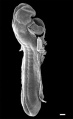
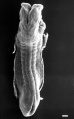
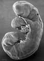
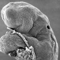
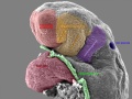
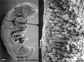
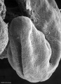
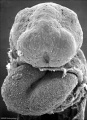
Human Embryo Carnegie stage 11
Week 4 Carnegie stage 11 25 days, 19 somite pairs. This is a scanning EM of the embryo fractured to show the neural tube, notochord and somites.
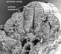
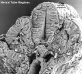
Neural tube level of the body (note somites are present) showing the location of future tube regions.
Neural tube regions
- roof plate - dorsal region
- alar plate - dorsal lateral region (sensory input fine touch, proprioception, and vibration)
- basal plate - ventral lateral region (motor output to skeletal muscle)
- floor plate - ventral region
Regional patterning
- roof plate region - signals from surface ectoderm
- alar plate region - signals from surface ectoderm
- basal plate region - signals from the notochord and floor plate
- floor plate region - signals from the notochord
Neural tube level of the head contains similar regions that differentiate to form other brain structures.
- Links: unlabeled version | labeled version | labeled neural
- Stage 11 SEM Images: dorsolateral whole embryo | dorsal embryo | lateral embryo | lateral head | lateral head with overlay | embryo cross-section | ventrolateral head | ventrolateral head with overlay | ventral head | buccopharyngeal membrane | neural crest | posterior neuropore | anterior neuropore | Carnegie stage 11
- Human Embryo (stage 11)
dorsolateral whole embryo
dorsal embryo
lateral embryo
lateral head
lateral head with overlay

embryo cross-section
embryo cross-section label
neural cross-section label

ventrolateral head
ventrolateral head with overlay

ventral head

buccopharyngeal membrane
neural crest
posterior neuropore
anterior neuropore


Image Source: Scanning electron micrographs of the Carnegie stages of the early human embryos are reproduced with the permission of Prof Kathy Sulik, from embryos collected by Dr. Vekemans and Tania Attié-Bitach. Images are for educational purposes only and cannot be reproduced electronically or in writing without permission.
Stage 22 Spinal Cord Links: image unlabeled | image labeled | image low resolution | ANAT3411 | spinal cord | meninges
| Virtual Slide Features - Stage 22 Spinal Cord | ||
|---|---|---|
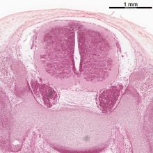
Spinal Cord Features |
The links shown are to specific features shown on the Human embryo (stage 22) Spinal Cord virtual slide. See also notes on Spinal Cord Development
Clicking the text will open the slide at a detailed view with the structure generally located in the centre of the view. The slide then can also be zoomed out from the set magnification using the controls in the upper left or the mouse. Use your browser back button to return to this table. Other Features
| |
| All Virtual Slides | Making your own Link - You can also make your own selected feature view. (See also Permalink help)
|
|
| Week: | 1 | 2 | 3 | 4 | 5 | 6 | 7 | 8 |
| Carnegie stage: | 1 2 3 4 | 5 6 | 7 8 9 | 10 11 12 13 | 14 15 | 16 17 | 18 19 | 20 21 22 23 |
Image version links: Large 1000px | 800px | Medium 600px | Small 400px
Cite this page: Hill, M.A. (2024, April 27) Embryology Stage11 sem101.jpg. Retrieved from https://embryology.med.unsw.edu.au/embryology/index.php/File:Stage11_sem101.jpg
- © Dr Mark Hill 2024, UNSW Embryology ISBN: 978 0 7334 2609 4 - UNSW CRICOS Provider Code No. 00098G
File history
Click on a date/time to view the file as it appeared at that time.
| Date/Time | Thumbnail | Dimensions | User | Comment | |
|---|---|---|---|---|---|
| current | 18:27, 26 May 2017 | | 1,000 × 898 (175 KB) | Z8600021 (talk | contribs) | |
| 18:03, 26 May 2017 |  | 1,000 × 898 (173 KB) | Z8600021 (talk | contribs) | ||
| 17:58, 26 May 2017 |  | 1,000 × 898 (171 KB) | Z8600021 (talk | contribs) |
You cannot overwrite this file.
File usage
The following 46 pages use this file:
- BGDA Lecture - Development of the Nervous System
- Human Embryo SEM
- File:Stage11 sem10.jpg
- File:Stage11 sem100.jpg
- File:Stage11 sem100a.jpg
- File:Stage11 sem100b.jpg
- File:Stage11 sem100c.jpg
- File:Stage11 sem101.jpg
- File:Stage11 sem10a.jpg
- File:Stage11 sem10b.jpg
- File:Stage11 sem10c.jpg
- File:Stage11 sem13.jpg
- File:Stage11 sem13a.jpg
- File:Stage11 sem13b.jpg
- File:Stage11 sem13c.jpg
- File:Stage11 sem2.jpg
- File:Stage11 sem21.jpg
- File:Stage11 sem2a.jpg
- File:Stage11 sem2b.jpg
- File:Stage11 sem2c.jpg
- File:Stage11 sem3.jpg
- File:Stage11 sem3a.jpg
- File:Stage11 sem3b.gif
- File:Stage11 sem3b.jpg
- File:Stage11 sem3c.jpg
- File:Stage11 sem4.jpg
- File:Stage11 sem4a.jpg
- File:Stage11 sem4b.jpg
- File:Stage11 sem4c.jpg
- File:Stage11 sem5.jpg
- File:Stage11 sem5a.jpg
- File:Stage11 sem5b.jpg
- File:Stage11 sem5c.jpg
- File:Stage11 sem6.jpg
- File:Stage11 sem7.jpg
- File:Stage11 sem7a.jpg
- File:Stage11 sem7b.jpg
- File:Stage11 sem8.jpg
- File:Stage11 sem81.jpg
- File:Stage11 sem82.jpg
- File:Stage11 sem8a.jpg
- File:Stage11 sem8b.jpg
- File:Stage11 sem9.jpg
- File:Stage11 sem9a.jpg
- File:Stage11 sem9b.jpg
- Template:Stage11SEM