File:Keibel Mall 220.jpg

Original file (646 × 1,000 pixels, file size: 93 KB, MIME type: image/jpeg)
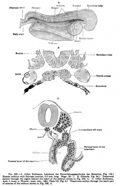
Fig. 220 Human Embryo Week 4
At a slightly later period the sheet of mesoderm on each side of the neural tube becomes longitudinally separated from the more laterally situated mesoderm (Fig. 220, A and C) and at the same time divided into a series of segments (mesoblastic somites). In the chick the first somites formed are the occipital somites (J. T. Patterson, Biological Bulletin, 1907, vol. xiii, p. 121), then follow in turn the cervical, thoracic, lumbar, sacral, and coccygeal. It is probable that the first somites formed in the human embryo belong to the occipital region. In the latter half of the first month of development in the human embryo there are found anterior to the cersdcal myotomes three incomplete occipital myotomes. The relations of these myotomes to the first somites differentiated have not yet been definitely determined. Etemod (Anat. Anz., 1899, p. 131) has described an embryo with eight somites, Kollmann (Anat. Anz., 1890, Arch. f. Anat. u. Physiol., Anat. Abt., 1891) one with fourteen (Fig. 220, A), and Mall (Journ. Morphol., 1897) likewise one with fourteen. Mall considers the first three somites in his embryo to be occipital somites. They probably correspond with the first three somites in the embryo described by Kollmann and possibly with the first three in the embryo described by Eternod.
| Week: | 1 | 2 | 3 | 4 | 5 | 6 | 7 | 8 |
| Carnegie stage: | 1 2 3 4 | 5 6 | 7 8 9 | 10 11 12 13 | 14 15 | 16 17 | 18 19 | 20 21 22 23 |
- KM Figure Links: The Germ Cells | Segmentation | First Primitive Segment | Gastrulation | External Form | Placenta | Axial Skeleton | Limb Skeleton | Skull | Muscular System
| Historic Disclaimer - information about historic embryology pages |
|---|
| Pages where the terms "Historic" (textbooks, papers, people, recommendations) appear on this site, and sections within pages where this disclaimer appears, indicate that the content and scientific understanding are specific to the time of publication. This means that while some scientific descriptions are still accurate, the terminology and interpretation of the developmental mechanisms reflect the understanding at the time of original publication and those of the preceding periods, these terms, interpretations and recommendations may not reflect our current scientific understanding. (More? Embryology History | Historic Embryology Papers) |
Glossary Links
- Glossary: A | B | C | D | E | F | G | H | I | J | K | L | M | N | O | P | Q | R | S | T | U | V | W | X | Y | Z | Numbers | Symbols | Term Link
Cite this page: Hill, M.A. (2024, April 27) Embryology Keibel Mall 220.jpg. Retrieved from https://embryology.med.unsw.edu.au/embryology/index.php/File:Keibel_Mall_220.jpg
- © Dr Mark Hill 2024, UNSW Embryology ISBN: 978 0 7334 2609 4 - UNSW CRICOS Provider Code No. 00098G
File history
Click on a date/time to view the file as it appeared at that time.
| Date/Time | Thumbnail | Dimensions | User | Comment | |
|---|---|---|---|---|---|
| current | 08:15, 24 August 2012 |  | 646 × 1,000 (93 KB) | Z8600021 (talk | contribs) |
You cannot overwrite this file.
File usage
The following 2 pages use this file:
