File:Human heart developmental functional networks.jpg

Original file (833 × 614 pixels, file size: 424 KB, MIME type: image/jpeg)
Human Heart Development Functional Networks
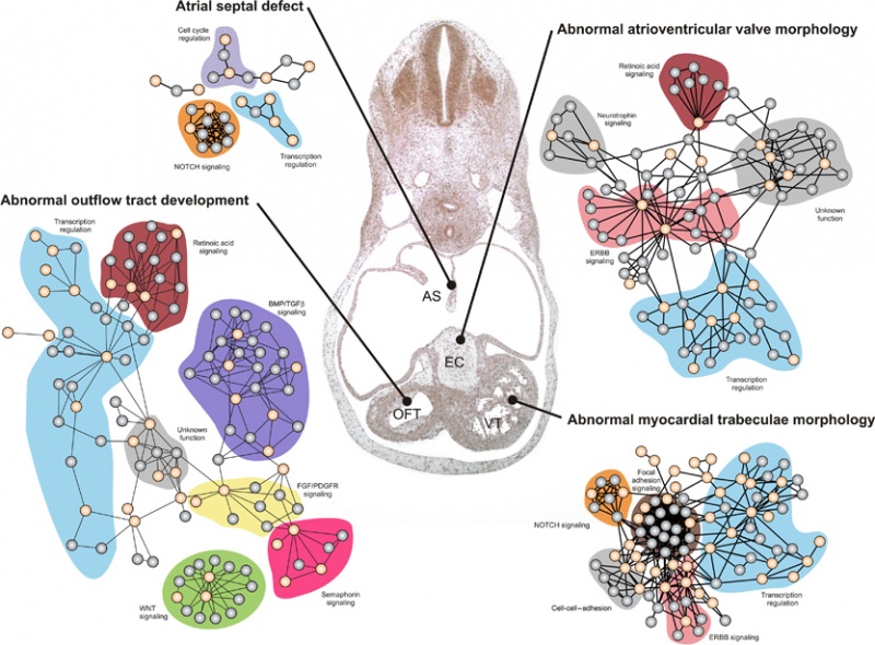
Examples of four functional networks driving the development of different anatomical structures in the human heart.
These four networks constructed by analyzing the interaction patterns of four different sets of cardiac development (CD) proteins corresponding to the morphological groups ‘atrial septal defects,’ ‘abnormal atrioventricular valve morphology,’ ‘abnormal myocardial trabeculae morphology,’ and ‘abnormal outflow tract development’ (Supplementary Table S1).
- CD proteins from the relevant groups are shown in orange and their interaction partners are shown in gray.
- Functional modules annotated by literature curation are indicated with a colored background.
- High-resolution figures (including protein names) can be seen in Supplementary Figures S2A, S2C, S2D, and S3B, respectively.
- Centrally in the figure is a haematoxylin-eosin stained frontal section of the heart from a 37-day human embryo.
Tissues affected by the four networks are marked;
- AS - developing atrial septum
- EC - endocardial cushions, which are anatomical precursors to the atrioventricular valves
- VT - developing ventricular trabeculae
- OFT - developing outflow tract (mislabeled OTF)
The entire set of 19 networks is shown in detail in Supplementary Figures S1, S2, S3 and S4, and can be downloaded from http://www.cbs.dtu.dk/suppl/dgf/.
Reference
Lage K, Møllgård K, Greenway S, Wakimoto H, Gorham JM, Workman CT, Bendsen E, Hansen NT, Rigina O, Roque FS, Wiese C, Christoffels VM, Roberts AE, Smoot LB, Pu WT, Donahoe PK, Tommerup N, Brunak S, Seidman CE, Seidman JG & Larsen LA. (2010). Dissecting spatio-temporal protein networks driving human heart development and related disorders. Mol. Syst. Biol. , 6, 381. PMID: 20571530 DOI.
Copyright
This is an open-access article distributed under the terms of the Creative Commons Attribution Noncommercial Share Alike 3.0 Unported License, which allows readers to alter, transform, or build upon the article and then distribute the resulting work under the same or similar license to this one. The work must be attributed back to the original author and commercial use is not permitted without specific permission.
Original file name: Msb201036-f1.jpg
Cite this page: Hill, M.A. (2024, April 27) Embryology Human heart developmental functional networks.jpg. Retrieved from https://embryology.med.unsw.edu.au/embryology/index.php/File:Human_heart_developmental_functional_networks.jpg
- © Dr Mark Hill 2024, UNSW Embryology ISBN: 978 0 7334 2609 4 - UNSW CRICOS Provider Code No. 00098G
File history
Click on a date/time to view the file as it appeared at that time.
| Date/Time | Thumbnail | Dimensions | User | Comment | |
|---|---|---|---|---|---|
| current | 14:12, 15 August 2011 |  | 833 × 614 (424 KB) | S8600021 (talk | contribs) | ==Human Heart Development Functional Networks== Examples of four functional networks driving the development of different anatomical structures in the human heart. These four networks constructed by analyzing the interaction patterns of four different |
You cannot overwrite this file.
File usage
The following page uses this file: