File:Gray0458.jpg: Difference between revisions
From Embryology
(==Diagram of the vascular channels in a human embryo of the second week== (After Eternod.) * The red lines are the dorsal aortæ continued into the umbilical arteries. * The red dotted lines are the ventral aortæ, and the blue dotted lines the vit...) |
mNo edit summary |
||
| (3 intermediate revisions by the same user not shown) | |||
| Line 1: | Line 1: | ||
==Diagram of the vascular channels in a human embryo of the second week== | ==Fig. 458. Diagram of the vascular channels in a human embryo of the second week== | ||
(After Eternod.) | (After Eternod.) | ||
* | * '''red lines''' - are the dorsal aortae continued into the umbilical arteries. | ||
* | * '''red dotted lines''' - are the ventral aortae. | ||
* '''blue dotted lines''' - are the vitelline veins. | |||
{{Heart Links}} | |||
{{Gray Cardiovascular}} | |||
{{Gray Anatomy}} | {{Gray Anatomy}} | ||
Latest revision as of 12:49, 4 March 2015
Fig. 458. Diagram of the vascular channels in a human embryo of the second week
(After Eternod.)
- red lines - are the dorsal aortae continued into the umbilical arteries.
- red dotted lines - are the ventral aortae.
- blue dotted lines - are the vitelline veins.
- Gray - Cardiovascular: 448 Artery and vein | 458 Human embryo week 2 vascular | 459 Human embryo 14 days with yolk-sac
- Gray's Images: Development | Lymphatic | Neural | Vision | Hearing | Somatosensory | Integumentary | Respiratory | Gastrointestinal | Urogenital | Endocrine | Surface Anatomy | iBook | Historic Disclaimer
| Historic Disclaimer - information about historic embryology pages |
|---|
 |
| iBook - Gray's Embryology | |
|---|---|

|
|
Reference
Gray H. Anatomy of the human body. (1918) Philadelphia: Lea & Febiger.
Cite this page: Hill, M.A. (2024, May 13) Embryology Gray0458.jpg. Retrieved from https://embryology.med.unsw.edu.au/embryology/index.php/File:Gray0458.jpg
- © Dr Mark Hill 2024, UNSW Embryology ISBN: 978 0 7334 2609 4 - UNSW CRICOS Provider Code No. 00098G
File history
Click on a date/time to view the file as it appeared at that time.
| Date/Time | Thumbnail | Dimensions | User | Comment | |
|---|---|---|---|---|---|
| current | 17:46, 23 August 2014 |  | 693 × 911 (83 KB) | Z8600021 (talk | contribs) | |
| 17:46, 23 August 2014 | 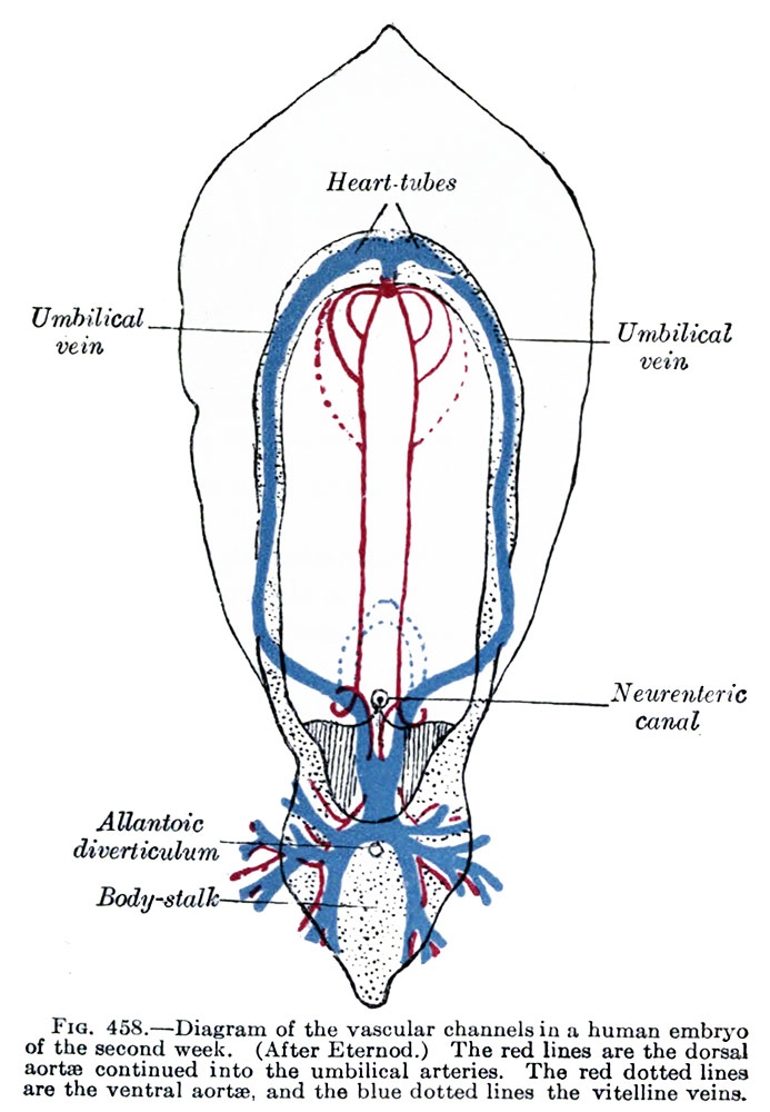 | 693 × 1,000 (109 KB) | Z8600021 (talk | contribs) | ==Diagram of the vascular channels in a human embryo of the second week== (After Eternod.) * The red lines are the dorsal aortæ continued into the umbilical arteries. * The red dotted lines are the ventral aortæ, and the blue dotted lines the vit... |
You cannot overwrite this file.
File usage
The following 2 pages use this file: