Fetal ECHO Meeting 2012: Difference between revisions
| Line 54: | Line 54: | ||
| Section | | Section | ||
| Name | | Name | ||
| Description | | Description (GA Week 10) | ||
|- | |- | ||
Revision as of 20:06, 4 October 2012
Fetal ECHO Meeting 5th-8th October 2012 ROYAL PRINCE ALFRED HOSPITAL, SYDNEY, AUSTRALIA

Human Embryo Development

Weeks shown are ovulation age (OA) for gestational age (GA) add 2 weeks.

Heart Development
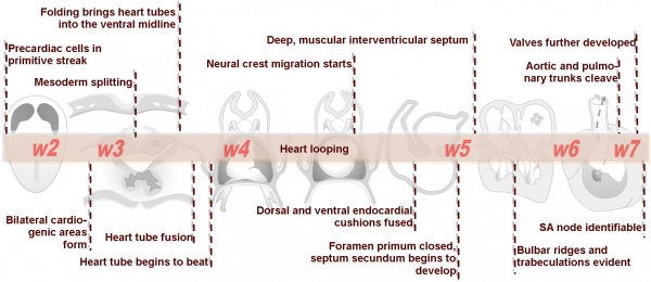
Weeks shown are ovulation age (OA) for clinical Gestational Age (GA) add 2 weeks.
- Links: PO timeline | GA timeline


Image day 21 to 25 (GA Week 5)
.jpg)

|

|

|
| Stage 11 | Stage 12 | Stage 13 |


Cardiovascular Movies
|
|
| Heart Fields |
| Page | Play |
| Stage 22 Embryo - Cardiovascular | ||
|---|---|---|
| Section | Name | Description (GA Week 10) |

|
C6L | L and R common carotid arteries. L and R internal jugular veins. |

|
C7L | L brachiocephalic vein (transverse anastomosis of L, R jugular veins). L common carotid artery. Brachiocephalic trunk. |

|
D1L | Aortic arch. Thymus (retrosternal). Trachea. Oesophagus. Large L and R jugular veins. |

|
D2L | Aortic arch. L jugular lymph sac lying laterally with hemiazygos joining it. Trachea. Oesophagus. Draining of azygos vein into R jugular vein. |

|
D3L | Ascending aorta attached cranial to transverse pericardial sinus. Superior vena cava expanding into R jugular vein. Trachea (bifurcation). Oesophagus. Azygos (R) and hemiazygos (L) veins. |

|
D4L | Ascending aorta. Pulmonary trunk and arteries. Thoracic aorta. Oesophagus. Bronchi. Note azygos vein on right, and precursor of hemiazygos vein on left side.
(Image excerpt different scale from previous images) |

|
D5L | Superior vena cava. Ascending arch of aorta. Pulmonary trunk with the other two semilunar valves.
Sinus above one valve and branching of pulmonary trunk into L and R pulmonary arteries. |

|
D6L | Superior vena cava. Ascending aorta with transverse pericardial sinus behind. Semilunar valve at origin of pulmonary trunk. L. atrium and auricle. Thoracic aorta. |

|
D7L | R atrium with R venous valve. R ventricle close to origin of pulmonary trunk. L.ventricular wall and three semilunar valves of aortic ostium. |

|
E1L | Section through all four chambers of heart.
left ventricle- outflow tract close to origin of ascending aorta. Deep coronary sulcus and transverse pericardial sinus. right atrium- R venous valve of the inferior vena cava, the small septum secundum, the aperture in the dorsal part of septum primum (ostium secundum). (The foramen ovale arises later with the elongation of the septum secundum). left atrium- entrance of the L and R pulmonary veins and the auricle. |

|
E2L | Section through all four chambers of heart.
Ventricles. Right venous valve. Between the tiny ridge of the septum secundum of the R atrium and the R venous valve. right atrium- drainage of the inferior vena cava (cf. E3). R atrioventricular canal. Septum primum. left atrium- drainage sites of R and L pulmonary veins. |

|
E3L | Section through all four chambers of heart.
Ventricles. Diaphragm and liver. Pericardium and cavity. R atrium and two cusps of the atrioventricular valve. L atrium and its auricle. Thoracic aorta. Inferior vena cava. Coronary sinus. |
Embryonic Heart Rate
In a 1996 study normal successful human gestations were defined by EHR criteria at different early embryonic (34-56 days GA, from last menstrual period) developmental stages (at the earliest stages when embryo length is difficult to measure gestational sac diameters are included). [1]
- Stage 9-10 2 mm embryo (gestational sac diameter of 20 mm) EHR at least 75 beats / minute
- Stage 11-12 5 mm embryo (gestational sac diameter of 30 mm) EHR at least 100 beats / minute
- Stage 16 10 mm embryo EHR at least 120 beats / minute
- Stage 18 15 mm embryo EHR at least 130 beats / minute
References
- ↑ <pubmed>8921130</pubmed>
Online Textbooks
- Developmental Biology (6th ed.) Gilbert, Scott F. Sunderland (MA): Sinauer Associates, Inc.; c2000. Figure 15.6 Cascade of heart development
Search Bookshelf heart development
Reviews
<pubmed>21940548</pubmed> <pubmed>17224285</pubmed>| PMC1858673 <pubmed></pubmed>
Articles
<pubmed>17384040</pubmed>| MC2190734 J Ultrasound Med.
Search Pubmed
Search Pubmed heart development
External Links
External Links Notice - The dynamic nature of the internet may mean that some of these listed links may no longer function. If the link no longer works search the web with the link text or name. Links to any external commercial sites are provided for information purposes only and should never be considered an endorsement. UNSW Embryology is provided as an educational resource with no clinical information or commercial affiliation.
- Cardiovascular Ultrasound is an open access, peer-reviewed, online journal covering clinical, technological, experimental, biological, and molecular aspects of ultrasound applications in cardiovascular physiology and disease. Search - Fetal