Category:Week 1: Difference between revisions
No edit summary |
No edit summary |
||
| Line 15: | Line 15: | ||
:Dr Mark Hill 2009, '''''UNSW Embryology''''' ISBN: 978 0 7334 2609 4 - UNSW CRICOS Provider Code No. 00098G | :Dr Mark Hill 2009, '''''UNSW Embryology''''' ISBN: 978 0 7334 2609 4 - UNSW CRICOS Provider Code No. 00098G | ||
[[Category:Human Embryo]] [[Category:Carnegie Stage 1]] | [[Category:Human Embryo]] [[Category:Carnegie Stage 1]] [[Category:Carnegie Stage 3]] | ||
Revision as of 16:43, 15 September 2009
Introduction

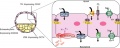
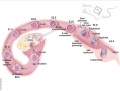
This page shows some key events of human development during the embryonic period of the first eight weeks (weeks 1 - 8) following fertilization. This period is also considered the organogenic period, when most organs within the embryo have begun to form. There are links to more detailed descriptions which can be viewed in a week by week format, by the historic Carnegie stages or integrated into a Timeline of human development.
Online resources available include: individual images of all Carnegie stages, scanning electron micrographs of the earlier stages, cross-sections showing internal structures at mid- and late-embryonic, 3D reconstructions of internal structures, animations of processes, ultrasound scans and information about abnormalites of development.
Note that there is variability in the actual timing of specific events and at the end of this period fetal development begins.
Week by Week
Carnegie Stages Link
- Dr Mark Hill 2009, UNSW Embryology ISBN: 978 0 7334 2609 4 - UNSW CRICOS Provider Code No. 00098G
Subcategories
This category has the following 12 subcategories, out of 12 total.
Pages in category 'Week 1'
The following 91 pages are in this category, out of 91 total.
B
C
- Carnegie stage 1
- Template:Carnegie stage 1 links
- Carnegie stage 2
- Template:Carnegie stage 2 links
- Carnegie stage 3
- Template:Carnegie stage 3 links
- Carnegie stage 4
- Template:Carnegie stage table week 1
- Template:CE8190
- Template:CE8260
- Template:CE8450
- Template:CE8452
- Template:CE8500.1
- Template:CE8630
- Template:CE8663
- Template:CE8698
- Template:CE8794
- Template:CE8904
- Template:CS1
- Template:CS2
- Template:CS3
- Template:CS4
E
I
M
R
Media in category 'Week 1'
The following 69 files are in this category, out of 69 total.
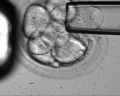 Blastomere isolation.jpg 1,200 × 961; 95 KB
 Blastomere mitosis 01 icon.jpg 150 × 102; 3 KB
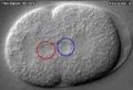
 Blastomere mitotic spindle orientation.jpg 600 × 414; 21 KB
 CSt3.jpg 500 × 377; 20 KB
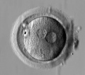
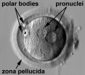
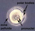 Early zygote labelled.jpg 500 × 441; 29 KB
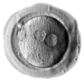
 Early zygote.jpg 500 × 441; 23 KB
 Human blastocyst day 1-5.jpg 500 × 450; 46 KB
 Human blastocyst day 1-6.jpg 708 × 338; 45 KB
 Human blastocyst day 3-6.mov ; 4.26 MB
- Human blastocyst day 5-6 small.mov ; 509 KB
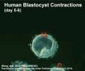 Human blastocyst day 5-6.jpg 498 × 414; 24 KB
- Human blastocyst day 5-6.mov ; 979 KB
- Human blastocyst hatching day 5-6.mov ; 1.11 MB
 Human Carnegie stage 1-23.jpg 1,000 × 563; 98 KB
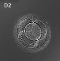 Human embryo day 2.jpg 400 × 409; 19 KB
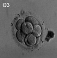 Human embryo day 3.jpg 400 × 409; 7 KB
 Human embryo day 5 label.gif 500 × 506; 243 KB
 Human embryo day 5 label.jpg 500 × 506; 37 KB
 Human embryo day 5 label2.jpg 500 × 506; 43 KB
 Human embryo day 5.jpg 400 × 409; 6 KB
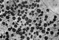
 Human oocyte-metaphase I.jpg 400 × 409; 32 KB
 Human oocyte-metaphase II.jpg 400 × 409; 12 KB
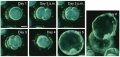
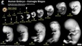
 Human preimplantation embryos 01.jpg 1,280 × 629; 261 KB
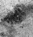
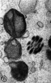
 Human pronuclear stage EM02.jpg 639 × 1,000; 194 KB
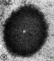
 Human pronuclear stage EM022.jpg 1,100 × 705; 225 KB
 Human pronuclear stage EM03-05.jpg 982 × 439; 104 KB
 Human pronuclear stage EM06.jpg 907 × 1,000; 245 KB
 Human pronuclear stage EM07.jpg 357 × 509; 52 KB
 Human pronuclear stage EM08.jpg 359 × 513; 56 KB
 Human pronuclear stage EM09.jpg 361 × 506; 53 KB
 Human pronuclear stage EM10.jpg 630 × 781; 140 KB
 Human pronuclear stage EM11.jpg 625 × 768; 130 KB
 Human pronuclear stage EM12.jpg 998 × 777; 265 KB
 Human pronuclear stage EM13.jpg 998 × 771; 247 KB
 Human pronuclear stage EM14-16.jpg 1,013 × 459; 141 KB
 Human pronuclear stage EM17.jpg 1,104 × 504; 156 KB
 Human pronuclear stage EM18.jpg 477 × 541; 66 KB
 Human pronuclear stage EM19.jpg 478 × 534; 67 KB
 Human pronuclear stage EM20.jpg 1,114 × 762; 237 KB
 Human pronuclear stage EM21.jpg 366 × 587; 58 KB
 Human pronuclear stage EM22.jpg 366 × 581; 59 KB
 Human pronuclear stage EM25.jpg 1,013 × 782; 201 KB
 Human pronuclear stage EM26.jpg 1,010 × 784; 187 KB
 Human pronuclear stage EM27.jpg 997 × 777; 227 KB
 Human pronuclear stage EM28.jpg 993 × 774; 239 KB
 Human pronuclear stage EM29.jpg 967 × 763; 183 KB
 Human pronuclear stage EM30.jpg 955 × 853; 192 KB
 Human zygote two pronuclei 01.jpg 528 × 472; 34 KB
 Human zygote two pronuclei 02.jpg 519 × 457; 28 KB
 Human zygote two pronuclei 02.png 433 × 422; 121 KB
 Human zygote two pronuclei 03.jpg 503 × 477; 33 KB
 Human zygote two pronuclei 22.jpg 519 × 457; 38 KB
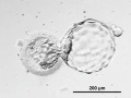
 Human-oocyte to blastocyst.jpg 600 × 402; 49 KB
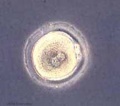
 Human-oocyte.jpg 400 × 409; 29 KB
 Implanting human conceptus 01.jpg 1,056 × 834; 246 KB
 Model human blastocyst development.jpg 946 × 726; 84 KB
 Mouse - blastocoel formation.jpg 800 × 319; 47 KB
 Mouse E0-E5.jpg 991 × 749; 90 KB
 Mouse- preimplantation gene expression.jpg 800 × 612; 106 KB
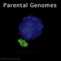 Parental genome mix 01 icon.jpg 320 × 320; 4 KB
- Pronuclear fusion 001.mov ; 115 KB
 Stage1 size with ruler.jpg 400 × 193; 9 KB
 Stage2 bf01.jpg 650 × 521; 49 KB
 Stage2.jpg 216 × 185; 4 KB
 Stage3 bf01.jpg 507 × 625; 39 KB
 Stage3 bf02.jpg 507 × 625; 31 KB
 Week 1 cartoon.jpg 800 × 598; 37 KB
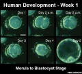
 Week1 summary.jpg 1,000 × 747; 107 KB
 Windle1940 fig01.jpg 1,280 × 894; 387 KB


