Category:Carnegie Stage 1: Difference between revisions
From Embryology
mNo edit summary |
mNo edit summary |
||
| Line 4: | Line 4: | ||
:'''Links:''' [[Carnegie stage 1|Carnegie Stage 1]] | {{zygote}} | [[Week 1]] | {{meiosis}} | [http://tiny.cc/Carnegie_Stage_1 Tinycc | :'''Links:''' [[Carnegie stage 1|Carnegie Stage 1]] | {{zygote}} | [[Week 1]] | {{meiosis}} | [http://tiny.cc/Carnegie_Stage_1 Tinycc Link] | ||
Revision as of 13:27, 15 May 2018
This Embryology category lists pages and media related to Carnegie Stage 1 occurring in week 1 (GA week 3) of human development.
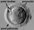
This stage occurs following fertilisation and is the first single cell, the zygote. The zygote forms when the male (spermatozoa) and female (oocyte) haploid pronuclei combine to form the single diploid zygote nucleus.
- Links: Carnegie Stage 1 | zygote | Week 1 | meiosis | Tinycc Link
| Week: | 1 | 2 | 3 | 4 | 5 | 6 | 7 | 8 |
| Carnegie stage: | 1 2 3 4 | 5 6 | 7 8 9 | 10 11 12 13 | 14 15 | 16 17 | 18 19 | 20 21 22 23 |
Subcategories
This category has the following 2 subcategories, out of 2 total.
Pages in category 'Carnegie Stage 1'
The following 11 pages are in this category, out of 11 total.
H
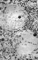
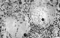
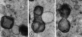
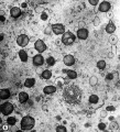
Media in category 'Carnegie Stage 1'
The following 47 files are in this category, out of 47 total.
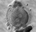
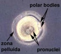 Early zygote labelled.jpg 500 × 441; 29 KB
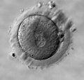
 Early zygote.jpg 500 × 441; 23 KB
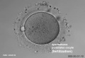
 Human fertilization movie 1 frame 01.jpg 600 × 409; 27 KB
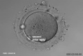
 Human fertilization movie 1 frame 02.jpg 600 × 409; 27 KB
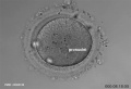
 Human fertilization movie 1 frame 03.jpg 600 × 409; 26 KB
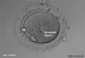
 Human fertilization movie 1 frame 04.jpg 600 × 409; 24 KB
 Human fertilization movie 1 frame 05.jpg 600 × 409; 25 KB
 Human fertilization movie 1 frame 06.jpg 600 × 409; 25 KB
 Human fertilization movie 1 frame 07.jpg 600 × 409; 24 KB
 Human fertilization movie 1 frame 08.jpg 600 × 409; 25 KB
 Human fertilization movie 1 frame 09.jpg 600 × 409; 24 KB
 Human fertilization movie 1 frame 10.jpg 600 × 409; 25 KB
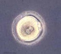
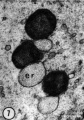
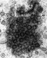
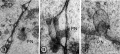
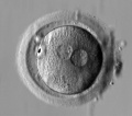
 Human pronuclear stage EM02.jpg 639 × 1,000; 194 KB
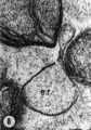
 Human pronuclear stage EM022.jpg 1,100 × 705; 225 KB
 Human pronuclear stage EM03-05.jpg 982 × 439; 104 KB
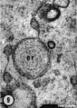
 Human pronuclear stage EM06.jpg 907 × 1,000; 245 KB
 Human pronuclear stage EM07.jpg 357 × 509; 52 KB
 Human pronuclear stage EM08.jpg 359 × 513; 56 KB
 Human pronuclear stage EM09.jpg 361 × 506; 53 KB
 Human pronuclear stage EM10.jpg 630 × 781; 140 KB
 Human pronuclear stage EM11.jpg 625 × 768; 130 KB
 Human pronuclear stage EM12.jpg 998 × 777; 265 KB
 Human pronuclear stage EM13.jpg 998 × 771; 247 KB
 Human pronuclear stage EM14-16.jpg 1,013 × 459; 141 KB
 Human pronuclear stage EM17.jpg 1,104 × 504; 156 KB
 Human pronuclear stage EM18.jpg 477 × 541; 66 KB
 Human pronuclear stage EM19.jpg 478 × 534; 67 KB
 Human pronuclear stage EM20.jpg 1,114 × 762; 237 KB
 Human pronuclear stage EM21.jpg 366 × 587; 58 KB
 Human pronuclear stage EM22.jpg 366 × 581; 59 KB
 Human pronuclear stage EM25.jpg 1,013 × 782; 201 KB
 Human pronuclear stage EM26.jpg 1,010 × 784; 187 KB
 Human pronuclear stage EM27.jpg 997 × 777; 227 KB
 Human pronuclear stage EM28.jpg 993 × 774; 239 KB
 Human pronuclear stage EM29.jpg 967 × 763; 183 KB
 Human pronuclear stage EM30.jpg 955 × 853; 192 KB
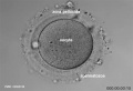
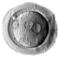
 Human zygote two pronuclei 01.jpg 528 × 472; 34 KB
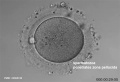
 Human zygote two pronuclei 02.jpg 519 × 457; 28 KB
 Human zygote two pronuclei 02.png 433 × 422; 121 KB
 Human zygote two pronuclei 03.jpg 503 × 477; 33 KB
 Human zygote two pronuclei 22.jpg 519 × 457; 38 KB
 Human-oocyte to blastocyst.jpg 600 × 402; 49 KB
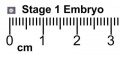 Stage1 size with ruler.jpg 400 × 193; 9 KB
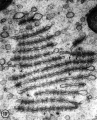
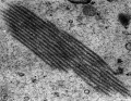
 Zamboni1966 fig02.jpg 1,556 × 1,000; 397 KB
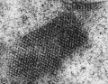
 Zamboni1966 fig06.jpg 1,280 × 1,158; 419 KB
 Zamboni1966 fig20.jpg 1,526 × 1,000; 429 KB
 Zamboni1966 fig29.jpg 1,280 × 1,003; 302 KB


