Carnegie Stages
Introduction
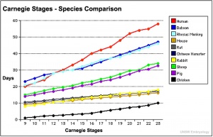
Carnegie stages are named after the famous US Institute which began collecting and classifying embryos in the early 1900's. Stages are based on the external and/or internal morphological development of the embryo, and are not directly dependent on either age or size. The human embryonic period proper is divided into 23 Carnegie stages. Criteria beyond morphological features include ranges of age in days, number of somites present, and embryonic crown rump lengths (CRL).
- Carnegie Stages: 1 | 2 | 3 | 4 | 5 | 6 | 7 | 8 | 9 | 10 | 11 | 12 | 13 | 14 | 15 | 16 | 17 | 18 | 19 | 20 | 21 | 22 | 23 | About Stages | Timeline
The following text and information about the collection is modifed from the original Carnegie Institute website.
Franklin P. Mall (1862-1917) is most remembered for his work done at the Department of Embryology at the Carnegie Institute of Washington. Mall began collecting human embryos while a postgraduate student in Lepzig with Wilhelm His, but didn't receive the first Carnegie specimen until his position at Johns Hopkins University. (More? Franklin Mall)
Surprizingly age and size proves a poor way to organize embryos. It is very difficult to accurately age an embryo, and it could shrink a full 50% in the preserving fluids. Mall took it upon himself to find a better way. He had more success basing his "staging" scheme on morphological characteristics. To that end, Mall and his colleagues not only prepared and preserved serial sections of the embryos, they also made hundreds of three-dimensional models at different stages of growth. According to Adrianne Noe, who now manages the collection at the National Museum of Health and Medicine, Mall gathered the most renowned scientists, scholars, artists, photographers, and craftspeople ever to apply their interests and skills to embryology. One of the first to be hired, in 1913, was modeler Osborne O. Heard, who spent 42 years at the department and made over 700 wax-based reconstructions. The results of this team effort still stand as the international standard by which human embryos are described and classified.
Carnegie Human Embryo Glossary
Age
Postovulatory age is one criterion for the overall staging of embryos.
It is the length of time since the last ovulation before fertilization took place and is estimated by assigning an embryo to a developmental stage and then referring to a standard table of norms. A range of +/- 1 day is expected. Postovulatory age is stated in days or weeks.
Case Number
The basis of the Carnegie Collection of Embryos was Franklin P. Mall's personal collection of 813 embryos, which he began in 1887 with the first specimen. Carnegie Embryo No. 1. was acquired by Mall while he was an assistant in Pathology at the Johns Hopkins Hospital. All subsequent specimens in the Collection were numbered sequentially at the time of acquisition, i.e. case number.
Crown-Rump (C-R)
One criterion for the overall classification of human embryonic development.
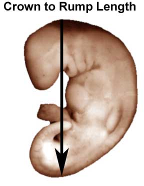
A measurement of prenatal length, from the vertex of the skull (crown), along the curvature of the spine to the midpoint between the apices of the buttocks (rump), of the developing embryo. This measurement was developed for smaller specimens, 35mm or less, so that their natural curved posture is not disturbed. C-R length is stated in millimeters.
An alternative measurement of prenatal length is the greatest length (G.L.), which some researchers find to be more useful in the assessment of length of an embryo. The G.L. is determined by measuring the embryo in a straight line, (i.e. caliper length) without any attempt to straighten the natural curvature of the specimen. G.L. is stated in millimeters.
Grade
Specimens in the Carnegie Collection have been graded Excellent, Good, Fair or Poor.
This reference is based on the total grade of the specimen, including both its original quality and the condition of the specimen.
Normal/Abnormal
Of the approximately 600 sectioned embryos in the Carnegie Collection assigned to the 23 stages, a majority have been classified as normal. However, variations in, and anomolies of, individual organs are known to occur.
Section Information
Plane of Section
The surface formed by extension through an axis of the embryo.
There are three primary descriptive terms referring to the planes of the embryo:
- Coronal A vertical plane dividing the body into anterior and posterior portions.
- Sagittal Any plane parallel to the median.
- Transverse A plane horizontal to the median.
Thinness of Section
The specified thinness of the cut embryonic section for mounting on a glass slide in serial order. Thinness is measured in micrometers. Some regions of a few of the specimens in the Collection were cut at various thinnesses; these instances are represented in the search results.
Slides
Total number of glass slides containing serial histologic sections of each specimen in the Collection.
Sections
Total number of serial histologic sections on any number of glass slides for each specimen in the Collection.
Sex
Gender identification, i.e. male or female, is noted where apparent. In the embryo, the gonads do not acquire male or female morphological characteristics until the 7th or 8th week of development (stages 18-23). Therefore, many specimens in the embryonic period are not identified by gender. This assignment applies mostly to very late embryonic period specimens in the Collection.
Somites
One criterion for the overall classification of human embryonic development.
Somites are paired segments of paraxial mesoderm appearing in longitudinal rows along the left and right side of the neural groove and notochord. They commence in the third or early fourth week of development (approximately the 20th day), appearing first in the cervical region of the embryo. Their formation proceeds in a craniocaudal direction. New somites appear approximately three per day, until at the end of the 5th week when 42 to 44 pairs are present. Differentiation of the somites leads to formation of the axial skeleton.
Stage of Development
The definitive classification of human embryos into developmental groups termed stages.
Stages are based on the external and/or internal morphological development of the embryo, and are not directly dependent on either age or size. The human embryonic period proper is divided into 23 Carnegie stages.
Criteria beyond morphological features include age in days, number of somites present, and embryonic length. This chart shows the relationship between Stage, Age and embryonic length.
Stain Type
The type of individual dye or staining substance, or combination of dyes and reagents, used in histologic technique to color the constituents of cells and tissues.
- Carnegie Stages: 1 | 2 | 3 | 4 | 5 | 6 | 7 | 8 | 9 | 10 | 11 | 12 | 13 | 14 | 15 | 16 | 17 | 18 | 19 | 20 | 21 | 22 | 23 | About Stages | Timeline
Cite this page: Hill, M.A. (2024, April 27) Embryology Carnegie Stages. Retrieved from https://embryology.med.unsw.edu.au/embryology/index.php/Carnegie_Stages
- © Dr Mark Hill 2024, UNSW Embryology ISBN: 978 0 7334 2609 4 - UNSW CRICOS Provider Code No. 00098G

