Carnegie stage 4
Introduction
Stage 4 sees the attached blastocyst begin implantation into the maternal uterine lining epithelium and underlying stroma. The entire cellular products of the original fertilisation event are now referred to as the conceptus, that will include embryonic and extra embryonic tissues of the fatal membranes and placenta.
- Links: Lecture - Week 1 and 2 Development | Implantation | Week 1 | Week 2
- Carnegie Stages: 1 | 2 | 3 | 4 | 5 | 6 | 7 | 8 | 9 | 10 | 11 | 12 | 13 | 14 | 15 | 16 | 17 | 18 | 19 | 20 | 21 | 22 | 23 | About Stages | Timeline
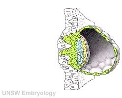 |
Facts: Week 1-2, size 0.1 - 0.2 mm Features: Adplantation and implantation commences, increase in hCG Links: Implantation | Week 2 |
|
<wikiflv height="220" width="248" autoplay="true" position="left">Week2_001.flv|File:Week2_001_icon.jpg</wikiflv> |
The animation (left) shows the process of implantation, occurring during week 2 of development in humans.
The beginning of the animation shows: the uterus lining (endometrium epithelium), the hatched blastocyst with a flat outer layer of trophoblast cells (green), the inner cell mass which has formed into the bilaminar embryo (epiblast and hypoblast) and the large fluid-filled space (blastocoel).
|
| Quicktime movie |
- Carnegie Stages: 1 | 2 | 3 | 4 | 5 | 6 | 7 | 8 | 9 | 10 | 11 | 12 | 13 | 14 | 15 | 16 | 17 | 18 | 19 | 20 | 21 | 22 | 23 | About Stages | Timeline
Cite this page: Hill, M.A. (2026, September 29) Embryology Carnegie stage 4. Retrieved from https://embryology.med.unsw.edu.au/embryology/index.php/Carnegie_stage_4
- © Dr Mark Hill 2026, UNSW Embryology ISBN: 978 0 7334 2609 4 - UNSW CRICOS Provider Code No. 00098G
