Carnegie stage 4
| Embryology - 28 Apr 2024 |
|---|
| Google Translate - select your language from the list shown below (this will open a new external page) |
|
العربية | català | 中文 | 中國傳統的 | français | Deutsche | עִברִית | हिंदी | bahasa Indonesia | italiano | 日本語 | 한국어 | မြန်မာ | Pilipino | Polskie | português | ਪੰਜਾਬੀ ਦੇ | Română | русский | Español | Swahili | Svensk | ไทย | Türkçe | اردو | ייִדיש | Tiếng Việt These external translations are automated and may not be accurate. (More? About Translations) |
Introduction
|
Stage 4 sees the attached blastocyst begin implantation into the maternal uterine lining epithelium and underlying stroma. The entire cellular products of the original fertilisation event are now referred to as the conceptus, this will include embryonic and extra-embryonic tissues of the fetal membranes and placenta. When conceptus implantation completes, a coagulation plug is formed at the site of implantation and the entire process of development will occur enclosed within the wall of the uterus. The process of attachment and implantation initiates the at first localised transformation of the uterine endometrium into the decidua (maternal decidua). This process is described as decidualization (decidualisation) that eventually spreads throughout the entire uterine lining. Summary
See also Events |
| Stage 4 Links: Week 2 | Implantation | | Trophoblast | Human Chorionic Gonadotropin | Lecture | Practical | Category:Carnegie Stage 4 | Next Stage 5 |
| Week: | 1 | 2 | 3 | 4 | 5 | 6 | 7 | 8 |
| Carnegie stage: | 1 2 3 4 | 5 6 | 7 8 9 | 10 11 12 13 | 14 15 | 16 17 | 18 19 | 20 21 22 23 |
- Carnegie Stages: 1 | 2 | 3 | 4 | 5 | 6 | 7 | 8 | 9 | 10 | 11 | 12 | 13 | 14 | 15 | 16 | 17 | 18 | 19 | 20 | 21 | 22 | 23 | About Stages | Timeline
Carnegie Collection
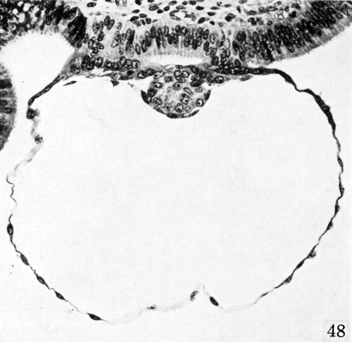
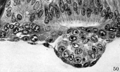
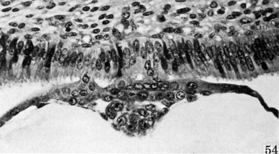
Note these are implanting monkey blastocysts[1] as there are no human examples of this stage in the collection.
| The uterus epithelium is shown at the top of the image.
The trophectoderm (trophoblast layer proliferates and invades the epithelium. The cytotrophoblast layer forms a squamous epithelium surrounding the blastocoel. The inner cell mass (embryoblast) is aligned towards the epithelium.
|

|
The images below are a serial serial sections of the embryo inner cell mass.

|

|

|

|

|

|
Stage 4 Animation
|
<html5media height="400" width="450">File:Week2_001.mp4</html5media> |
The animation (left) shows the process of implantation, occurring during week 2 of development in humans.
The beginning of the animation shows: the uterus lining (endometrium epithelium), the hatched blastocyst with a flat outer layer of trophoblast cells (green), the inner cell mass which has formed into the bilaminar embryo (epiblast and hypoblast) and the large fluid-filled space (blastocoel).
|
Events
- mitosis - cells on surface of blastocyst (trophoblast cells) divide more rapidly than the inner cells (inner cell mass, embryoblast)
- implantation - adhesion to endometrium epithelium and implantation through this layer into the underlying uterine stroma.
- Endocrine signalling - (hCG)trophoblast cells secrete hCG (human Chorionic Gonadotropin/Gonadotrophin) the signal of pregnancy.
- corpus luteum - signal from trophoblasts begins to maintain this endocrine tissue in the maternal ovary that secretes progesterone.
References
- ↑ Heuser CH. and Streeter GL. Development of the macaque embryo. (1941) Contrib. Embryol., Carnegie Inst. Wash. Publ. 525, 29: 15-55.
Additional Images
- Carnegie Stages: 1 | 2 | 3 | 4 | 5 | 6 | 7 | 8 | 9 | 10 | 11 | 12 | 13 | 14 | 15 | 16 | 17 | 18 | 19 | 20 | 21 | 22 | 23 | About Stages | Timeline
Cite this page: Hill, M.A. (2024, April 28) Embryology Carnegie stage 4. Retrieved from https://embryology.med.unsw.edu.au/embryology/index.php/Carnegie_stage_4
- © Dr Mark Hill 2024, UNSW Embryology ISBN: 978 0 7334 2609 4 - UNSW CRICOS Provider Code No. 00098G