Gastrointestinal Tract - Abnormalities
| Embryology - 26 Jun 2024 |
|---|
| Google Translate - select your language from the list shown below (this will open a new external page) |
|
العربية | català | 中文 | 中國傳統的 | français | Deutsche | עִברִית | हिंदी | bahasa Indonesia | italiano | 日本語 | 한국어 | မြန်မာ | Pilipino | Polskie | português | ਪੰਜਾਬੀ ਦੇ | Română | русский | Español | Swahili | Svensk | ไทย | Türkçe | اردو | ייִדיש | Tiếng Việt These external translations are automated and may not be accurate. (More? About Translations) |
| ICD-11 |
|---|
|
Structural developmental anomalies of the digestive tract - LB10 Structural developmental anomalies of salivary glands or ducts | LB11 Congenital diverticulum of pharynx | LB12 Structural developmental anomalies of oesophagus | LB13 Structural developmental anomalies of stomach | LB14 Structural developmental anomalies of duodenum | LB15 Structural developmental anomalies of small intestine | LB16 Structural developmental anomalies of large intestine | LB17 Structural developmental anomalies of anal canal | LB18 Congenital anomalies of intestinal fixation Structural developmental anomalies of the liver, biliary tract, pancreas or spleen |
Introduction

The "simple tube" of the gastrointestinal tract and its associated organs have many different tract and organ specific gastrointestinal abnormalities. Due to the complex nature (different germ layer contributions, organogenisis) of the growth, elongation and folding of the tract, there are also several mechanical disorders of folding (rotation). Musculoskeletal abnormalities of the anterior body wall can also result in gastrointestinal abnormalities.
Note that as this system begins function (digestively) postnatally, unless there is a determined genetic history within the family, several abnormalities only become evident postnatally, in particular, metabolic disorders often identified by the Guthrie test.
International Classification of Diseases
| ICD10 Other congenital malformations of the digestive system (Q38-Q45) | ||||||
|---|---|---|---|---|---|---|
| XVII Congenital Malformations - Other congenital malformations of the digestive system (Q38-Q45) | ||||||
| Q38 Other congenital malformations of tongue, mouth and pharynx
Excl.: macrostomia (Q18.4) microstomia (Q18.5)
| ||||||
Q39 Congenital malformations of oesophagus
| ||||||
Q40 Other congenital malformations of upper alimentary tract
| ||||||
| Q41 Congenital absence, atresia and stenosis of small intestine
Incl.: congenital obstruction, occlusion and stricture of small intestine or intestine NOS Excl.: meconium ileus (E84.1)
| ||||||
| Q42 Congenital absence, atresia and stenosis of large intestine
Incl.: congenital obstruction, occlusion and stricture of large intestine
| ||||||
Q43 Other congenital malformations of intestine
| ||||||
Q44 Congenital malformations of gallbladder, bile ducts and liver
| ||||||
| Q45 Other congenital malformations of digestive system
Excl.: congenital: diaphragmatic hernia (Q79.0) hiatus hernia (Q40.1)
| ||||||
|
World Health Organisation. International Statistical Classification of Diseases and Related Health Problems. (1992) 10th Revision (ICD-10). Geneva: WHO ICD-10 - 2016 Online (English) | ||||||
Links: Gastrointestinal Abnormalities
| ||||||
| ICD10 - Gastrointestinal | Genital | Renal | Integumentary |
Some Recent Findings
|
| More recent papers |
|---|
 This table allows an automated computer search of the external PubMed database using the listed "Search term" text link.
More? References | Discussion Page | Journal Searches | 2019 References | 2020 References Search term: Abnormal Development Gastrointestinal Tract <pubmed limit=5>Abnormal Development Gastrointestinal Tract</pubmed> |
Movies
|
|
|
|


Statistics
Australian

|
The pie diagram shows the relative contribution of major gastrointestinal tract abnormalities as a percentage of the total number of congenital abnormalities in Australia beween 1981 - 92.
Note that the digestive system represents approximately 6% of all major congenital abnormalities. One of the most common abnormalities occurring in (2% - 3% population) is Meckel's diverticulum. The mouth (cleft lip, cleft palate) is part of the digestive tract, but more accurately reflects an abnormality of face formation. |
| Data shown as a percentage of all major abnormalities based upon published statistics using the same groupings as Congenital Malformations Australia 1981-1992 P. Lancaster and E. Pedisich ISSN 1321-8352. |
| Australian GIT Abnormalities (2002-2003) |
|---|
Oesophageal atresia/stenosis - (2.0 per 10,000 births) ICD-10 Q39.0–Q39.3
|
Small intestinal atresia/stenosis - (2.4 per 10,000 births) ICD-10 Q41.0-Q41.2
|
Anorectal atresia/stenosis - ( 3.1 per 10,000 births) ICD-10 Q42.0–Q42.3
|
Hirschsprung’s disease - (1.3 per 10,000 births) ICD-10 Q43.1
|
Exomphalos - (Omphalocele) (2.1 per 10,000 births) ICD-10 Q79.2
|
Gastroschisis - (2.6 per 10,000 births) ICD-10 Q79.3
|
|
| Australian Palate Abnormalities (2002-2003) |
|---|
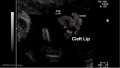
| Cleft lip with or without cleft palate (9.2 per 10,000 births) ICD-10 Q36.0, Q36.1, Q36.9, Q37.0–Q37.5, Q37.8, Q37.9 |
A congenital anomaly characterised by a partial or complete clefting of the upper lip, with or without clefting of the alveolar ridge or the hard palate. Excludes a midline cleft of the upper or lower lip and an oblique facial fissure (going towards the eye).
|
| Cleft palate without cleft lip (8.1 per 10,000 births) ICD-10 Q35.0–Q35.9 |
A congenital anomaly characterised by a closure defect of the hard and/or soft palate behind the foramen incisivum without a cleft lip. This anomaly includes sub-mucous cleft palate, but excludes cleft palate with a cleft lip, a functional short palate and high narrow palate.
|
|
USA Selected
CDC National estimates for selected GIT related major birth defects (2004–2006).
| USA Selected Statistics | ||||||||||||||||||||||||||||||||||||||||||||||||||||||||||||||||||||||||
|---|---|---|---|---|---|---|---|---|---|---|---|---|---|---|---|---|---|---|---|---|---|---|---|---|---|---|---|---|---|---|---|---|---|---|---|---|---|---|---|---|---|---|---|---|---|---|---|---|---|---|---|---|---|---|---|---|---|---|---|---|---|---|---|---|---|---|---|---|---|---|---|---|
| ||||||||||||||||||||||||||||||||||||||||||||||||||||||||||||||||||||||||
Some Recent Findings
|
| More recent papers |
|---|
This table allows an automated computer search of the external PubMed database using the listed "Search term" text link.
More? References | Discussion Page | Journal Searches | 2019 References | 2020 References Search term: Abnormal Development Gastrointestinal Tract <pubmed limit=5>Abnormal Development Gastrointestinal Tract</pubmed> |
Lumen Abnormalities
There are several types of abnormalities (atresia, stenosis and duplication) that impact upon the continuity of the gastrointestinal tract lumen.
Atresia
ICD-11 LB12.1 Atresia of oesophagus | LB14 Structural developmental anomalies of duodenum | LB20.21 Biliary atresia | LB15.1 Atresia of small intestine | LB17.0 Anorectal malformations
An interruption of the lumen where the naming is based upon anatomical location.
Oesophageal Atresia
ICD-11 LB12.1 Atresia of oesophagus
See oesophageal atresia review[7]
| X-ray | Classification[8] |

|

|
The 1962 Waterston classification is a useful tool for predicting post-operative morbidity associated with independent risk factors such as; birth weight, cardiac anomalies, and pre-operative pneumonia.[9] There are alternative prognostic classification systems.[10]
Atresia of oesophagus with tracheo-oesophageal fistula
Atresia of oesophagus with broncho-oesophageal fistula, OA/TOF)
"Oesophageal atresia encompasses a group of congenital anomalies with an interruption in the continuity of the oesophagus, with or without persistent communication with the trachea. In 86% of cases there is a distal tracheooesophageal fistula, in 7% of cases there is no fistulous connection, while in 4% of cases there is a tracheooesophageal fistula without atresia. The remaining cases are made up of patients with OA with proximal, or both proximal and distal, tracheooesophageal fistula."
This abnormality has been shown to be associated with Tbx1 mutations that also include DiGeorge syndrome.[11]
Search PubMed: Oesophageal Atresia
Pyloric Atresia
Pyloric atresia (PA) - a very rare condition (incidence 1 in 100,000 newborns) and about 1% of all intestinal atresias.
Search PubMed: Pyloric Atresia
Duodenal Atresia
ICD-11 LB14 Structural developmental anomalies of duodenum
Search PubMed: Duodenal Atresia
Biliary Atresia
ICD-11 LB20.21 Biliary atresia
Search PubMed: Biliary Atresia
Small Intestine Atresia
ICD-11 LB15.1 Atresia of small intestine
Search PubMed: Small Intestine Atresia
Anorectal Atresia
ICD-11 LB17.0 Anorectal malformations
Search PubMed: Anorectal Atresia
Stenosis
A narrowing of the lumen (esophageal stenosis, duodenal stenosis, pyloric stenosis)
Oesophageal stenosis[12]
Duplication

An incomplete recanalization resulting in parallel lumens, this is really a specialized form of stenosis.
Oesophagus
| ICD-11 LB12 Structural developmental anomalies of oesophagus - Any congenital defect of oesophagus that results from interference with the normal growth and differentiation of the fetus.
LB12.0 Congenital oesophageal web or ring | LB12.1 Atresia of oesophagus | LB12.2 Oesophageal fistula without atresia | LB12.3 Congenital stenosis or stricture of oesophagus | LB12.4 Congenital diverticulum of oesophagus | LB12.5 Congenital dilatation of oesophagus |
LB12.0 Congenital oesophageal web or ring
ICD-11 LB12.0 Congenital oesophageal web or ring - A rare form of incomplete oesophageal obstruction due to a developmental defect of the primitive foregut that presents as a mucosal lesion forming an incomplete diaphragm. Symptoms (apparent from birth) include dysphagia, regurgitation, and choking.
LB12.1 Atresia of oesophagus
ICD-11 LB12.1 Atresia of oesophagus - Oesophageal atresia encompasses a group of congenital anomalies with an interruption in the continuity of the oesophagus, with or without persistent communication with the trachea. In 86% of cases there is a distal tracheooesophageal fistula, in 7% of cases there is no fistulous connection, while in 4% of cases there is a tracheooesophageal fistula without atresia. The remaining cases are made up of patients with OA with proximal, or both proximal and distal, tracheooesophageal fistula.
LB12.2 Oesophageal fistula without atresia
ICD-11 LB12.2 Oesophageal fistula without atresia - This is a birth defect (congenital anomaly) of oesophagus, and one type of EA/TEF, namely isolated "H"-shaped atresia. Tracheoesophageal fistula in which there is no esophageal atresia because the esophagus is continuous to the stomach. Fistula is present between the esophagus and the trachea. Incidence of TE fistula without atresia varies between 1 -11% of esophageal malformations.
LB12.3 Congenital stenosis or stricture of oesophagus
ICD-11 LB12.3 Congenital stenosis or stricture of oesophagus - A form of incomplete oesophageal obstruction due to a developmental defect of the primitive foregut. Abnormal narrowing of the oesophagus occurs most often at the junction of the middle and lower thirds. Clinical manifestations, apparent 2 to 3 weeks after birth, include dysphagia and progressive vomiting.
LB12.4 Congenital diverticulum of oesophagus
ICD-11 LB12.4 Congenital diverticulum of oesophagus - A very rare congenital diverticulum which is typically located just above the cricopharyngeal junction. It is usually asymptomatic unless complicated by an inflammatory process. If the diverticulum compresses the trachea or is associated with oesophageal stenosis or fistula, the symptoms of stridor, progressive dysphagia, respiratory distress, severe choking, and regurgitation may be present from birth.
LB12.5 Congenital dilatation of oesophagus
ICD-11 LB12.5 Congenital dilatation of oesophagus - This is a congenital abnormal enlargement of the lower portion of the esophagus, as seen in patients with achalasia.
Stomach
| ICD-11 LB13 Structural developmental anomalies of stomach - Any congenital defect of stomach that results from interference with the normal growth and differentiation of the fetus.
LB13.0 Congenital hypertrophic pyloric stenosis | LB13.1 Congenital hiatus hernia | LB13.2 Congenital antral web | DA40.2 Gastric volvulus |
LB13.0 Congenital hypertrophic pyloric stenosis
ICD-11 LB13.0 Congenital hypertrophic pyloric stenosis
A not uncommon congenital malformation of the stomach of unknown cause in which there is hypertrophy and hyperplasia of the circular muscle of the pylorus. Symptoms of gastric outlet obstruction usually appear between the third and sixth weeks of life. The anomaly is manifested by intermittent vomiting (which increases in frequency and becomes projectile), regurgitation, weight loss, dehydration, electrolyte imbalance, sometimes a small palpable pyloric mass, and visible peristaltic contractions across the epigastrium; there may also be jaundice. Some cases appear to be familial (possibly of autosomal dominant inheritance).
LB13.1 Congenital hiatus hernia
ICD-11 LB13.1 Congenital hiatus hernia - Congenital diaphragmatic hernia is an embryopathy which is defined by the absence of development of all or part of the diaphragmatic dome that results in the presence of abdominal viscera in the thorax, whit compression of the homolateral lung and impaired development of the controlateral lung.
LB13.2 Congenital antral web
ICD-11 LB13.2 Congenital antral web
DA40.2 Gastric volvulus
ICD-11 DA40.2 Gastric volvulus - Gastric volvulus is an uncommon clinical entity defined as an abnormal rotation (twisting) of all or part of the stomach by more than 180 degrees, creating a closed-loop obstruction of the flow of material through the stomach. It can result in incarceration and strangulation, with variable loss of blood supply. Although rare in childhood, a wandering spleen may also be associated with gastric volvulus, because they share a common etiology: congenital absence of intraperitoneal visceral attachments.
Duodenum
ICD-11 LB14 Structural developmental anomalies of duodenum
Small Intestine
| ICD-11 LB15 Structural developmental anomalies of small intestine - LB15.0 Meckel diverticulum | LB15.1 Atresia of small intestine | LB15.2 Congenital short bowel | LB15.3 Congenital diverticulitis of small intestine | LB15.4 Congenital diverticulosis of small intestine | LB15.5 Congenital diverticulum of small intestine |
LB15.0 Meckel diverticulum
ICD-11 LB15.0 Meckel diverticulum - A congenital abnormality characterized by the outpouching or sac formation in the ILEUM. It is a remnant of the embryonic YOLK SAC in which the VITELLINE DUCT failed to close. During early gestation, the ompahlomesenteric or vitelline duct connects the fetal yolk sac to the primitive gut. By 7-8 weeks of gestation, this duct is normally completely obliterated. A Meckel diverticulum results when this structure fails to resorb completely.
LB15.1 Atresia of small intestine
ICD-11 LB15.1 Atresia of small intestine - Jejunoileal atresias and stenoses are major causes of neonatal intestinal obstruction. Atresia refers to a congenital obstruction with complete occlusion of the intestinal lumen. It accounts for 95% of obstructions. Four types of jejunoileal atresias are described. They can range from having a small area of blockage or web to missing large sections of the intestines. Intestinal atresia is one of the most frequent causes of bowel obstruction in the newborn. The ileal atresia is more common than jejunal atresia, and multiple foci are more common than isolated atresia. The most accepted theory regarding the etiology of jejunoileal atresia is that of an intrauterine vascular accident resulting in necrosis of the affected segment. Stenosis, on the other hand, refers to a partial occlusion with incomplete obstruction and accounts for the remaining 5% of cases. A stenosis has an intact mesentery and is a localized narrowing of the bowel. No loss of continuity of the lumen exists.
LB15.2 Congenital short bowel
ICD-11 LB15.2 Congenital short bowel - Short bowel syndrome is a condition in which nutrients are not properly absorbed due to a congenital defect where a large part of the small intestine is missing.
LB15.3 Congenital diverticulitis of small intestine
ICD-11 LB15.3 Congenital diverticulitis of small intestine - This refers to a clinical entity characterized by the presence of sac-like congenital herniations in the wall of the small intestine, in which the pouches of small intestine (diverticula) become infected or inflamed.
LB15.4 Congenital diverticulosis of small intestine
ICD-11 LB15.4 Congenital diverticulosis of small intestine - This refers to a condition characterized by the presence of congenital multiple sack-like mucosal herniations called diverticula through weak points in the wall or lining of the small intestine. Most people with diverticulosis do not have any discomfort or symptoms. However, some people may experience pain or discomfort in the abdomen, bloating, and bleeding.
LB15.5 Congenital diverticulum of small intestine
ICD-11 LB15.5 Congenital diverticulum of small intestine - This refers to a morphological condition in which there is single small congenital pouch in the lining of the small intestine, bulging outward through a weak spot.
Large Intestine
| ICD-11 LB16 Structural developmental anomalies of large intestine - LB16.0 Congenital absence, atresia or stenosis of large intestine | LB16.1 Hirschsprung disease | LB16.2 Immature ganglionosis of large intestine | LB16.3 Congenital hypoganglionosis of large intestine
LB16.0 Congenital absence, atresia or stenosis of large intestineICD-11 LB16.0 Congenital absence, atresia or stenosis of large intestine - Colonic atresia is a congenital intestinal malformation resulting in a non-latent segment of the colon and characterized by lower intestinal obstruction manifesting with abdominal distention and failure to pass meconium in newborns. LB16.1 Hirschsprung diseaseICD-11 LB16.1 Hirschsprung disease - This is a developmental anomaly affecting the intestinal tract characterized by congenital absence of myenteric ganglion cells (aganglionosis) in a segment of the large bowel. Due to the absence of intrinsic innervation of the muscle layers of the affected segment, there is a loss of motor function. This results in an abnormally large or dilated colon (congenital megacolon) with intestinal occlusion or constipation. This condition becomes evident shortly after birth. LB16.2 Immature ganglionosis of large intestineICD-11 LB16.2 Immature ganglionosis of large intestine - When the number of ganglion cells is normal but the ganglion cells are prominently immature, the disease is referred to as immature ganglionosis or immaturity of ganglia. LB16.3 Congenital hypoganglionosis of large intestineICD-11 LB16.3 Congenital hypoganglionosis of large intestine - The number and size of ganglion cells are small at birth. The size of ganglion cells tends to increase over time, but because their numbers do not increase the symptoms of dysmotility do not improve.Taguchi T, Masumoto K, Ieiri S, Nakatsuji T & Akiyoshi J. (2006). New classification of hypoganglionosis: congenital and acquired hypoganglionosis. J. Pediatr. Surg. , 41, 2046-51. PMID: 17161202 DOI.
Intestinal MalrotationA recent study[14] has suggested that malrotation may result from the stunted embryonic development of intestinal secondary loops.
VolvulusICD-11 DA91.1 Volvulus of small intestine | DB30.1 Volvulus of large intestine Twisting of the midgut (bowel) which causes obstruction to the flow of material. Can include a variable loss of local blood supply which leads to tissue death.
Diagnosis is generally by upper gastrointestinal radiologic examination or less frequently by barium enema or CT scan. Corrective surgery is generally by the Ladd's procedure, even with surgical treatment there is still significant associated complications and long-term morbidity. What abnormal embryological processes could interfere with normal rotation and fixation of the gut? Search PubMed: intestinal+malrotation OMIM: Volvulus of Midgut Links: Medlineplus - childhood volvulus | AAFP - Bilious Vomiting in the Newborn | Pediatric education - Neonatal Bilious Emesis | Situs Inversus VisceraDisturbance of the lateralisation of the liver may produce transposition of some or all of the foregut and its derivatives.
Heterotaxia is a term used to describe the rare congenital defect where the major visceral organs are distributed left-right abnormally within the chest and abdomen.
Meckel's DiverticulumICD-11 LB15.0 Meckel diverticulum
This GIT abnormality is a very common (incidence of 1–2% in the general population) and results from improper closure and absorption of the omphalomesenteric duct (vitelline duct) in development. This transient developmental duct connects the yolk to the primitive gastrointestinal tract. In addition to Meckel's diverticulum there are a range of other vitelline duct abnormalities, which depend on the degree from a completely patent duct at the umbilicus to lesser remnants (cysts, fibrous cords connecting umbilicus to distal ileum, granulation tissue at umbilicus, or umbilical hernias).
Intestinal AganglionosisICD-11 LB16.1 Hirschsprung disease (intestinal aganglionosis, Hirschsprung's disease, aganglionic colon, megacolon, congenital aganglionic megacolon, congenital megacolon) A condition caused by the lack of enteric nervous system (neural ganglia) in the intestinal tract responsible for gastric motility (peristalsis). In general, its severity is dependent upon the amount of the GIT that lacks intrinsic ganglia, due to developmental lack of neural crest migration into those segments. (More? Neural Crest System - Abnormalities) Historically, Hirschsprung's disease takes its name from Dr Harald Hirschsprung (1830-1916) a Danish pediatrician (of German extraction). In 1886, he presented at the German Society of Pediatrics conference in Berlin a case of 2 infants who died of complications of bowel obstruction (H. Hirschsprung, Stuhltragheit Neugeborener in Folge von Dilatation und Hypertrophie des Colons, Jhrb f Kinderh 27 (1888), pp. 1-7). Later autopsies identified a dilatation and hypertrophy of large intestine, and the rectum appeared normally narrow. Hirschsprung suggested that the condition was an inborn disease and named it congenital megacolon. The first indication in newborns is an absence of the first bowel movement, other symptoms include throwing up and intestinal infections. Clinically this is detected by one or more tests (barium enema and x ray, manometry or biopsy) and can currently only be treated by surgery. A temoporary ostomy (Colostomy or Ileostomy) with a stoma is carried out prior to a more permanent pull-through surgery.
GastroschisisBy definition, gastroschisis is a body wall musculoskeletal defect, not a gastrointestinal tract defect, which in turn impacts upon GIT development.
Gastroschisis Ultrasound
Gastroschisis ClassificationThere has been a recent attempt to classify gastroschisis in order to measure clinical outcomes.[20]
Omphalocele Omphalocele ruptured during birth exposing liver and small intestine.[21]  The omphalocele, pubic diastasis, loss of pubic bones and polydactyly in Gli3Xt; Alx4Lst; Shh combinatorial mutants.[22] The abnormality omphalocele appears similar to gastroschisis, though involves "covered by membranes" and a lack of normal return of the bowel to the abdominal cavity and has a different position relative to the umbilical cord. The abnormality origin differs, as this is a failure of midgut loops to return to the body cavity after initial herniation into the umbilical cord during week 6 - 10.
Short-Bowel SyndromeICD-11 KB89.1 Short bowel syndrome Not generally a developmental abnormality, but related to therapeutic intervention in GIT abnormalities or disease. Short bowel syndrome is a group of problems affecting people who have had half or more of their small intestine removed. The most common reason for removing part of the small intestine is to treat Crohn's disease. Short bowel syndrome is treated through changes in diet, intravenous feeding, vitamin and mineral supplements, and medicine to relieve symptoms. (NDDIC)
Obstetric CholestasisA recent paper in the British Medical Journal discusses this pregnancy associated disease. "Obstetric cholestasis (or intrahepatic cholestasis of pregnancy) remains widely disregarded as an important clinical problem, with many obstetricians still considering its main symptom, pruritus, a natural association of pregnancy. Obstetric cholestasis is associated with cholesterol gallstones. It may be extremely stressful for the mother but also carries risks for the baby." Piotr Milkiewicz, Elwyn Elias, Catherine Williamson, and Judith Weaver BMJ 2002; 324: 123-124 Small Bowel ObstructionThe are two major forms of small bowel obstruction are from either external (extrinsic) or internal (intrinsic) causes. Listed below are a few examples of both causes.
Necrotizing EnterocolitisNecrotizing enterocolitis (NE) is the death of intestinal tissue that occurs postnatally in mainly in premature and low birth weight infants (1 in 2,000 - 4,000 births). The underdeveloped gastointestinal tract appears to be susceptible to bacteria, normally found within the tract,to spread widely to other regions where they damage the tract wall and may enter the bloodstream. Those with a higher risk for this condition include:
Meconium Plug Syndrome(functional immaturity of the colon) Term used to describe a transient disorder of the newborn colon, which is characterized by delayed passage of meconium (more than 24 to 48 h), intestinal dilatation and yellow/green vomiting. More common in premature infants and can be determined by radiological dye study. A recent study[23] by looked at thecorrelation of meconium plug as identified radiologically covering 1994 to 2007, of 77 patients (mean gestational age 37.4 weeks, birth weight, 2977 g) Hirschsprung's disease was found in 10 patients (13%). "Although all patients with plugs and persistent abnormal stooling patterns should prompt a rectal biopsy and genetic probe, the incidence of Hirschsprung's and cystic fibrosis may not be as high as previously reported."
Meconium IleusIs a similar meconium obstruction occurring within the small intestine ileum due to abnormal meconium properties possibly associated with abnormalities of small intestine function.
Intestinal PerforationUsually identified in neonates a disorder due to to one or a combination of:
Appendix DuplicationAppendix duplication is an extremely rare congenital anomaly (0.004% to 0.009% of appendectomy specimens) first classified according to their anatomic location by Cave in 1936[24] and a later modified by Wallbridge in 1963[25], subsequently two more types of appendix abnormalities have been identified.[26][27] Modified Cave-Wallbridge Classification (table from[28])
Anorectal Malformations Persistent cloaca perineum[29] (ARMs) A group of many different abnormalities that can involve the distal anus and rectum as well as the urinary and genital tracts, for review see[29]. Occurring with an incidence of approximately 1 in 5000 live births.
Anal AtresiaAnal atresia or imperforate anus is an abnormality of incomplete anorectal region development occurring in about 1 in 5,000 infants. Resulting in accumulation of stool within the colon.
Congenital CloacaAnal muscles and vagina wall do not form leading to a variable opening composing all or some of the rectum, vagina and bladder. Surgically requires a colostomy and other procedures to transfers a muscle from another part of the body to create a functioning sphincter at the anus. References
ReviewsMartin V & Shaw-Smith C. (2010). Review of genetic factors in intestinal malrotation. Pediatr. Surg. Int. , 26, 769-81. PMID: 20549505 DOI. Strouse PJ. (2004). Disorders of intestinal rotation and fixation ("malrotation"). Pediatr Radiol , 34, 837-51. PMID: 15378215 DOI. Levy AD & Hobbs CM. (2004). From the archives of the AFIP. Meckel diverticulum: radiologic features with pathologic Correlation. Radiographics , 24, 565-87. PMID: 15026601 DOI. Chitkara DK, Nurko S, Shoffner JM, Buie T & Flores A. (2003). Abnormalities in gastrointestinal motility are associated with diseases of oxidative phosphorylation in children. Am. J. Gastroenterol. , 98, 871-7. PMID: 12738470 DOI. D'Agostino J. (2002). Common abdominal emergencies in children. Emerg. Med. Clin. North Am. , 20, 139-53. PMID: 11826631 Boudiaf M, Soyer P, Terem C, Pelage JP, Maissiat E & Rymer R. (2001). Ct evaluation of small bowel obstruction. Radiographics , 21, 613-24. PMID: 11353110 DOI. ArticlesMammadov E. (2018). Patent Omphalomesenteric Duct with Protruding Bowels through a Ruptured Omphalocele. Balkan Med J , 35, 118-119. PMID: 29400311 DOI. Jones KL, Benirschke K & Chambers CD. (2009). Gastroschisis: etiology and developmental pathogenesis. Clin. Genet. , 75, 322-5. PMID: 19419414 DOI. Feldkamp ML, Carey JC & Sadler TW. (2007). Development of gastroschisis: review of hypotheses, a novel hypothesis, and implications for research. Am. J. Med. Genet. A , 143A, 639-52. PMID: 17230493 DOI. Cassart M, Massez A, Lingier P, Absil AS, Donner C & Avni F. (2006). Sonographic prenatal diagnosis of malpositioned stomach as a feature of uncomplicated intestinal malrotation. Pediatr Radiol , 36, 358-60. PMID: 16465538 DOI. Ashraf A, Abdullatif H, Hardin W & Moates JM. (2005). Unusual case of neonatal diabetes mellitus due to congenital pancreas agenesis. Pediatr Diabetes , 6, 239-43. PMID: 16390394 DOI. Beaudoin S, Mathiot-Gavarin A, Gouizi G & Bargy F. (2005). Familial malrotation: report of three affected siblings. Pediatr. Surg. Int. , 21, 856-7. PMID: 16205928 DOI. Drewett M, Michailidis GD & Burge D. (2006). The perinatal management of gastroschisis. Early Hum. Dev. , 82, 305-12. PMID: 16563666 DOI. Vegunta RK, Wallace LJ, Leonardi MR, Gross TL, Renfroe Y, Marshall JS, Cohen HS, Hocker JR, Macwan KS, Clark SE, Ramiro S & Pearl RH. (2005). Perinatal management of gastroschisis: analysis of a newly established clinical pathway. J. Pediatr. Surg. , 40, 528-34. PMID: 15793730 DOI. Salomon LJ, Mahieu-Caputo D, Jouvet P, Jouannic JM, Benachi A, Grebille AG, Dumez Y & Dommergues M. (2004). Fetal home monitoring for the prenatal management of gastroschisis. Acta Obstet Gynecol Scand , 83, 1061-4. PMID: 15488122 DOI. Langer JC. (2003). Abdominal wall defects. World J Surg , 27, 117-24. PMID: 12557047 DOI. Search PubMedSearch Pubmed: gastrointestinal tract abnormalities | intestinal malrotation | Situs Inversus Viscera | Gastroschisis |
Intestinal Aganglionosis
External LinksExternal Links Notice - The dynamic nature of the internet may mean that some of these listed links may no longer function. If the link no longer works search the web with the link text or name. Links to any external commercial sites are provided for information purposes only and should never be considered an endorsement. UNSW Embryology is provided as an educational resource with no clinical information or commercial affiliation.
Glossary Links
Cite this page: Hill, M.A. (2024, June 26) Embryology Gastrointestinal Tract - Abnormalities. Retrieved from https://embryology.med.unsw.edu.au/embryology/index.php/Gastrointestinal_Tract_-_Abnormalities
|
||||||||||||||||||||||||||||||||||||||||||||||||||||||||||||||||||||||||||||||||||||||||||||


![97 Short Meckel's Diverticulum]](/embryology/index.php?title=File:Cullen1916_fig97.jpg)


