Uterus Development

Introduction
This page introduces the uterus as part of the internal female reproductive tract development. Two paramesonephric ducts form from coelomic epithelium extending beside the mesonephric ducts. In the absence of Mullerian Inhibitory Factor these ducts proliferate and grow extending from the vaginal plate on the wall of the urogenital sinus to lie beside the developing ovary. The paired ducts begin to fuse from the vaginal plate end, forming the primordial body of the uterus and the unfused lateral arms form the uterine tubes.
--Mark Hill 17:46, 12 April 2010 (EST)Page transferred from original site, still under construction.
Johannes Peter Muller (1801 - 1858) in 1830 was the first to described the duct named after him, the "Mullerian duct" also called the paramesonephric duct.
| Uterus Development Animation | original page
| Menstrual Cycle Links: Introduction | menstrual histology | ovary | corpus luteum | oocyte | uterus | Uterine Gland | estrous cycle | pregnancy test | ||
|
Some Recent Findings
- "In vertebrates the female reproductive tracts derive from a pair of tubular structures called Mullerian ducts, which are composed of three elements: a canalised epithelial tube, mesenchymal cells surrounding the tube and, most externally, coelomic epithelial cells. ... We show that all Mullerian duct components derive from the coelomic epithelium in both species. Our data support a model of a Mullerian epithelial tube derived from an epithelial anlage at the mesonephros anterior end, which then segregates from the epithelium and extends caudal of its own accord, via a process involving rapid cell proliferation. This tube is surrounded by mesenchymal cells derived from local delamination of coelomic epithelium." [1]
Deutscher E, Hung-Chang Yao H. Essential roles of mesenchyme-derived beta-catenin in mouse Mullerian duct morphogenesis. Dev Biol. 2007 May 3;
Paramesonephric Duct
The Mullerian duct (= paramesonephric duct, preferred terminology) paired ducts that form the epithelial lining of female reproductive organs: utererine tube, uterus, upper vaginal canal. The term "paramesonephric" duct means beside the mesonephric (Wolffian) duct, which is its anatomical location in early development. Mullerian refers to Johannes Peter Müller (1801-1858) a German scientist who specialised in comparative anatomy. These ducts initially form and then degenerate in the male.
A recent study using both chicken and mouse embryos has shown that these initially paired tubular structures derive from the coelomic epithelium.[2]
- "Mullerian epithelial tube derived from an epithelial anlage at the mesonephros anterior end, which then segregates from the epithelium and extends caudal of its own accord, via a process involving rapid cell proliferation. This tube is surrounded by mesenchymal cells derived from local delamination of coelomic epithelium."
Mullerian ducts have three elements:
- a canalised epithelial tube
- mesenchymal cells surrounding the tube
- coelomic epithelial cells
Uterine Development Movie
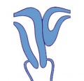
| Anterior view of development of the female uterus and vagina between Week 9 and 20.
The paramesonephric ducts (red) fuse in the midline to form the genital canal. The urogenital sinus (yellow), in contact with the paramesonephric duct, thickens to form the sinusal tubercle which extends as a solid vaginal plate, then becomes hollow as the sinovaginal bulb, finally forming the vagina.
|
Development Overview

|

|
| Internal Genital Tract Differentiation |
The data below gives an overview of the timecourse of embryonic human uterine development.[3]
- Carnegie stage 18 - Mullerian duct to the coelomic cavity was formed as the result of an invagination of the coelomic epithelium - stage 18
- Carnegie stages 19 - 23 - duct grows independently from the invagination - stage 19
- Week 20 - uterine horn fimbrial development begins and continues after birth - second trimester
- Carnegie Stages: 1 | 2 | 3 | 4 | 5 | 6 | 7 | 8 | 9 | 10 | 11 | 12 | 13 | 14 | 15 | 16 | 17 | 18 | 19 | 20 | 21 | 22 | 23 | About Stages | Timeline
Fetal Uterus
|
|

|
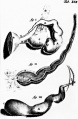
| Urogenital sinus of female human embryo of eight and a half to nine weeks old (From model by Keibel) (Image: Gray's Anatomy) | (Image modified from: Drews U, Sulak O, Schenck PA. Androgens and the development of the vagina.Biol Reprod. 2002 Oct;67(4):1353-9. PMID: 12297555) |
Fetal Uterus Growth

Graph shows the growth during the fetal period of the uterus between week 19 and 38.[4] During this time the uterine circumferunce increases from about 20 mm to just under 60mm and the width increases from less than 10mm to just over 20 mm.
Uterine horn fimbrial development begins after week 20 and continues after birth.
Uterine growth continues postnatally, increasing outer muscle thickness and cyclic changes in the lining with puberty.
Adult external uterine orifice to the fundus is approximately 6.25 cm.
Newborn Uterus

Uterine Tubes


The unfused portion of the paramesonephric ducts will form the uterine tubes. Note that there are several synonyms used for the paired uterine tubes or Fallopian tubes or oviducts or uterine horns.
In the adult, the uterine tube has been described in 4 anatomical regions.
- Infundibulum - funnel-shaped open end of the uterine tube with fimbriae (finger-like extensions), which are closely associated with the ovary. Opens into the peritoneal cavity (abdominal ostium, ostium abdominale)
- Ampulla - uterine tube with highly folded structure with plicae (mucosal folds) and secondary folds dividing the lumen, usual site for fertilization.
- Isthmus - narrow portion of the uterine tube with fewer mucosal folds and a thick muscularis layer.
- Intramural - uterine tube which passes through the muscular wall of the uterus. (an alternative interpretation is that it is an extension of the body of the uterus)
Mucosa
- formed by a ciliated and secretory epithelium resting on a very cellular lamina propria.
- The number of ciliated cells and non-ciliated secretory cells varies along the oviduct.
- Secretory activity varies during the menstrual cycle, and resting secretory cells are also referred to as peg-cells.
- Some of the secreted substances are thought to nourish the oocyte and the very early embryo.
Muscularis
- inner circular muscle layer and an outer longitudinal layer.
- An inner longitudinal layer is present in the isthmus and the intramural part of the oviduct.
- Peristaltic muscle action seems to be more important for the transport of sperm and oocyte than the action of the cilia.
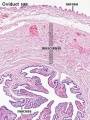
Uterine tube (monkey) histology overview
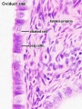
Uterine tube (monkey) epithelium and underlying histology


Uterine Blood Supply
Uterus Histology
See also Menstrual Cycle - Histology
Uterine tube histology overview showing epithelium and underlying muscular layers
Uterine tube epithelium histology showing secretory and ciliated cells

Uterine body endometrium and myometrium during the proliferative phase of the menstrual cycle overview

Uterine body endometrium during the proliferative phase of the menstrual cycle

Uterine body endometrium during the secretory phase of the menstrual cycle overview

Uterine body endometrium during the secretory phase of the menstrual cycle


Abnormalities

|
A range of uterine and vaginal anatomical anomalies based upon the abnormal development and fusion of the paramesonephric ducts and vaginal plate development.
|

|
Unicornate Uterus - failure of the paramesonephric ducts to fuse. A single paramesomnephric duct has fused with the vaginal plate and now opens into the vagina, while the other forms a diverticulum. |
Uterine Duplication
(uterus didelphys, double uterus, uterus didelphis) A rare uterine developmental abnormality where the paramesonephric ducts (Mullerian ducts) completely fail to fuse generating two separate uterus parts each connected to the cervix and having an ovary each.
Uterus/Vaginal
Mayer-Rokitansky-Kuster-Hauser syndrome (MRKH, MRK anomaly, Rokitansky-Kuster-Hauser syndrome, RKH syndrome, RKH) consists of congenital aplasia of the uterus and the upper part of vagina due to anomalous development of Müllerian ducts, either isolated or associated with other congenital malformations, including renal, skeletal, hearing and heart defects. Has an incidence of approximately 1 in 4500 newborn girls and has been associated with a microdeletion at 17q12.[5]
Septate Uterus
Cervical: cervical agenesis, cervical duplication
Environmental Abnormalities
DES Diethylstilbestrol or diethylstilbetrol, is a drug that was prescribed to women from 1938-1971 to prevent miscarriage in high-risk pregnancies. The drug acted as a potent estrogen (mimics natural hormone) and therefore could also act as a potential endocrine disruptor. This led to a number of developing fetal reproductive tract and other abnormalities. In the female fetus, it increased risk of abnormal reproductive tract and also carcinogenic (cancer forming). In the male fetus, it increased the occurance of abnormal genitalia. The drug was banned by FDA (USA) in 1979 as a teratogen, it had previously also been used as livestock growth promoter and could have potentially entered the human food chain. (More? [endocrine2.htm Endocrine Abnormalities] | Abnormal Development - Drugs)
Links: Endocrine Abnormalities | Abnormal Development - Drugs | Childrens Hospital Boston - Congenital Anomalies of the Uterus | Medical Education Image Link - Cervical agenesis | OMIM - Rokitansky-Kuster-Hauser syndrome |
Broad Ligament
| The broad ligament is found associated with the internal human female genital tract. It forms a mesentery consisting of a double fold of the peritoneum that connects the uterus to the peritoneal floor and walls.
Anatomically it has three parts:
Abnormalities include peritoneal endometriosis. |
File:Image1161.gif |
Molecular
Wnt genes - Wnt4, Wnt5a, and Wnt7a implicated in the formation and morphogenesis of the Müllerian duct.
Wnt7a - mediates the patterning of the oviduct and differentiation of the uterus.
beta-catenin - manufactured in the mesenchyme is a downstream effector of Wnt7a.
Bmp2 - decidualization regulator of gene expression and function (shown in mouse uterus).
Lim1, Lhx9, Emx, Pax-2, Hox-A9, Hox-A10, Hox-A11, Hox-A13, WT1, SF-1, GATA-4. TGF-beta
References
- ↑ Guioli S, Sekido R, Lovell-Badge R. The origin of the Mullerian duct in chick and mouse. Dev Biol. 2007 Feb 15;302(2):389-98. PMID: 17070514
- ↑ The origin of the Mullerian duct in chick and mouse. Guioli S, Sekido R, Lovell-Badge R. Dev Biol. 2006 Oct 3 PMID: 17070514
- ↑ Development of the human Mullerian duct in the sexually undifferentiated stage. Hashimoto R. Anat Rec A Discov Mol Cell Evol Biol. 2003 Jun;272(2):514-9. PMID: 12740945
- ↑ Development of the fetal uterus between 19 and 38 weeks of gestation: in-utero ultrasonographic measurements. Soriano D, Lipitz S, Seidman DS, Maymon R, Mashiach S, Achiron R. Hum Reprod. 1999 Jan;14(1):215-8. PMID: 10374123
- ↑ Recurrent microdeletion at 17q12 as a cause of Mayer-Rokitansky-Kuster-Hauser (MRKH) syndrome: two case reports. Bernardini L, Gimelli S, Gervasini C, Carella M, Baban A, Frontino G, Barbano G, Divizia MT, Fedele L, Novelli A, Béna F, Lalatta F, Miozzo M, Dallapiccola B. Orphanet J Rare Dis. 2009 Nov 4;4:25. PMID: 19889212
Reviews
- Lifetime changes in the vulva and vagina. Farage M, Maibach H. Arch Gynecol Obstet. 2006 Jan;273(4):195-202. PMID: 16208476
- Function of sexual glands and mechanism of sex differentiation. Kavlock R, Cummings AJ Toxicol Sci. 2004 Aug;29(3):167-78. Review. PMID: 15467266 | See Related Articles
Articles
- Essential roles of mesenchyme-derived beta-catenin in mouse Mullerian duct morphogenesis. Deutscher E, Hung-Chang Yao H. Dev Biol. 2007 May 3; PMID: 17532316
- Guioli S, Sekido R, Lovell-Badge R. The origin of the Mullerian duct in chick and mouse. Dev Biol. 2007 Feb 15;302(2):389-98.
- Hashimoto R. Development of the human Mullerian duct in the sexually undifferentiated stage. Anat Rec A Discov Mol Cell Evol Biol. 2003 Jun;272(2):514-9.
Search PubMed
Search May 2007 "embryonic uterine development" 3,025 reference articles of which 491 were reviews.
Search Pubmed: Uterus Development | embryonic uterine development | Paramesonephric Duct | Mullerian Duct | Endocrine Disruptors
Additional Images
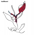
Urogenital indifferent
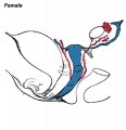
Urogenital female
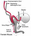
Mouse - paramesonephric duct
Unicornate uterus
Historic drawing of the uterine tube (Reinier De Graaf)
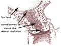
Cervical mucus plug


Internet Links
- Uterus Histology UWA Blue Histology - Female Reproductive Tract
Glossary Links
- Glossary: A | B | C | D | E | F | G | H | I | J | K | L | M | N | O | P | Q | R | S | T | U | V | W | X | Y | Z | Numbers | Symbols | Term Link
Genital Links: genital | Lecture - Medicine | Lecture - Science | Lecture Movie | Medicine - Practical | primordial germ cell | meiosis | endocrine gonad | Genital Movies | genital abnormalities | Assisted Reproductive Technology | puberty | Category:Genital
| ||||
|
Cite this page: Hill, M.A. (2024, June 23) Embryology Uterus Development. Retrieved from https://embryology.med.unsw.edu.au/embryology/index.php/Uterus_Development
- © Dr Mark Hill 2024, UNSW Embryology ISBN: 978 0 7334 2609 4 - UNSW CRICOS Provider Code No. 00098G