Chicken Development
| Embryology - 15 Jun 2024 |
|---|
| Google Translate - select your language from the list shown below (this will open a new external page) |
|
العربية | català | 中文 | 中國傳統的 | français | Deutsche | עִברִית | हिंदी | bahasa Indonesia | italiano | 日本語 | 한국어 | မြန်မာ | Pilipino | Polskie | português | ਪੰਜਾਬੀ ਦੇ | Română | русский | Español | Swahili | Svensk | ไทย | Türkçe | اردو | ייִדיש | Tiếng Việt These external translations are automated and may not be accurate. (More? About Translations) |
Introduction

The chicken (taxon -Gallus gallus) embryo develops and hatches in 20 to 21 days and has been extensively used in embryology studies. Historically, the chicken embryo was one of the first embryos studied, readily available and easy to incubate, embryo development can be directly observed by cutting a small window in the egg shell. A key to this model organism study was the establishment of a staging atlas by Hamburger & Hamilton in 1951 [1], which allowed specifc developmental landmarks to be seen and correlated with experimental manipulations of development. This much cited paper included images of all key stages and was more recently republished in the journal Developmental Dynamics (1993), for a new generation of avian researchers. Probably just as important has been the recent chicken genome sequencing, providing a resource to extend our knowledge of this excellent developmental model.
Fertilized eggs can be easily maintained in humidified incubators and during early stages of development the embryo floats on to of the egg yolk that it is using for nutrition. As the embryo grows it sinks into, or below the, yolk. The regular appearance of somites allowed early experimenters to acurately stage the embryo. The embryo was accessible and easy to manipulate (limb grafts/removal etc) that were informative about developmental processes. Chicken cells and tissues (neural ganglia/fragments) are also easy to grow in tissue culture. The discovery that quail cells have a different nuclear appearance meant that transplanted cells (chick/quail chimeras) could be tracked during development. For example, LeDourian's studies showed how neural crest cells migrate widely throughout the embryo.
The table below compares the chicken with other bird species.
| Bird | Days |
|---|---|
| Budgerigar | 18 |
| Chicken | 21 |
| Duck | 28 |
| Finch | 14 |
| Goose | 28 |
| Guinea fowl | 28 |
| Muscovy duck | 35 |
| Parrot | 26 |
| Pheasant | 24 |
| Pigeon | 18 |
| Quail | 16 |
| Swan | 35 |
| Turkey | 28 |
Chicken Stages
Chicken stages - Hamburger & Hamilton staged the chicken embryo in 1951. The original paper had approx 25 citations between 1955 - 59, while in the year 1991 alone there were over 300 citations. Series of Embryonic Chicken Growth. J. Morphology, 88 49 - 92 (1951). Atlas recently republished by J.R. Sanes in Developmental Dynamics 195 229-275 (1993).
The Hamburger Hamilton Stages are most commonly used series for chicken staging. Note that there was also an earlier Witschi staging, an older 1900 Normentafeln zur Entwicklungsgeschichte der Wirbeltiere - Gallus domesticus (Normal Plates of the Development of the Chicken Embryo} by Franz Keibel and Karl Abraham, and an even older (1883) series in The Elements of Embryology by Foster, Balfour, Sedgwick, and Heape.
Normal Plates of the Development of the Chicken Embryo} (1900)


- Links: Chicken Stages | Hamburger Hamilton | Witschi | 1900 | 1883 | PDF Poster- Hamburger Hamilton Stages | 2006 reproduction of the original paper
Some Recent Findings
|
| More recent papers |
|---|
 This table allows an automated computer search of the external PubMed database using the listed "Search term" text link.
More? References | Discussion Page | Journal Searches | 2019 References | 2020 References Search term: Chicken Embryology <pubmed limit=5>Chicken Embryology</pubmed> |
Gallus gallus
Taxonomy Id: 9031
Preferred common name: chicken
Rank: species
Genetic code: Translation table 1 (Standard) Mitochondrial genetic code: Translation table 2
Other names: dwarf Leghorn chickens (includes), red jungle fowl (includes), chickens (common name), Gallus domestics (misnomer), Gallus galls domesticus (misnomer)
Lineage (abbreviated ): Eukaryota; Metazoa; Chordata; Craniata; Vertebrata; Archosauria; Aves; Neognathae; Galliformes; Phasianidae; Phasianinae; Gallus
Other Chicken Atlases
Vertebrate and Invertebrate Embryos (7th Edition) G.C. Schoenwolf, Prentice Hall, New Jersey
An Atlas of Embryology (1975) W.H. Freeman and B. Bracegirdle, Heinemann Educational Books, UK.
This is an ATLAS (no description of development) , basically reprinted from the original 1963 edition.
Photos with labelled diagrams covering Amphioxus (worm) Frog, Chicken.
An Atlas for Staging Mammalian and Chick Embryos (1987) H. Bultler and B.H. Juurlink, CRC Press Inc., Florida
This ATLAS is not a complete series of development but has interesting comparisons of species.
Mostly photos of embryos with a few drawn diagrams and a series of staging correlation graphs.
Bird Evolution
Birds and Dinosaurs? as quoted in a Curent Biology review "...abundant and ever increasing evidence places birds as one surviving lineage of the diverse clade Dinosauria"[3][4]
Chicken Genomics
The first draft of the chicken genome was publicly released in March, 2004. There are a number of sites that have begun looking into establishing chicken genomics partly due to its powerful history as a model of vertebrate development that is easy to observe, manipulate and is also cheap. (see also NIH Proposal for Chicken Genomics | NCBI Chicken Genome Resources)
A summary of chicken genome resources has recently been identified in a review in Developmental Dynamics by Antin PB and Konieczka JH.[5]
Chicken Sex Determination
In chicken development sex determination depends on a ZZ male/ZW female mechanism.
This differs from mammalian sex determination which is based upon testis expression of an Sry gene in somatic supporting Sertoli cells.
In the gonad, the coelomic epithelium contributes only to non-steroidogenic interstitial cells and nephrogenous mesenchyme contributes both Sertoli cells and steroidogenic cells.
Genital

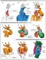
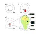
Primordial Germ Cell Migration Model[6]
| HH12–13 - yolk sac circulation courses in loop (red arrows) to enter the embryo via the heart. The majority of PGCs (green dots) localized axially at the border between the area opaca and pellucida, where the sinus terminalis converged in the anterior vitelline veins. | HH14–16 - PGCs (green dots) circulated effectively towards the embryo via the sinus terminalis and the anterior vitelline veins towards the heart. Then PGCs traffic via the aorta to the caudal part of the embryo and become lodged in the genital ridges. |
Chicken Heart
Note these are Hamburger Hamilton Stages of chicken development, see also Heart 3D reconstruction.
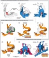
Chicken (day 2, Stage 12)
Chicken (day 3, Stage 16)
Chicken (day 4, Stage 21)
Chicken (day 5, Stage 25)


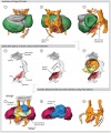
Chicken Somitogenesis

Chick somitogenesis oscillator[7] |
|

Chicken body elongation model[8]
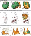
Chicken Limb

Limb Hairy2 Expression Model[9]
Hairy2 is a "molecular oscillator" involved in both somite and limb development.
Historic Studies
The Elements of Embryology - Volume 1 by Foster, M., Balfour, F. M., Sedgwick, A., & Heape, W. (1883)
The History of the Chick: Egg structure and incubation beginning | Summary whole incubation | First day | Second day - first half | Second day - second half | Third day | Fourth day | Fifth day | Sixth day to incubation end
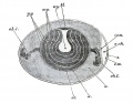
Fig. 1. Diagrammatic section of an unincubated fowl's egg
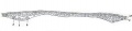
Fig. 3. Section of a blastoderm of a fowl's egg at the commencement of incubation
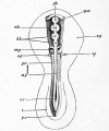
Fig. 27. Embryo of the chick between thirty and thirty-six hours, viewed from above as an opaque object.
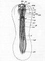
Fig. 28. an embryo chick of about thirty-six hours, viewed from below as a transparent object.
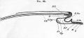
Fig. 29. Diagrammatic longitudinal section through the axis of an embryo.
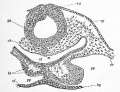
Fig. 30. Transverse section through the posterior part of the head of an embryo chick of thirty hours.
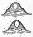
Fig. 31. Two sections of a thirty-six hours' embryo heart shortly after its formation. a is the anterior section.
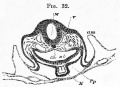
Fig. 32. Transverse section at the end of the second day through the bulbus arteriosus. (Copied from His.)
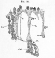
Fig. 33. Surface view from below of the posterior end pellucid area of a thirty-six hours' chick.


Elements of Embryology - Volume 1 - Figures
References
- ↑ <pubmed>1304821</pubmed>
- ↑ <pubmed>22046043</pubmed>
- ↑ <pubmed>16713939</pubmed>
- ↑ <pubmed>16893476</pubmed>
- ↑ <pubmed>15739221</pubmed>| Developmental Dynamics
- ↑ <pubmed>23213395</pubmed>| PMC3507194 | Biol Open
- ↑ <pubmed>20184730</pubmed>
- ↑ <pubmed>23118616</pubmed>| PLoS Biol.
- ↑ <pubmed>23213390</pubmed>| PMC3507187 | Biol Open
Search Pubmed
Search Pubmed: chicken development
Additional Images
Chicken endoderm origin

| Animal Development: axolotl | bat | cat | chicken | cow | dog | dolphin | echidna | fly | frog | goat | grasshopper | guinea pig | hamster | horse | kangaroo | koala | lizard | medaka | mouse | opossum | pig | platypus | rabbit | rat | salamander | sea squirt | sea urchin | sheep | worm | zebrafish | life cycles | development timetable | development models | K12 |
External Links
External Links Notice - The dynamic nature of the internet may mean that some of these listed links may no longer function. If the link no longer works search the web with the link text or name. Links to any external commercial sites are provided for information purposes only and should never be considered an endorsement. UNSW Embryology is provided as an educational resource with no clinical information or commercial affiliation.
- Developmental Dynamics - Chicken Special Issue (2004) | Poster- Hamburger Hamilton Stages | Republished Hamburger Hamilton Stages Paper
- Developmental Biology - Quail-Chick Chimeras
- Nicole Le Douarin pioneered the use of quail-chick chimeras to study the developmental fate of cells in the bird embryo. The videotape Nicole Le Douarin gave us permission to digitize is titled, "Quail-Chick Chimeras in Development of the Nervous System and Immune System" and it was made in 1987. These digital video sequences and still images come from the first part of her videotape. These chimeras were a key to our understanding cell migration (eg neural crest) in the embryo.
- Quicktime movie sequence 1 (477k) showing newly hatched quail-chick chimeras; white feathers are chick and dark, pigmented feathers are quail.
- Quicktime movie sequence 2 (1.3 MB) Sequence showing the preparation of the chick host; removing a portion of host's neural tube and neural crest.
- Quicktime movie sequence 3 (1.4 MB) Sequence showing the removal and "cleaning off" of donor quail neural tube and neural crest.
- Quicktime movie sequence 4 (1.5 MB) Sequence showing transplantation and grafting of donor quail neural tube and neural crest into the chick host; at the end of this sequence, you see the host chick embryo 5 hours later with its healed in graft.
- Developmental Biology- Laurie Iten's Serially Sectioned Frog and Chick Embryos
- Chicken genomic websites
- AvianNet http://www.ri.bbsrc.ac.uk/chickmap
- NCBI Chicken Genome Resources
- Genome browser - Washington University Genome Sequencing Center (WUGSC)
- Genome browser -  Ensembl
- http://www.ri.bbsrc.ac.uk/chickmap
- http://poultry.mph.msu.edu/mapsnew.html
- http://ag.ansc.purdue.edu/poultry/
- http://www.zod.wau.nl/vf/chickensite/chicken.html
- http://tetra.gig.usda.gov:8400/chickgbase/manager.html
Glossary Links
- Glossary: A | B | C | D | E | F | G | H | I | J | K | L | M | N | O | P | Q | R | S | T | U | V | W | X | Y | Z | Numbers | Symbols | Term Link
Cite this page: Hill, M.A. (2024, June 15) Embryology Chicken Development. Retrieved from https://embryology.med.unsw.edu.au/embryology/index.php/Chicken_Development
- © Dr Mark Hill 2024, UNSW Embryology ISBN: 978 0 7334 2609 4 - UNSW CRICOS Provider Code No. 00098G