User:Z3462474: Difference between revisions
No edit summary |
|||
| Line 28: | Line 28: | ||
'''''What animal models are available for muscular dystrophy?''''' | '''''What animal models are available for muscular dystrophy?''''' | ||
The mouse is probably the most widely used animal model for muscular dystrophy. Other animal models that have been utilised include the golden retriever dog and pigs. <ref name="PMID26140716"><pubmed>26140716</pubmed></ref> <ref name="PMID22968479"><pubmed>22968479</pubmed></ref> <ref name="PMID27634466"><pubmed>27634466</pubmed></ref> | The mouse is probably the most widely used animal model for muscular dystrophy. Other animal models that have been utilised include the golden retriever dog and pigs. <ref name="PMID26140716"><pubmed>26140716</pubmed></ref> <ref name="PMID22968479"><pubmed>22968479</pubmed></ref> <ref name="PMID27634466"><pubmed>27634466</pubmed></ref> | ||
==Lab 6 Assessment== | ==Lab 6 Assessment== | ||
Revision as of 16:44, 22 September 2016
| Student Information (expand to read) | ||||||||||||||||||||||||||||||||||||||||||||||||||||||||||||||||
|---|---|---|---|---|---|---|---|---|---|---|---|---|---|---|---|---|---|---|---|---|---|---|---|---|---|---|---|---|---|---|---|---|---|---|---|---|---|---|---|---|---|---|---|---|---|---|---|---|---|---|---|---|---|---|---|---|---|---|---|---|---|---|---|---|
| Individual Assessments | ||||||||||||||||||||||||||||||||||||||||||||||||||||||||||||||||
|
Please leave this template on top of your student page as I will add your assessment items here. Beginning your online work - Working Online in this course
Click here to email Dr Mark Hill | ||||||||||||||||||||||||||||||||||||||||||||||||||||||||||||||||
| Lab 1 Assessment - Researching a Topic | ||||||||||||||||||||||||||||||||||||||||||||||||||||||||||||||||
In the lab I showed you how to find the PubMed reference database and search it using a topic word. Lab 1 assessment will be for you to use this to find a research reference on "fertilization" and write a brief summary of the main finding of the paper.
| ||||||||||||||||||||||||||||||||||||||||||||||||||||||||||||||||
| Lab 2 Assessment - Uploading an Image | ||||||||||||||||||||||||||||||||||||||||||||||||||||||||||||||||
OK you are now in a group
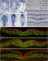
Initially the topic can be as specific or as broad as you want. Chicken embryo E-cad and P-cad gastrulation[1] References
| ||||||||||||||||||||||||||||||||||||||||||||||||||||||||||||||||
| Lab 4 Assessment - GIT Quiz | ||||||||||||||||||||||||||||||||||||||||||||||||||||||||||||||||
|
ANAT2341 Quiz Example | Category:Quiz | ANAT2341 Student 2015 Quiz Questions | Design 4 quiz questions based upon gastrointestinal tract. Add the quiz to your own page under Lab 4 assessment and provide a sub-sub-heading on the topic of the quiz. An example is shown below (open this page in view code or edit mode). Note that it is not just how you ask the question, but also how you explain the correct answer. | ||||||||||||||||||||||||||||||||||||||||||||||||||||||||||||||||
| Lab 5 Assessment - Course Review | ||||||||||||||||||||||||||||||||||||||||||||||||||||||||||||||||
| Complete the course review questionnaire and add the fact you have completed to your student page. | ||||||||||||||||||||||||||||||||||||||||||||||||||||||||||||||||
| Lab 6 Assessment - Cleft Lip and Palate | ||||||||||||||||||||||||||||||||||||||||||||||||||||||||||||||||
| ||||||||||||||||||||||||||||||||||||||||||||||||||||||||||||||||
| Lab 7 Assessment - Muscular Dystrophy | ||||||||||||||||||||||||||||||||||||||||||||||||||||||||||||||||
| ||||||||||||||||||||||||||||||||||||||||||||||||||||||||||||||||
| Lab 8 Assessment - Quiz | ||||||||||||||||||||||||||||||||||||||||||||||||||||||||||||||||
| A brief quiz was held in the practical class on urogenital development. | ||||||||||||||||||||||||||||||||||||||||||||||||||||||||||||||||
| Lab 9 Assessment - Peer Assessment | ||||||||||||||||||||||||||||||||||||||||||||||||||||||||||||||||
| ||||||||||||||||||||||||||||||||||||||||||||||||||||||||||||||||
| Lab 10 Assessment - Stem Cells | ||||||||||||||||||||||||||||||||||||||||||||||||||||||||||||||||
As part of the assessment for this course, you will give a 15 minutes journal club presentation in Lab 10. For this you will in your current student group discuss a recent (published after 2011) original research article (not a review!) on stem cell biology or technology.
| ||||||||||||||||||||||||||||||||||||||||||||||||||||||||||||||||
| Lab 11 Assessment - Heart Development | ||||||||||||||||||||||||||||||||||||||||||||||||||||||||||||||||
| Read the following recent review article on heart repair and from the reference list identify a cited research article and write a brief summary of the paper's main findings. Then describe how the original research result was used in the review article.
<pubmed>26932668</pubmed>Development | ||||||||||||||||||||||||||||||||||||||||||||||||||||||||||||||||
| ||||||||||||||||||||||||||||||||||||||||||||||||||||||||||||||||
Lab 7 Assessment
Muscular Dystrophy
What is/are the dystrophin mutation(s)?
The dystrophin gene mutations cause both Duchenne (DMD) and Becker (BMD) muscular dystrophies. The dystrophin gene is the longest known human gene of 2.4 Mb’s on chromosome X, and it codes for the protein dystrophin which is expressed in all striated skeletal, smooth and cardiac muscle cells. There are also isoforms expressed in the retina and brain cells. The dystrophin mutations are usually either: deletions of one or more exons (approximately 65%); duplications of exons (approximately 10%); or single point mutations (approximately 15%). If such mutations are out-of-frame they lead to severe deficiency of dystrophin causing DMD disease. The less severe BMD is usually a result of in-frame mutations. [1] These mutations are recessive and affect approximately 1 in 3500 to 5000 males born worldwide. [2]
What is the function of dystrophin?
The dystrophin protein is a component of the large protein complex: the dystrophin-glycoprotein complex (DGC). The DGC spans the sarcolemma (the cell membrane of skeletal muscle fibre cells) binds to surrounding ligands and to dystrophin inside the cell. [3] This complex is responsible for stabilising the sarcolemma by integrating the components of the cytoskeleton of muscle cells. These interactions of the dystrophin protein with components such as the actin filaments and microtubules of the muscle cell are mediated by dystrophin’s four main functional domains. It has also been found that dystrophin may bind membrane phospholipids to further support its stabilisation of the sarcolemma. [2]
What other tissues/organs are affected by this disorder?
The heart has been found to be affected by some forms muscular dystrophy, including cardiac complications such as heart failure and/or arrhythmias. [4] Moreover, respiratory failure due to the breathing muscles becoming weak severely limits the lifespan of DMD patients. [2]
What therapies exist for DMD?
Extensive research surrounds the use of gene replacement therapy for all mutation-types of DMD. However, the extremely long nature of the dystrophin gene renders it difficult to replace and many other challenges remain such as with the nature of the viral vectors used in gene replacement. A number of other genetics-based approaches are being investigated such as gene product modifiers (exon-skipping) and utrophin modulation strategies. [5] [2] Pharmacological treatment for DMD involves administration of corticosteroids for suppression of the associated inflammation in the muscles, however this has significant side effects and questionable therapeutic efficacy. [6]
What animal models are available for muscular dystrophy?
The mouse is probably the most widely used animal model for muscular dystrophy. Other animal models that have been utilised include the golden retriever dog and pigs. [2] [7] [8]
Lab 6 Assessment
Identify a known genetic mutation that is associated with cleft lip or palate
The SATB2 gene mutation has been shown to be associated with Non-syndromic cleft lip with or without cleft palate.
Identify a recent research article on this gene
How does this mutation affect developmental signalling in normal development
SATB2 is located at the chromosome position 2q33.1 and encodes an AT-rich sequence binding protein of 733 amino acids. This protein (Satb2) helps to regulate the transcription of large domains of chromatin and is the first cell-type-specific transcription factor to do so. [9] More specifically, Satb2 carries out transcriptional regulation directly through modulating chromatin remodelling by binding AT-rich sequences in nuclear matrix-attachment regions (MARs) of DNA. It also indirectly, through association with other transcription regulators, modulates cis-regulation elements to control the expression of target genes and downstream biological processes. SATB2 plays a crucial role in development and tissue regeneration, especially in craniofacial development and patterning, formation of the palate, and differentiation and maturation of osteoblasts. It also appears to contribute to development of the central nervous system -particularly the formation of the corpus callosum and pons-, as well as cancer prognosis and development, and the regulation of immune function. [10] Thus mutation of this gene will lead to perturbations in these functional roles it plays in development.
Lab 5 Assessment
Completed ANAT2341 Lab 5 - Course Feedback Questionnaire
Lab 4 Assessment
Gastrointestinal Tract Quiz
Lab 3 Assessment
| Mark Hill 31 August 2016 - Lab 3 Assessment Quiz - Mesoderm and Ectoderm development. All correct, well done! | Assessment 5/5 |
Lab 2 Assessment
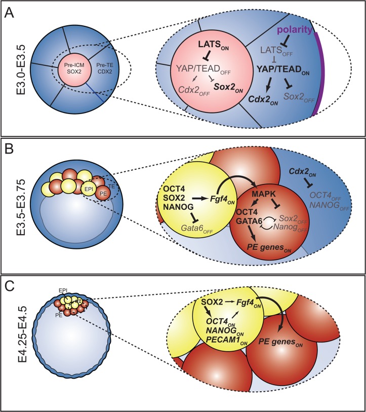
Roles and Regulation of SOX2 in Blastocyst Formation[11]
| Mark Hill 29 August 2016 - All information Reference, Copyright and Student Image template correctly included with the file and referenced on your page here. Note though, that the reference subheading in the file summary box should have just <pubmed>25340657</pubmed> to display the reference correctly. You only include the ref name for a citation as shown correctly on your page here. | Assessment 5/5 |
Lab 1 Assessment
<pubmed>25818081</pubmed>
Main Findings: This investigation compared 121 infertile patients, who were diagnosed with Inflammatory Bowel Disease (IBD) preceding their first IVF cycle, with 470 non-IBD infertile patients receiving IVF. Of the women with IBD, 71 had ulcerative colitis, 49 had Crohn’s disease, and 1 was unclassified. A majority of the IBD patients were not taking any medications during their IVF treatment. Non-IBD and IBD patients had similar age, parity and follicle-stimulating hormone levels on cycle day three. One characteristic that differed slightly was BMI, which was lower in those with ulcerative colitis.
Overall it was found that infertile women with IBD had similar rates of pregnancy and live births after IVF as those of non-IBD infertile women. Cumulative live birth rates were also similar. The study also concluded that it was more common among IBD patients, particularly those with Crohn’s disease, to experience tubal factor infertility as compared to non-IBD patients. Furthermore, live birth rates after IVF treatment were not influenced by prior surgery in patients with Crohn’s disease or ulcerative colitis. The results of this study were impacted by several limitations such as: its retrospective approach; confounding factors due to unmeasured variables like activity status; inability to obtain information regarding tobacco usage; and limited generalisability due to the cohort selection procedure.
| Mark Hill 18 August 2016 - You have added the citation correctly and written a good brief summary of the article findings. An interesting paper looking at any possible associations between IVF success and other existing medical conditions, note also that the authors have noted the limitations of their research study findings. | Assessment 5/5 |
Lab Attendance
Z3462474 (talk) 14:34, 5 August 2016 (AEST)
Z3462474 (talk) - I made a mistake in the second lab and did not do four squiggles (only three)
Z3462474 (talk) 13:13, 19 August 2016 (AEST)
Z3462474 (talk) 13:07, 26 August 2016 (AEST)
Z3462474 (talk) 13:06, 2 September 2016 (AEST)
Z3462474 (talk) 13:08, 9 September 2016 (AEST)
New Sub-Heading
Internal Link
https://embryology.med.unsw.edu.au/embryology/index.php/ANAT2341_Lab_1
Referencing
PMID 27486480
- ↑ <pubmed>26295289</pubmed>
- ↑ 2.0 2.1 2.2 2.3 2.4 <pubmed>26140716</pubmed>
- ↑ <pubmed>25086336</pubmed>
- ↑ <pubmed>27340611</pubmed>
- ↑ <pubmed>26140505</pubmed>
- ↑ <pubmed>27621596</pubmed>
- ↑ <pubmed>22968479</pubmed>
- ↑ <pubmed>27634466</pubmed>
- ↑ <pubmed>26605140</pubmed>
- ↑ <pubmed>24411565</pubmed>
- ↑ <pubmed>25340657</pubmed>


