Neural - Vascular Development: Difference between revisions
mNo edit summary |
mNo edit summary |
||
| Line 18: | Line 18: | ||
|-bgcolor="F5FAFF" | |-bgcolor="F5FAFF" | ||
| | | | ||
* '''Formation of the circle of Willis during human embryonic development'''<ref name=PMID27037515><pubmed>27037515</pubmed></ref> "The circle of Willis (CW) is a circulatory anastomosis that supplies blood to the brain and adjacent structures. We examined the timing of formation of CW in 20 Japanese human embryo samples by using 3-dimensional reconstruction of serial histological sections. The CW was closed in 1 (n = 6), 2 (n = 8), 2 (n = 3) and 2 (n = 3) samples at Carnegie stages 20, 21, 22, and 23, respectively. The CW was unclosed in 13 samples (unclosed at ACOM alone, 6 samples; ACOM and bilateral P1, 4; left PCOM and right P1, 1; right PCOM and right P1, 1; ACOM and left PCOM, 1). It was difficult to predict whether the circle would close during further development, as such variations frequently exist in adults." | |||
* '''Foxc1 is required for early stage telencephalic vascular development'''<ref name=PMID25733312><pubmed>25733312</pubmed></ref> "The brain vascular system arises from the perineural vascular plexus (PNVP) which sprouts radially into the neuroepithelium and subsequently branches off laterally to form a secondary plexus in the subventricular zone (SVZ), the subventricular vascular plexus (SVP). The process of SVP formation remains to be fully elucidated. We investigated the role of Foxc1 in early stage vascular formation in the ventral telencephalon. Results: The Foxc1 loss of function mutant mouse, Foxc1ch/ch , showed enlarged telencephalon and hemorrhaging in the ventral telencephalon by E11.0. The mutant demonstrated blood vessel dilation and aggregation of endothelial cells in the SVZ after the invasion of endothelial cells through the radial path, which lead to failure of SVP formation. During this early stage of vascular development, Foxc1 was expressed in endothelial cells and pericytes, as well as in cranial mesenchyme surrounding the neural tube." | * '''Foxc1 is required for early stage telencephalic vascular development'''<ref name=PMID25733312><pubmed>25733312</pubmed></ref> "The brain vascular system arises from the perineural vascular plexus (PNVP) which sprouts radially into the neuroepithelium and subsequently branches off laterally to form a secondary plexus in the subventricular zone (SVZ), the subventricular vascular plexus (SVP). The process of SVP formation remains to be fully elucidated. We investigated the role of Foxc1 in early stage vascular formation in the ventral telencephalon. Results: The Foxc1 loss of function mutant mouse, Foxc1ch/ch , showed enlarged telencephalon and hemorrhaging in the ventral telencephalon by E11.0. The mutant demonstrated blood vessel dilation and aggregation of endothelial cells in the SVZ after the invasion of endothelial cells through the radial path, which lead to failure of SVP formation. During this early stage of vascular development, Foxc1 was expressed in endothelial cells and pericytes, as well as in cranial mesenchyme surrounding the neural tube." | ||
* '''Review - The human brain intracerebral microvascular system: development and structure.'''<ref name=PMID22993505><pubmed>22993505</pubmed>| [http://www.ncbi.nlm.nih.gov/pmc/articles/PMC3440694 PMC3440694] | [http://journal.frontiersin.org/article/10.3389/fnana.2012.00038/abstract Front Neuroanat.]</ref> "The capillary from the meningeal inner pial lamella play a crucial role in the development and structural organization of the cerebral cortex extrinsic and intrinsic microvascular compartments. Only pial capillaries are capable of perforating through the cortex external glial limiting membrane (EGLM) to enter into the nervous tissue, although incapable of perforating the membrane to exit the brain. Circulatory dynamics and functional demands determine which capillaries become arterial and which capillaries become venous." | * '''Review - The human brain intracerebral microvascular system: development and structure.'''<ref name=PMID22993505><pubmed>22993505</pubmed>| [http://www.ncbi.nlm.nih.gov/pmc/articles/PMC3440694 PMC3440694] | [http://journal.frontiersin.org/article/10.3389/fnana.2012.00038/abstract Front Neuroanat.]</ref> "The capillary from the meningeal inner pial lamella play a crucial role in the development and structural organization of the cerebral cortex extrinsic and intrinsic microvascular compartments. Only pial capillaries are capable of perforating through the cortex external glial limiting membrane (EGLM) to enter into the nervous tissue, although incapable of perforating the membrane to exit the brain. Circulatory dynamics and functional demands determine which capillaries become arterial and which capillaries become venous." | ||
Revision as of 13:20, 13 April 2016
| Embryology - 15 Jun 2024 |
|---|
| Google Translate - select your language from the list shown below (this will open a new external page) |
|
العربية | català | 中文 | 中國傳統的 | français | Deutsche | עִברִית | हिंदी | bahasa Indonesia | italiano | 日本語 | 한국어 | မြန်မာ | Pilipino | Polskie | português | ਪੰਜਾਬੀ ਦੇ | Română | русский | Español | Swahili | Svensk | ไทย | Türkçe | اردو | ייִדיש | Tiếng Việt These external translations are automated and may not be accurate. (More? About Translations) |
Introduction

Draft Page
See the historic articles on human vascular development by:
- Mall FP. On the Development of the Blood-Vessels of the Brain in the Human Embryo. (1905) Amer. J. of Anat. 4; 1–18.
- Streeter GL. The Developmental Alterations in the Vascular System of the Brain of the Human Embryo (1921)

Some Recent Findings
|
| More recent papers |
|---|
 This table allows an automated computer search of the external PubMed database using the listed "Search term" text link.
More? References | Discussion Page | Journal Searches | 2019 References | 2020 References Search term: Neural Vascular System Development <pubmed limit=5>Neural Vascular System Development</pubmed> |
Cerebral Blood Supply Development

|
Embryonic stage
|
|
(above text modified from reference[4])
Cerebral Veins
Figures from the 1905 study by Mall.[5]
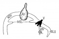
Fig 14. Veins of the head of an embryo four weeks old.
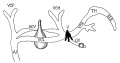
Fig 15. Veins of the head during the fifth week.
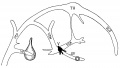
Fig 16. Veins of the head beginning of third month.
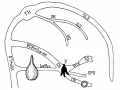
Fig 17. Veins of the brain of an older foetus.


Molecular
Foxc1
- required for early stage telencephalic vascular development[2]
References
- ↑ <pubmed>27037515</pubmed>
- ↑ 2.0 2.1 <pubmed>25733312</pubmed>
- ↑ <pubmed>22993505</pubmed>| PMC3440694 | Front Neuroanat.
- ↑ <pubmed>26060802</pubmed>| J Stroke.
- ↑ Mall FP. On the Development of the Blood-Vessels of the Brain in the Human Embryo. (1905) Amer. J. of Anat. 4; 1–18.
Online Textbooks
Reviews
<pubmed></pubmed> <pubmed></pubmed> <pubmed></pubmed> <pubmed></pubmed> <pubmed>22993505</pubmed> <pubmed>20561492</pubmed>
Articles
<pubmed></pubmed> <pubmed></pubmed> <pubmed></pubmed> <pubmed></pubmed> <pubmed>468705</pubmed>
Search PubMed
Search Pubmed: neural vascular system development |
External Links
External Links Notice - The dynamic nature of the internet may mean that some of these listed links may no longer function. If the link no longer works search the web with the link text or name. Links to any external commercial sites are provided for information purposes only and should never be considered an endorsement. UNSW Embryology is provided as an educational resource with no clinical information or commercial affiliation.
Additional Images
Historic
| Historic Disclaimer - information about historic embryology pages |
|---|
|
The Developmental Alterations in the Vascular System of the Brain of the Human Embryo (1921)
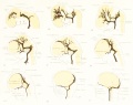
Plate 1
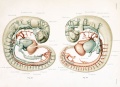
Plate 2
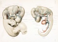
Plate 3
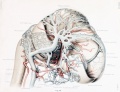
Plate 4
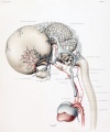
Plate 5


Gillilan, LA. Significant superficial anastomoses in the arterial blood supply to the human brain. J Comp Neurol. 1959 Jun;112:55-74. PMID 13850118
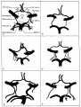
Fig. 1. Different patterns of the Circle of Willis
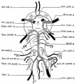
Fig. 2. Complex of arteries on the Ventral Surface of the Brain
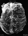
Fig. 3. Fetal Brain (6 month) Large Superficial Arteries


Gray, Henry. Anatomy of the Human Body Philadelphia: Lea & Febiger, 1918.
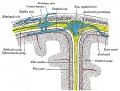
Fig. 769. Diagrammatic representation of a section across the top of the skull

Terms
Glossary Links
- Glossary: A | B | C | D | E | F | G | H | I | J | K | L | M | N | O | P | Q | R | S | T | U | V | W | X | Y | Z | Numbers | Symbols | Term Link
Cite this page: Hill, M.A. (2024, June 15) Embryology Neural - Vascular Development. Retrieved from https://embryology.med.unsw.edu.au/embryology/index.php/Neural_-_Vascular_Development
- © Dr Mark Hill 2024, UNSW Embryology ISBN: 978 0 7334 2609 4 - UNSW CRICOS Provider Code No. 00098G