Carnegie stage 16: Difference between revisions
| Line 34: | Line 34: | ||
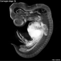
View: This is a dorsolateral view of embryo. Amniotic membrane removed. | View: This is a dorsolateral view of embryo. Amniotic membrane removed. | ||
Image source: | Image source: Embryology page Created: 19.03.1999 | ||
[[Human stage16 face 01.jpg]] | |||
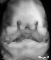
Ventral view of head region. | |||
{{Kyoto collection}} | {{Kyoto collection}} | ||
Revision as of 18:27, 15 May 2013
Introduction

Facts
Week 6, 37 - 42 days, 8 - 11 mm
Gestational Age - week 8
Events
Ectoderm: sensory placodes, lens pit, otocyst,nasal pits moved ventrally, fourth ventricle of brain
Mesoderm: heart prominence
Head: 1st, 2nd and 3rd pharyngeal arch, forebrain, eye, auricular hillocks
Body: heart, liver, umbilical cord, mesonephric ridge
Limb: upper and lower limb buds, hand plate, developing arm
Features
Eye showing retinal pigment, nasolacrimal groove, nasal pit, fourth ventricle of brain, umbilical cord, 1st and 2nd pharyngeal arches, cervical sinus, heart, developing arm with hand plate, foot plate
Identify: nasal pit, nasolacrimal groove, eye, 1st, 2nd and 3rd pharyngeal arches, 1st pharyngeal groove, maxillary and mandibular components of 1st pharyngeal arch, auricular hillocks, fourth ventricle of brain, heart prominence, upper limb bud, mesonephric ridge, lower limb bud, umbilical cord
- Links: Week 6 | Head | Lecture - Limb | Lecture - Gastrointestinal | Lecture - Head Development | Science Practical - Gastrointestinal | Science Practical - Head | Category:Carnegie Stage 16 | Stage 17
- Carnegie Stages: 1 | 2 | 3 | 4 | 5 | 6 | 7 | 8 | 9 | 10 | 11 | 12 | 13 | 14 | 15 | 16 | 17 | 18 | 19 | 20 | 21 | 22 | 23 | About Stages | Timeline
Kyoto Collection

View: This is a dorsolateral view of embryo. Amniotic membrane removed.
Image source: Embryology page Created: 19.03.1999
Ventral view of head region.
Image source: The Kyoto Collection images are reproduced with the permission of Prof. Kohei Shiota and Prof. Shigehito Yamada, Anatomy and Developmental Biology, Kyoto University Graduate School of Medicine, Kyoto, Japan for educational purposes only and cannot be reproduced electronically or in writing without permission.
Scanning EM

Ventral view of head showing upper lip, maxilla and nasal region.
Image Source: Prof Virginia Diewert
Carnegie Collection


| iBook - Carnegie Embryos | |
|---|---|

|
|
Additional Images
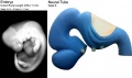
Neural tube model
Stage 16 Optical Projection Tomography
Stage16 cleft palate

External ear Stages 14-23 and adult
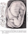
Historic drawing embryo 10mm CRL


- Carnegie Stages: 1 | 2 | 3 | 4 | 5 | 6 | 7 | 8 | 9 | 10 | 11 | 12 | 13 | 14 | 15 | 16 | 17 | 18 | 19 | 20 | 21 | 22 | 23 | About Stages | Timeline
Cite this page: Hill, M.A. (2024, June 14) Embryology Carnegie stage 16. Retrieved from https://embryology.med.unsw.edu.au/embryology/index.php/Carnegie_stage_16
- © Dr Mark Hill 2024, UNSW Embryology ISBN: 978 0 7334 2609 4 - UNSW CRICOS Provider Code No. 00098G