File:Adult heart CT01.jpg: Difference between revisions
(Z8600021 uploaded a new version of File:Adult heart CT01.jpg) |
mNo edit summary |
||
| (3 intermediate revisions by the same user not shown) | |||
| Line 1: | Line 1: | ||
==Adult Human Heart Computed Tomography== | |||
Heart anterior view. Image from a data set obtained using multidetector computed tomography in a patient undergoing investigation for coronary arterial disease. | |||
The pericardial reflections divide the ascending aorta into intrapericardial and extrapericardial components. The pulmonary trunk branches at the margins of the pericardial cavity into the right and left pulmonary arteries. | |||
Yellow shows the boundary of the pericardial cavity. | |||
{{Heart Links}} | |||
===Reference=== | |||
<pubmed>27587491</pubmed> | |||
https://www.ncbi.nlm.nih.gov/pmc/articles/PMC5011314/ | |||
http://journals.sagepub.com/doi/abs/10.1177/2150135116651114 | |||
PMID 27587491 | PMID 27587491 | ||
Figure 10. | ====Copyright==== | ||
© The Author(s) 2016 | |||
https://creativecommons.org/licenses/by/3.0/ | |||
Figure 10. cropped and resized, labeled with PMID. Text above modified from figure legend. | |||
{{Footer}} | |||
[[Category:Cardiovascular]][[Category:Heart]][[Category:Adult]] | |||
[[Category:Computed Tomography]] | |||
Latest revision as of 15:47, 29 Ocak 2017
Adult Human Heart Computed Tomography
Heart anterior view. Image from a data set obtained using multidetector computed tomography in a patient undergoing investigation for coronary arterial disease.
The pericardial reflections divide the ascending aorta into intrapericardial and extrapericardial components. The pulmonary trunk branches at the margins of the pericardial cavity into the right and left pulmonary arteries.
Yellow shows the boundary of the pericardial cavity.
Reference
<pubmed>27587491</pubmed>
https://www.ncbi.nlm.nih.gov/pmc/articles/PMC5011314/
http://journals.sagepub.com/doi/abs/10.1177/2150135116651114
PMID 27587491
Copyright
© The Author(s) 2016
https://creativecommons.org/licenses/by/3.0/
Figure 10. cropped and resized, labeled with PMID. Text above modified from figure legend.
Cite this page: Hill, M.A. (2026, October 6) Embryology Adult heart CT01.jpg. Retrieved from https://embryology.med.unsw.edu.au/embryology/index.php/File:Adult_heart_CT01.jpg
- © Dr Mark Hill 2026, UNSW Embryology ISBN: 978 0 7334 2609 4 - UNSW CRICOS Provider Code No. 00098G
File history
Yi efo/eka'e gwa ebo wo le nyangagi wuncin ye kamina wunga tinya nan
| Gwalagizhi | Nyangagi | Dimensions | User | Comment | |
|---|---|---|---|---|---|
| current | 15:43, 29 Ocak 2017 | 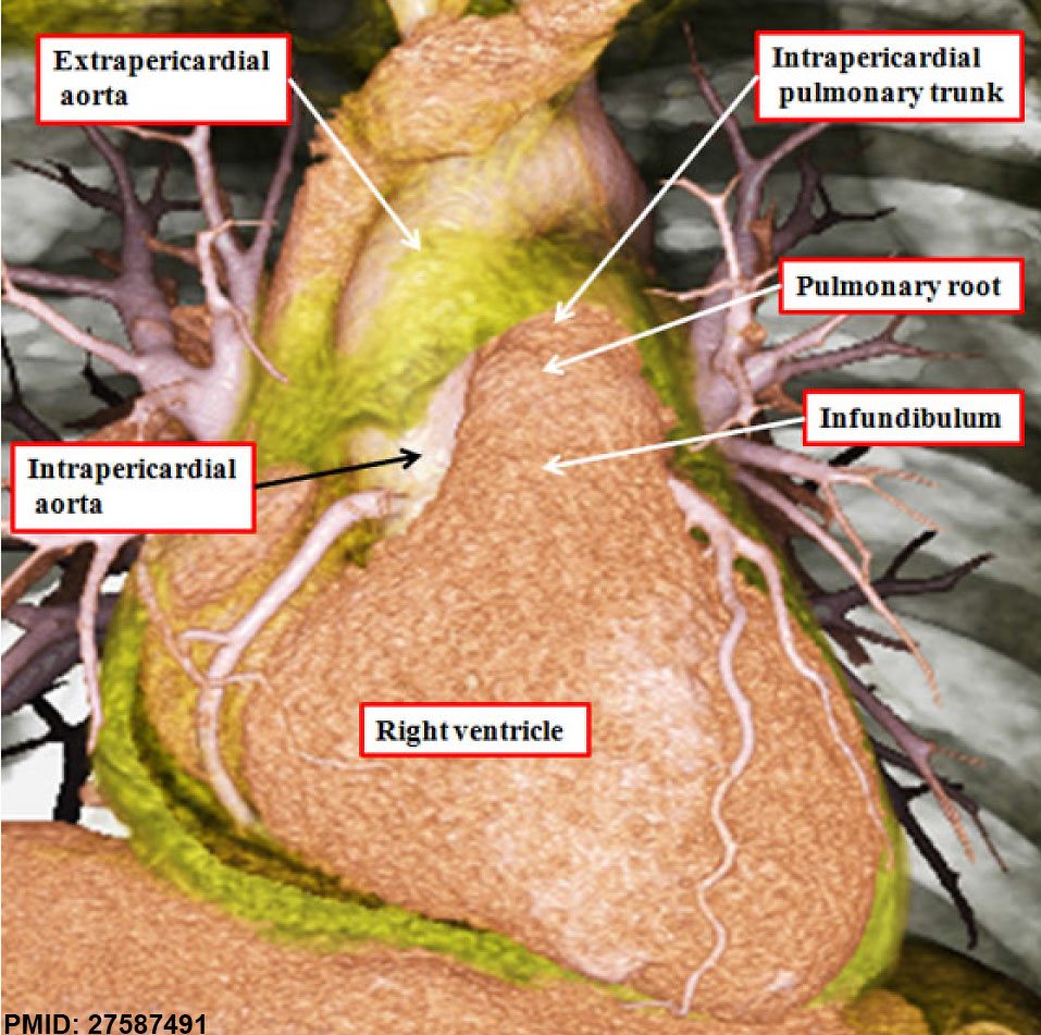 | 957 × 951 (212 KB) | Z8600021 (talk | contribs) | |
| 15:42, 29 Ocak 2017 | 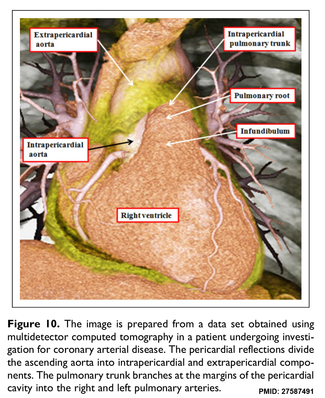 | 1,108 × 1,357 (307 KB) | Z8600021 (talk | contribs) | The image is prepared from a data set obtained using multidetector computed tomography in a patient undergoing investigation for coronary arterial disease. The pericardial reflections divide the ascending aorta into intrapericardial and extrapericardia... |
You cannot overwrite this file.
File usage
There are no pages that use this file.


