Vision - Extraocular Muscle Development: Difference between revisions
mNo edit summary |
mNo edit summary |
||
| (13 intermediate revisions by the same user not shown) | |||
| Line 4: | Line 4: | ||
A study by Gilbert (1957)<ref> | A study by Gilbert (1957)<ref name="Gilbert1957">{{Ref-Gilbert1957}}</ref> described the origin and development of the human extrinsic ocular muscles. | ||
| Line 14: | Line 14: | ||
|-bgcolor="F5FAFF" | |-bgcolor="F5FAFF" | ||
| | | | ||
* ''' | |||
* '''Eye alignment changes caused by sustained GDNF treatment of an extraocular muscle in infant non-human primates'''{{#pmid:32681083|PMID32681083}} "The ability of sustained treatment of a single extraocular muscle with glial cell line-derived neurotrophic factor (GDNF) to produce a strabismus in infant non-human primates was tested. Six infant non-human primates received a pellet containing GDNF, releasing 2 µg/day for 90 days, on one medial rectus muscle. Eye alignment was assessed up to 6 months. Five of the six animals showed a slow decrease in eye misalignment from the significant exotropia present at birth, ending with approximately 10° of exotropia. Controls became orthotropic. Misalignment averaged 8° three months after treatment ended. After sustained GDNF treatment, few changes were seen in mean myofiber cross-sectional areas compared to age-matched naïve controls. Neuromuscular junction number was unaltered in the medial rectus muscles, but were significantly reduced in the untreated lateral recti. Neuromuscular junctions on slow fibers became multiply innervated after this sustained GDNF treatment. Pitx2-positive cells significantly decreased in treated and contralateral medial rectus muscles. Our study suggests that balanced GDNF signaling plays a role in normal development and maintenance of orthotropia. Sustained GDNF treatment of one medial rectus muscle resulted in a measurable misalignment largely maintained 3 months after treatment ended. Structural changes suggest mechanisms for producing an imbalance in muscle function." | |||
* '''Twist3 is required for dedifferentiation during extraocular muscle regeneration in adult zebrafish'''{{#pmid:32320444|PMID32320444}} "Severely damaged adult zebrafish extraocular muscles (EOMs) regenerate through dedifferentiation of residual myocytes involving a muscle-to-mesenchyme transition. Members of the Twist family of basic helix-loop-helix transcription factors (TFs) are key regulators of the epithelial-mesenchymal transition (EMT) and are also involved in craniofacial development in humans and animal models. During zebrafish embryogenesis, twist family members (twist1a, twist1b, twist2, and twist3) function to regulate craniofacial skeletal development. Because of their roles as master regulators of stem cell biology, we hypothesized that twist TFs regulate adult EOM repair and regeneration. In this study, utilizing an adult zebrafish EOM regeneration model, we demonstrate that inhibiting twist3 function using translation-blocking morpholino oligonucleotides (MOs) impairs muscle regeneration by reducing myocyte dedifferentiation and proliferation in the regenerating muscle. This supports our hypothesis that twist TFs are involved in the early steps of dedifferentiation and highlights the importance of twist3 during EOM regeneration." | |||
|} | |} | ||
{| class="wikitable mw-collapsible mw-collapsed" | {| class="wikitable mw-collapsible mw-collapsed" | ||
! More recent papers | ! More recent papers | ||
|- | |- | ||
| [[File:Mark_Hill.jpg|90px|left]] {{Most_Recent_Refs}} | | [[File:Mark_Hill.jpg|90px|left]] {{Most_Recent_Refs}} | ||
Search term: ''Extraocular Muscle Embryology'' | Search term: [http://www.ncbi.nlm.nih.gov/pubmed/?term=Extraocular+Muscle+Development ''Extraocular Muscle Development''] | [http://www.ncbi.nlm.nih.gov/pubmed/?term=Extraocular+Muscle+Embryology ''Extraocular Muscle Embryology''] | [http://www.ncbi.nlm.nih.gov/pubmed/?term=superior+rectus+development ''superior rectus development''] | [http://www.ncbi.nlm.nih.gov/pubmed/?term=lateral+rectus+development ''lateral rectus development''] | [http://www.ncbi.nlm.nih.gov/pubmed/?term=medial+rectus+development ''medial rectus development''] | [http://www.ncbi.nlm.nih.gov/pubmed/?term=superior+oblique+development ''superior oblique development''] | [http://www.ncbi.nlm.nih.gov/pubmed/?term=inferior+oblique+development ''inferior oblique development''] | | ||
|} | |||
{| class="wikitable mw-collapsible mw-collapsed" | |||
! Older papers | |||
|- | |||
| {{Older papers}} | |||
* '''Palisade Endings Are a Constant Feature in the Extraocular Muscles of Frontal-Eyed, But Not Lateral-Eyed, Animals'''{{#pmid:26830369|PMID26830369}} "To test whether palisade endings are a general feature of mammalian extraocular muscles (EOMs). Palisade endings are not a universal feature of mammalian EOMs. So, if they are proprioceptors, not all species require them. Because in frontal-eyed species, the medial rectus muscle has the highest number of palisade endings, they likely play a special role in convergence." | |||
* '''Eyelid closure in embryogenesis is required for ocular adnexa development'''{{#pmid:25377219|PMID25377219}} "Mammalian eye development requires temporary fusion of the upper and lower eyelids in embryogenesis. Failure of lid closure in mice leads to an eye open at birth (EOB) phenotype. ...In addition to providing a protective barrier for the ocular surface, eyelid closure in embryogenesis is required for the development of ocular adnexa, including eyelid and extraocular muscles." | |||
|} | |} | ||
==Extraocular Muscles== | ==Extraocular Muscles== | ||
Extraocular muscles are required to move the eye within the orbit. Their embryonic origin requires an interaction between the cranial mesoderm and the migrating neural crest cells. | Extraocular muscles are required to move the eye within the orbit. Their embryonic origin requires an interaction between the cranial mesoderm and the migrating neural crest cells. | ||
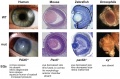
The following is from a recent paper comparing human to zebrafish muscle development. | The following is from a recent paper comparing human to zebrafish muscle development.{{#pmid:22132088|PMID22132088}} | ||
{| | {| | ||
! About the Muscles | ! About the Muscles | ||
| Line 47: | Line 57: | ||
| valign=top| [[File:Human_extraocular_muscles_01.jpg|200px]] | | valign=top| [[File:Human_extraocular_muscles_01.jpg|200px]] | ||
|} | |} | ||
Extraocular muscles (CAT scan back view of the eye)<ref>{{Ref-KolbFernandezNelson2012}}</ref> | |||
[[File:Extraocular-muscles-scan.jpg|600px]] | |||
==Innervation== | |||
Different cranial nerves supple the extraocular muscles.<ref name="Gilbert1957">{{Ref-Gilbert1957}}</ref> | |||
All the vertebrates: | |||
* third cranial nerve ({{CN III}}) oculomotor nerve supplies - superior rectus, medial rectus, inferior rectus, and inferior oblique | |||
* fourth cranial nerve ({{CN IV}}) trochlear nerve supplies - superior oblique | |||
* sixth cranial nerve ({{CN VI}}) abducens nerve supplies - lateral rectus | |||
In several groups of vertebrates: | |||
* sixth cranial nerve ({{CN VI}}) abducens nerve supplies - retractor bulbi (a seventh eyeball muscle) | |||
==Ciliary Muscles== | ==Ciliary Muscles== | ||
| Line 79: | Line 105: | ||
===Carnegie Stages - Eye=== | ===Carnegie Stages - Eye=== | ||
The following data is from a study of human embryonic carnegie stages | The following data is from a study of human embryonic carnegie stages{{#pmid:7364662|PMID7364662}} and other sources. | ||
* [[Carnegie_stage_10|Stage 10]] - optic primordia appear. | * [[Carnegie_stage_10|Stage 10]] - optic primordia appear. | ||
* [[Carnegie_stage_11|Stage 11]] - right and left optic primordia meet at the optic chiasma forming a U-shaped rim. | * [[Carnegie_stage_11|Stage 11]] - right and left optic primordia meet at the optic chiasma forming a U-shaped rim. | ||
| Line 98: | Line 124: | ||
| [[File:Mouse eye neural crest.jpg|400px]] | | [[File:Mouse eye neural crest.jpg|400px]] | ||
Mouse eye neural crest | Mouse eye neural crest{{#pmid:16403239|PMID16403239}} | ||
| [[File:Mouse_eye_TGF-beta_model.jpg|400px]] | | [[File:Mouse_eye_TGF-beta_model.jpg|400px]] | ||
Mouse eye TGF-beta model | Mouse eye TGF-beta model{{#pmid:16403239|PMID16403239}} | ||
|} | |} | ||
| Line 166: | Line 191: | ||
===Reviews=== | ===Reviews=== | ||
{{#pmid:20855501}} | |||
{{#pmid:18214786}} | |||
{{#pmid:17945333}} | |||
The International Journal of Developmental Biology [http://www.ijdb.ehu.es/web/contents.php?vol=48&issue=8-9 Vol. 48 Nos. 8/9 (2004) Eye Development] | The International Journal of Developmental Biology [http://www.ijdb.ehu.es/web/contents.php?vol=48&issue=8-9 Vol. 48 Nos. 8/9 (2004) Eye Development] | ||
===Articles=== | ===Articles=== | ||
Latest revision as of 11:22, 9 August 2020
Introduction

These notes introduce the development of the eye muscles. The adult eye has contributions from several different embryonic layers eventually forming neuronal, supportive connective tissue, optical structures, and muscular tissues. Additional pages are being developed to cover specific issues of this anatomical structure.
A study by Gilbert (1957)[1] described the origin and development of the human extrinsic ocular muscles.
| Senses Links: Introduction | placode | Hearing and Balance hearing | balance | vision | smell | taste | touch | Stage 22 | Category:Sensory |
Some Recent Findings
|
| More recent papers |
|---|
 This table allows an automated computer search of the external PubMed database using the listed "Search term" text link.
More? References | Discussion Page | Journal Searches | 2019 References | 2020 References Search term: Extraocular Muscle Development | Extraocular Muscle Embryology | superior rectus development | lateral rectus development | medial rectus development | superior oblique development | inferior oblique development | |
| Older papers |
|---|
| These papers originally appeared in the Some Recent Findings table, but as that list grew in length have now been shuffled down to this collapsible table.
See also the Discussion Page for other references listed by year and References on this current page.
|
Extraocular Muscles
Extraocular muscles are required to move the eye within the orbit. Their embryonic origin requires an interaction between the cranial mesoderm and the migrating neural crest cells.
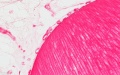
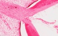
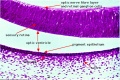
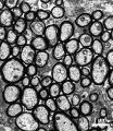
The following is from a recent paper comparing human to zebrafish muscle development.[6]
| About the Muscles | Legend | |
|---|---|---|
|
|
|
Extraocular muscles (CAT scan back view of the eye)[7]

Innervation
Different cranial nerves supple the extraocular muscles.[1]
All the vertebrates:
- third cranial nerve (CN III) oculomotor nerve supplies - superior rectus, medial rectus, inferior rectus, and inferior oblique
- fourth cranial nerve (CN IV) trochlear nerve supplies - superior oblique
- sixth cranial nerve (CN VI) abducens nerve supplies - lateral rectus
In several groups of vertebrates:
- sixth cranial nerve (CN VI) abducens nerve supplies - retractor bulbi (a seventh eyeball muscle)
Ciliary Muscles

The lens focusses by refracting light as it passes through the biconvex lens, which can be altered in shape (accommodation) by surrounding ciliary muscles. These ciliary muscles are activated (contracted) by parasympathetic innervation from the ciliary ganglion itself innervated by the oculomotor nerve (Cranial Nerve III) (More? Cranial Nerves).
surface ectoderm -> lens placode -> lens pit -> lens vesicle -> lens fibres -> lens capsule and embryonic/fetal nucleus.
Eyelids
Muscles of the eyelids are the Levator palpebræ superiors, Orbicularis oculi and Corrugator. The Orbicularis oculi and Corrugator are both supplied by the facial nerve.
(modified from Gray's Anatomy) |

|
Timeline
|
Embryonic Development
|

|
Carnegie Stages - Eye
The following data is from a study of human embryonic carnegie stages[8] and other sources.
- Stage 10 - optic primordia appear.
- Stage 11 - right and left optic primordia meet at the optic chiasma forming a U-shaped rim.
- Stage 12 - optic neural crest reaches its maximum extent and the optic vesicle becomes covered by a complete sheath,
- Stage 13 - By the end of the fourth week the optic vesicle lies close to the surface ectoderm. Optic evagination differentiation allows identification of optic part of retina, future pigmented layer of retina, and optic stalk. The surface ectoderm overlying the optic vesicle, in response to this contact, has thickened to form the lense placode.
- Stage 14 - (about 32 days) the lens placode is indented by the lens pit, cup-shaped and still communicates with the surface by a narrowing pore.
- Stage 15 - (about 33 days) the lens pit is closed. The lens vesicle and optic cup lie close to the surface ectoderm and appear to press against the surface.
- Stage 16 - (37 days) Growth of the lens body results in a D-shaped lens cavity. Perilental blood vessels (tunica vasculosa lentis) are visible. Prior to the development of the eyelids, one small sulcus or groove forms above the eye (eyelid groove) and another below it.
- Stages 17 - 19 - Retinal pigment is visible and the retinal fissure is largely closed. Eyelids grooves deepen, eyelid folds develop, first below, and then above, the eye.
- Stages 18 - Mesenchyme invades the region between the lens epithelium and the surface ectoderm.
- Stages 19 - 22 - the eyelid folds develop into the eyelids and cover more of the eye as the palpebral fissure takes shape. The upper and the lower eyelids meet at the outer canthus in Stage 19.
- Stage 20 - The lens cavity is lost and a lens suture begins to form. The inner canthus is established.
- Stage 23 - The retina comprises the pigmented layer, external limiting membrane, proliferative zone, external neuroblastic layer, transient fiber layer, internal neuroblastic layer, nerve fiber layer, and internal limiting membrane. Eyelids closure is complete (Note - shown as still open in the Kyoto embryo).
Neural Crest

Mouse eye neural crest[9] |

Mouse eye TGF-beta model[9] |
- Links: Image - Mouse eye neural crest | Image - Mouse eye TGF-beta model | Vision Development | Neural Crest Development | Head Development
Additional Images
Human stage 22 developing iris region
Human stage 22 developing iris region
Human stage 22 overview of optic nerve
Human stage 22 overview of eye
Human stage 22 lens and hyaloid vessels
Human stage 22 optic nerve (stalk)
Human stage 22 retina
Mouse adult optic nerve axons
Pax6 eye phenotypes


Historic Images
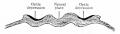
Fig. 456. Location of optic areas before the closure of the neural groove.
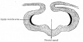
Fig. 457. Location of areas shown in Fig. 456 after the formation of the neural canal.
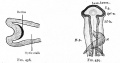
Fig. 458. Location of the optic area after the beginning of the formation of the optic cup and optic stalk. Fig. 459. Dorsal view of head of chick of 58 hours' incubation.
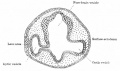
Fig. 460. Section through head of chick of two days' incubation.
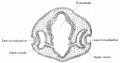
Fig. 461. Section through head of chick of three days' incubation.
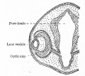
Fig. 462. Later stage in development of optic cup and lens than is shown in Fig. 461.
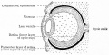
Fig. 463. Developing lens and optic cup.
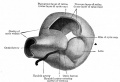
Fig. 464. Model showing lens and formation of optic cup.

Fig. 465. Stages in the development of the lens in the rabbit embryo.
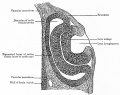
Fig. 466. Section through optic cup and lens invagination of chick of fifty-four hours' incubation.
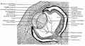
Fig. 467. Section through eye of human embryo of 13-14 weeks.
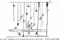
Fig. 468. Development of the retinal cells.
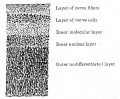
Fig. 469. Vertical section through retina of a four months' human embryo.
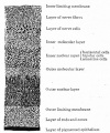
Fig. 470. Vertical section through retina of a five and one-half months' human embryo.
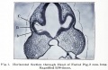
Fig. 1. Section through head of pig, 2 mm long.
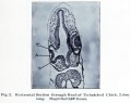
Fig. 2. Section through head of chick, 2 mm long.
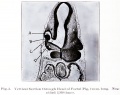
Fig. 3. Section through head of Foetal Pig, 2 mm long.
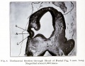
Fig. 4. Section through head of Foetal Pig, 3 mm long.
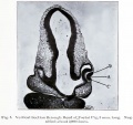
Fig. 5. Section through head of Foetal Pig, 3 mm long.
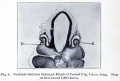
Fig. 6. Section through head of Foetal Pig, 4 mm long.
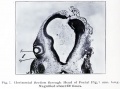
Fig. 7. Section through head of Foetal Pig, 7 mm long.
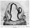
Fig. 8. Section through head of pig, 8 mm long.
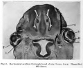
Fig. 9. Section through head of pig, 9 mm long.
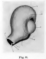
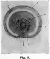
Fig. 11. colobomba of the fundus in the adult and means a lack of development.


References
- ↑ 1.0 1.1 Gilbert PW. The origin and development of the human extrinsic ocular muscles. (1957) Carnegie Instn. Wash. Publ. 611, Contrib. Embryol., Carnegie Inst. Wash. 36: 59-78.
- ↑ Fleuriet J, Willoughby CL, Kueppers RB, Mustari MJ & McLoon LK. (2020). Eye alignment changes caused by sustained GDNF treatment of an extraocular muscle in infant non-human primates. Sci Rep , 10, 11927. PMID: 32681083 DOI.
- ↑ Zhao Y, Louie KW, Tingle CF, Sha C, Heisel CJ, Unsworth SP, Kish PE & Kahana A. (2020). Twist3 is required for dedifferentiation during extraocular muscle regeneration in adult zebrafish. PLoS ONE , 15, e0231963. PMID: 32320444 DOI.
- ↑ Blumer R, Maurer-Gesek B, Gesslbauer B, Blumer M, Pechriggl E, Davis-López de Carrizosa MA, Horn AK, May PJ, Streicher J, de la Cruz RR & Pastor ÁM. (2016). Palisade Endings Are a Constant Feature in the Extraocular Muscles of Frontal-Eyed, But Not Lateral-Eyed, Animals. Invest. Ophthalmol. Vis. Sci. , 57, 320-31. PMID: 26830369 DOI.
- ↑ Meng Q, Mongan M, Carreira V, Kurita H, Liu CY, Kao WW & Xia Y. (2014). Eyelid closure in embryogenesis is required for ocular adnexa development. Invest. Ophthalmol. Vis. Sci. , 55, 7652-61. PMID: 25377219 DOI.
- ↑ Kasprick DS, Kish PE, Junttila TL, Ward LA, Bohnsack BL & Kahana A. (2011). Microanatomy of adult zebrafish extraocular muscles. PLoS ONE , 6, e27095. PMID: 22132088 DOI.
- ↑ Kolb H. Fernandez E. and Nelson R. Webvision: The Organization of the Retina and Visual System. (2012) (Internet) NCBI Bookshelf. PMID 21413389
- ↑ Pearson AA. (1980). The development of the eyelids. Part I. External features. J. Anat. , 130, 33-42. PMID: 7364662
- ↑ 9.0 9.1 Ittner LM, Wurdak H, Schwerdtfeger K, Kunz T, Ille F, Leveen P, Hjalt TA, Suter U, Karlsson S, Hafezi F, Born W & Sommer L. (2005). Compound developmental eye disorders following inactivation of TGFbeta signaling in neural-crest stem cells. J. Biol. , 4, 11. PMID: 16403239 DOI.
Online Textbooks
- Developmental Biology (6th ed.) Gilbert, Scott F. Sunderland (MA): Sinauer Associates, Inc.; c2000. Evolution of the mammalian middle ear bones from the reptilian jaw | Chick embryo rhombomere neural crest cells | Some derivatives of the pharyngeal arches | Formation of the Neural Tube | Differentiation of the Neural Tube | Tissue Architecture of the Central Nervous System | Neuronal Types | Snapshot Summary: Central Nervous System and Epidermis
- Neuroscience Purves, Dale; Augustine, George J.; Fitzpatrick, David; Katz, Lawrence C.; LaMantia, Anthony-Samuel; McNamara, James O.; Williams, S. Mark. Sunderland (MA): Sinauer Associates, Inc. ; c2001 The Auditory System | The Inner Ear | The Middle Ear | The External Ear | Early Brain Development | Construction of Neural Circuits | Modification of Brain Circuits as a Result of Experience
- Molecular Biology of the Cell (4th Edn) Alberts, Bruce; Johnson, Alexander; Lewis, Julian; Raff, Martin; Roberts, Keith; Walter, Peter. New York: Garland Publishing; 2002. Neural Development | The three phases of neural development
- Clinical Methods 63. Cranial Nerves IX and X: The Glossopharyngeal and Vagus Nerves | The Tongue | 126. The Ear and Auditory System | An Overview of the Head and Neck - Ears and Hearing | Audiometry
- Health Services/Technology Assessment Text (HSTAT) Bethesda (MD): National Library of Medicine (US), 2003 Oct. Developmental Disorders Associated with Failure to Thrive
- Eurekah Bioscience Collection Cranial Neural Crest and Development of the Head Skeleton
Reviews
Sung CH & Chuang JZ. (2010). The cell biology of vision. J. Cell Biol. , 190, 953-63. PMID: 20855501 DOI.
Heidary G, Engle EC & Hunter DG. (2008). Congenital fibrosis of the extraocular muscles. Semin Ophthalmol , 23, 3-8. PMID: 18214786 DOI.
Shih HP, Gross MK & Kioussi C. (2008). Muscle development: forming the head and trunk muscles. Acta Histochem. , 110, 97-108. PMID: 17945333 DOI. The International Journal of Developmental Biology Vol. 48 Nos. 8/9 (2004) Eye Development
Articles
Bookshelf Extraocular Muscle Development
Search Pubmed
Search Pubmed: Extraocular Muscle Development
Search Entrez: Extraocular Muscle Development
Terms
External Links
External Links Notice - The dynamic nature of the internet may mean that some of these listed links may no longer function. If the link no longer works search the web with the link text or name. Links to any external commercial sites are provided for information purposes only and should never be considered an endorsement. UNSW Embryology is provided as an educational resource with no clinical information or commercial affiliation.
Glossary Links
- Glossary: A | B | C | D | E | F | G | H | I | J | K | L | M | N | O | P | Q | R | S | T | U | V | W | X | Y | Z | Numbers | Symbols | Term Link
Cite this page: Hill, M.A. (2024, June 27) Embryology Vision - Extraocular Muscle Development. Retrieved from https://embryology.med.unsw.edu.au/embryology/index.php/Vision_-_Extraocular_Muscle_Development
- © Dr Mark Hill 2024, UNSW Embryology ISBN: 978 0 7334 2609 4 - UNSW CRICOS Provider Code No. 00098G