Paper - The origin of blood cells (1916)
| Embryology - 30 Apr 2024 |
|---|
| Google Translate - select your language from the list shown below (this will open a new external page) |
|
العربية | català | 中文 | 中國傳統的 | français | Deutsche | עִברִית | हिंदी | bahasa Indonesia | italiano | 日本語 | 한국어 | မြန်မာ | Pilipino | Polskie | português | ਪੰਜਾਬੀ ਦੇ | Română | русский | Español | Swahili | Svensk | ไทย | Türkçe | اردو | ייִדיש | Tiếng Việt These external translations are automated and may not be accurate. (More? About Translations) |
Danchakoff V. The origin of blood cells. (1916) Anat. Rec. 10(5): 397-414.
| Historic Disclaimer - information about historic embryology pages |
|---|
| Pages where the terms "Historic" (textbooks, papers, people, recommendations) appear on this site, and sections within pages where this disclaimer appears, indicate that the content and scientific understanding are specific to the time of publication. This means that while some scientific descriptions are still accurate, the terminology and interpretation of the developmental mechanisms reflect the understanding at the time of original publication and those of the preceding periods, these terms, interpretations and recommendations may not reflect our current scientific understanding. (More? Embryology History | Historic Embryology Papers) |
Origin of the Blood Cells. Development of the Haematopoetic Organs and Regeneration of the Blood Cells from the standpoint of the Monophyletic School
Vera Danchakoff The Rockefeller Institute for Medical Research
One Plate
Lecture delivered before the College of Physicians and Surgeons, Columbia University, New York, on November 24, 1915.
Introduction
The last century was marked by very intensive work in the field of haematology, including, as it does, erythropoesis, lymph- and leucopoesis. Many investigators have endeavored to throw light on such obscure problems as the embryonic and phylogenetic development of the blood, the structure of the haematopoetic organs, and the development of the vessels in which the differentiated products are conveyed into body tissues. Finally, the function of the different white blood cells has formed a new and interesting problem.
International cooperation has contributed certain results to this field of research, a part of which will have permanent value and form the basis for further investigations. I do not need to emphasize the merits of the investigators, to whom We owe an elucidation of the development of the lymphatics. Most of the students of blood embryogeny and blood phylogeny made their investigations inEurope. From what I can judge, America presents good opportunities for a rational understanding of the function of the lymphatic tissue, as is exhibited in some of the Work now being done at the Rockefeller Institute.
The student of biology is engaged in a study of the development and function of living matter, but function is inseparably connected with structure. A correct understanding of the function of the different blood cells could not even be approached some ten years ago. At that time the mutual relationship of the different blood cells was not established and there existed very few indications concerning the structure and localization of the haematopoetic organs. It is my intention to present here the results of those investigations which are principally concerned with the development and structure of the haematopoetic organs and of the elements which are differentiated in these organs.
A brief résumé of the knowledge of haematopoesis during the last century may be permitted. The zoologists, embryologists and histologists considered the blood islands of the area opaca as the source from whence arose the first red blood cells. The concurrence of views disappeared, however, as soon as the question of the origin of the blood islands was considered.
Very definite indications have shown that the blood development in mammals is transferred in early embryonic life from the area vasculosa to the liver and finally to the bone marrow; but the search of a haematopoetic organ,homologous to the mammalian liver, was fruitless in the meroblastic eggs of the birds and reptiles.
The problem of the genesis of a new haematopoetic organ was readily answered. Blood cells, capable of mitosis, were ejected at certain times from the existing and functioning organ, as for instance, from the area vasculosa into the liver. Here they found favorable conditions for their development, multi~ plication and differentiation, and the new haematopoetic organ began to function. How radically this interpretation has changed will be shown in this communication.
The facts, reported above, concerned chiefly the erythropoesis. The localization, the time and the source of the development of the white blood corpuseles were sometimes accounted for in at very peculiar way. The marked differences in the structure of the red and white bloodcorpuseles led to the assertion that these different cells must have a separate origin. The fact that white blood cells were not found in the vessels of young embryos and the presumably short duration of the haematopoetic function of the area vasculosa led many investigators to look for a development of leucocytes within the embryo body. Of course a separate genesis of the red and White blood cells under such circumstances was taken for granted. The presumption that the erythrocytes developed exclusively in the blood islands and that the leucocytes differentiated later in the embryo body itself seemed to be unshakable. The observations of V. der Stricht, describing the presence of leucocytes in the area vasculosa of the chick embryo with two segments, stood isolated. The opinion of Saxer and Bryce recognizing a common stem-cell for the white and the red blood corpuscles, seemed to be in contradiction to all known facts.
The conception of the lymphatic cells of the thymus, as deriving from the epithelium, shows how paradoxical was the judgment about certain forms of the white blood corpuscles. The same origin was conceived for the spleen cells in the reptiles. This latter assumption again prompted the admission of different stemcells in the case of the lymphocytes and the polymorphonuclear leucocytes.
In speaking of T definite stem-cells I have in mind such kinds of cells generally, as may be differentiated and identified morphologically. Up to the present, a definite morphological structure has been required as indispensable for an admission of difierences in the stem-cells. One has but to remember the elaborate, but vain efforts of Schridde to put in evidence a difference between the leucoblasts and the lymphoblasts.
In order to avoid somewhat irrelevant details, I shall take as support for my position the meroblastic eggs of reptiles and birds. All the blood cells of these animals are real cells, containing both cytoplasm and nucleus. This fact is not unimportant for a true conception of the principle of haematopoesis. The statement that the erythrocytes arise in the early stages of embryonic development from the blood-islands cells in the area opaca has remained unshaken. Recent studies of the origin of the blood islands has established definitely their mesodermic origin. The mesoderm contributes to the development of both,—the bloodisland cells and the endothelium. The mesenchymal cells of the mesoderm (MSCH.) appear to be a source from which develop various specific differentiation products depending on the gradual assertions of the environment.
We may, with our present knowledge, define at least some of the conditions necessary for an arrangement and difierentiation of the mesenchyme into haematopoetic organs (namely, those comprising the differentiation of red and granular cells). The area opaca, as is known, lies above the yolk, and the connection between the development of a haematopoetic organ and an abundance of nutritive material is still more obvious if the results of studies on the development of other haematopoetic organs are compared therewith.
Still another condition seems to be of equal value to the above. This consists of the localization of the developing haematopoetic organ in a place, where neither extensive growth, replacement of different organs nor muscle contraction takes place. Only in such regions of the embryo body will the mesenchyme under normal conditions develop into a haematopoetic organ, always characterized by a highly developed network of capillary veins.
The formation of the blood-islands is accompanied by the development of an endothelial membrane. A vascular wall surrounds the blood-islands, regardless of the fact that a communication with the heart may or may not be effected (Loeb, Stockard). I must emphasize here that the endothelial capillaries develop not only around the blood-islands, but also in regions where these are lacking, and p that some of the bloodislands remain outside of the haematopoetic capillaries.
Just before the differentiation of the endothelial membrane takes place, all the cells of the blood—islands have a similar structure (BLISC. (H BL.L.LM'C.) ), regardless of whether they appear as aggregates of numerous cells, merely as small groups or even as isolated cells. These cells possess an intensely basophilic, slightly vacuolized cytoplasm, a spherical nucleus with a well pronounced nucleolus, and they are potentially spherical, but very amoeboid in character. This structure, characterizing, as it does, the stem-cells of the blood elements from the earliest stages of their appearance, remains unchanged in the development of new haematopoetic organs (compare ET/.HBL., Il/.HBL. and HBL. in I, II and III).
The differentiation of the endothelial membrane (I, EN DC.) separates the major portion of the blood—islands from some of these cells, as well as from other amoeboid cells, which still continue to split off from the mesenchyme (I, E V.HBL.). This separation of similar elements establishes very different environ—ments for different groups of cells. The physical and chemical conditions Within the vessels, which are lined with a continuous endothelial membrane, must differlargely from the conditions offered by the tissue outside the vessels. This difference in environmental conditions necessarily affects the further development of previously like stem-cells. However, the reproduction of the stem—cells is evidently not influenced by the different environmental conditions, for the mother cell multiplies within as Well as outside the vessels (I, E’V.HBL., IV.HBL.). The differentiation, on the contrary, is subject to strong influences of the environment and manifests itself in very different ways. Within the vessels the stem—cells produce in their cytoplasm haemoglobin (I, ERBL.), outside of the vessels the same young undifferentiated cells give rise to acidophilic granules (I, GRBL.).
Changes in the nuclear structure keep pace with the differentiation in the cytoplasm.
In the early development of the blood cells in reptiles and birds (Danchakoff), and partly in fishes (Maximoff), many similar features may be noticed during the simultaneous differentiation of the granuloblasts and erythroblasts from a morphologically and genetically identical mother-cell. This stem-cell main tains its own existence (I, ET/'.HBL., IT/.HBL.) by uninterrupted multiplication, on the one hand, and on the other it differentiates into red cells (ERCC) Within the vessels and into granular leuco— cytes (LCC.) outside the vessels. 5
The first development, differentiation and multiplication of the blood cells occurs in mammals (Maximoff), birds, reptiles and in some fishes on the surface of the yolk. As is known the fundamental difference existing between the eggs of reptiles and birds and those of mammals, ‘consists in the fact that the latter have lost the yolk so characteristic of the eggs of reptiles and birds. This fundamental difference necessarily requires a difference in the development of the haematopoetic organs in these groups of animals.
The rapid appearance of anlagen and the development of different organs in the embryo body naturally attracted the attention of the embryologists to the body of the embryo itself. This fact accounts for a comparatively early recognition of the haematopoetic function of the liver. The first development of the blood cells in the area vasculosa, the embryonic localization of the haematopoesis in the liver and its final establishment in the bone marroW,-—~such are the data fixed for the erythro— and granulopoesis in the mammals.
The most minute and laborious study of the Whole embryo body in birds has failed to show the localization of the erythropoesis before a comparatively late stage. By the 13-14 days of incubation it has started in the bone marrow. A cursory glance at the liver of the meroblastic eggs was sufficient to establish an absence of any noticeable blood development in this organ. Neither the thymus, the spleen, nor the tissue of the Wolflian body (mesonephros) shows any indication of erythropoesis. All these organs contain vessels and capillary nets but they are all merely thoroughfares.
Although in regions far removed from the embryo body in the annexes of the yolk-sac there Was demonstrated by injection (Popoff) an amazingly extensive and exuberant net—Work of large venous capillaries, its development and existence seemed to be fully explained by the resorptionof the enormous quantity of yolk, and no one thought of thoroughly analyzing the cell elements present Within the capillaries. Whereas a closer study (Danchakoff) of this capillary net and of its contents would show that the large meshes of this net are distended by young undifferentiated blood cells, and it would be easy to observe that outside of the capillaries the basophilic amoeboid cells are also rather numerous. The structure of all these cells, both Within and Without the capillaries would permit of ascribing to this capillary net a real haematopoetic (erythropoetic and granulepoetic) function, homologous to that of the mammalian liver.
Both within and without the vessels the young mother-cells bear the same structure as the blood-island cells after their isolation (I, E’V.H.BL., I V.HBL.). This morphological cell unit was most unhappily termed the large lymphocyte. The efforts of the last 7 to 8 years have not yet been able to render conclusively the dictum, that the morphological cell unit, formerly known as the large lymphocyte, appears first by the dissociation of the bloodislands and is a young, undifferentiated cell. This cell differentiates in accordance with given environmental conditions. Briefly, it corresponds morphologically to the large lymphocyte and is physiologically the haemoblast.
It appears only natural that the young stem-cells, bbserved in the haematopoetic net of the yolk sac annexes, corresponds exactly to the stem-cells which were first found in the bloodislands; for they appear to be the direct result of an uninterrupted reproduction of the latter.
The erythropoesis and the granulopoesis now remain distinctly separated, as in the first stages of their development. The distribution of the different cell elements within the haematopoetic capillary net in the annexes is always very characteristic. Every cell is free and does not manifest any continuity with its neighbor. The cells lie very closely together in the zones adjacent to the endothelial membrane, and their reciprocal pressure flattens them and renders them polygonal. Even a slight fluid current could hardly be supposed to be present in these parts of the vessels. In the centre of the vascular lumen the cells are arranged more loosely and are free from pressure. The position of the cells shows clearly, that they are removed from here and directed by the current into the body of the embryo.
It is easy to notice, that the most highly differentiated cells, rich in haemoglobin, lie in the centre of the vascular lumen, just where the current is strongest. Besides these already differentiated cells the capillaries include numerous cells in various stages of differentiation, beginning with the youngest haemoglobin-free stem-cells, and including the whole range of cells which gradually accumulate haemoglobin in their cytoplasm and equally gradually change their structure into that characteristic of an erythrocyte.
The youngest stem-cells, which I will call lymphoid haemc cytoblasts, are found in the zones immediately adjacent to the endothelial membrane, and these young blood cells within the vessels are so much like the white blood cells lying outside the vessels, that Bizzozero regarded them in the bone marrow as a layer of white blood corpuscles. In the embryonic haematopoetic organs these cells are very numerous and sometimes form a continuous layer of basophilic cells, becoming gradually substituted towards the centre by cells, which accumulate haemoglobin in their cytoplasm.
The structure of the haematopoetic net and the distribution of the cells in their different stages of development reminds one strongly of the conditions under which spermatogenesis takes place. Here is seen the same uninterrupted‘differentiation of the younger cells into highly differentiated cells, which thereby lose much of their usual cell appearance. We also find the same location of the young undifferentiated elements, which secures their existence by uninterrupted multiplication and continuous splitting off of cell generations which undergo further differentiation. Finally, we see the presence of a current in the centre of the vessels, which removes the differentiated products.
As mentioned above, the area vasculosa of birds and reptiles contains outside the vessels numerous cells, which give rise to the granular leucopoesis. Both in the earliest stages and now in the yolk sac annexes the leucocytes develop extravascularly. They nevertheless show very close connection with the vessels, occasionally surrounding them by a continuous layer. Their differentiation here follows the same principles as elsewhere and from a common morphologically and genetically identical stemcell (I, EV.HBL.) leads to the development of numerous special (acidophilic) leucocytes (I, LCC.). The differentiation of blood cells in the area vasculosa and in the annexes of the yolk sac is completed by the development of the cell elements described.
Another question now arises, viz., the question of how a new haematopoetic organ originates, whether it is the mammalian liver or the definitive haematopoetic organ —the bone marrow, or finally, one of the organs assigned to the lymphopoesis.
I have mentioned above how easily this problem was solved not long ago. The first red cell (the erythroblast) differentiated from the blood-island cell; it supplied the organism, by multiplication and differentiation, with erythrocytes. At a given time the current transported the erythroblasts into definite regions of the embryo body. They stopped there, multiplied and differentiated further. Shortly the development of a new haematopoetic organ appeared as a strictly localized metastasis. It was difficult even to expect any other conclusion from studies made upon haematopoetic organs, the development of which was more or less advanced. Indeed, such organs offer a complete parallel between the differentiating blood cells in the new organ and those, which were observed in the earlier organ. Only studies of the very earliest stages in the development of a new haematopoetic organ will show by definite evidence, that the development of a new organ is due, not to metastasis from a former existing organ, but to a local differentiation and multiplication of the mesenchymal cells, still preserving their faculty of polyvalent differentiation (see II and III).
The young undifferentiated mesenchymal cell (MSCHJ maintains itself for many days, if not for the whole duration of the life of the organism, and evidently retains its potency for multiplication and differentiation. Similar mesenchymal cells give rise to accumulations of amoeboid cells around the embryonic double aorta, described by Professor Huntington. The blood-island-like agglomerations of similar cells in the development of the bone marrow arise also by a splitting off from the mesenchyme. An endothelial vascular membrane, partly formed in loco, partly growing from outside the bone marrow cavity, surrounds some of these cell groups and marks them at the same time as erythroblasts. Similar cells, remaining between the capillaries, become the source of the granulopoesis (Maximoff, Danchakoff).
That being the case, we have to conclude, that the whole development of the new haematopoetic organ f. ex. of the bone marrow ( see plate III) takes place locally and leads to the double differentiation of a common mother—cell into red and granular cells, the young stem-cell itself being most tenaciously retained in the precincts of the haematopoetic organ.
Nevertheless, it is possible to notice certain features of the haematopoesis in the bone marrow, characteristic for this organ, which appear already in the embryo and are very conspicuous in the adult animal. This is a reduction in number of the young undifferentiated stem-cells, both in the erythropoesis and the leucopoesis regions. The development of the highly differentiated erythrocytes and leucocytes is effected by the further development of cells which already contain respectively ' haemoglobin (erythroblasts) or acidophilic granules (granulocytoblasts). The haematopoesis now develops in a more homoplastic way, differing from that seen in the early embryonic stages in which the haematopoesis develops principally in a heteroplastic way. This difference is, however, not essential; one has but to bleed the animal, whereupon the stem-cell promptly produces by multiplication a whole generation of similar stem-cells (Danchakoff), which are very numerous in the haematopoetic tissue of the embryonic organism.
Some essential differences may still be observed in the structure of the bone marrow, in comparison with the former haematopoetic organ, namely new directions in the differentiation of the blood cells, leading to the development of small lymphocytes and of Mast cells. Since the latter find their origin on a larger scale in the connective tissue and in special organs, lymph glands, thymus and spleen, the mode of their differentiation is more conveniently studied in theclatter organs.
Finally, I have to emphasize the striking analogy seen in the general conditions which accompany the development of every new erythro-granulopoetic organ: an abundance of food supply and the localization distant from any rapidly growing organs; these conditions are met with both in the yolk sac and in the cavities of the bones and seem to be so essential that in reptiles which have no extremities, haematopoesis finds its localization in the cavities of the vertebra, thus developing a haematopoetic organ in the form of a whole series of isolated centres (Danchakoff). Even the localization of haematopoesis in the liver of mammals, which have lost the yolk in their eggs, correspond in the highest degree to the conditions cited and which, at this time, the embryo offers.
Some of the environmental conditions for the differentiation of the mesenchyme into an erythro—leucopoetic organ, as well as those for the development of a haemocytoblast into an erythrocyte or into a leucocyte could be, at least partly, explained. However, the differentiation of the blood stem-cells into erythrocytes and leucocytes does not present all the possible inherent potentialities of the haemocytoblast. New differentiation products result under new conditions in the mesenchyme of the embryo body (see plate II).
The mesenchyme cells are disseminated through the whole organism and occupy all the interspaces between various organs. It is difficult at present to define the conditions under which the mesenchymal cell undergoes differentiation into a fibroblast. This ubiquitous differentiation has for long overshadowed, by its omnipresence and its conspicuousness, all the other lines of development which find place in the so-called connective tissue. Meanwhile certain other directions in the differentiation of the mesenchymal cells arealmost as general as the differentiation into the fibroblasts; they convert the connective tissue into a seat of most interesting diffuse lymphopoetic, and, in early embryonic stages and in lower animals, into granulopoetic differentiation. The development of the lymphatic glands, of the thymus and of the spleen thus becomes merely localized hypertrophic centres of a similar diffuse differentiation, spread out through the loose connective tissue.
In speaking of the lympho-granulopoesis, I therefore associate the differentiation phenomena in all the regions above mentioned in one common account. These lines of differentiation are very simple and similar. The first isolationof the mesenchymal cells from the general ties takes place as a rule in the form of large lymphoid haemocytoblasts (II, HBI/., L.LMC.),which sometimes arrange themselves as blood-island-like groups of cells. Their rapid, uninterrupted multiplication produces numerous smaller cells (II, SM.HBL.). These cells become a source of the true small lymphocytes (II, SM.LMC.) and of numerous Wandering cells (II, W.C.), so characteristic of the loose connective tissue. The latter may also arise directly from the mesenchymal cells, especially in later embryonic stages. With the appearance of the small lymphocytes and Wandering cells the differentiation possibilities of the lymphoid—haemocytoblast in the connective tissue are still not exhausted. Throughout the regions of the embryo body in birds and especially in reptiles, but less evidently in mammals, the splitting off of granuloblasts is very conspicuous (II, GRBL.).i Most of them contain acidophilic granules. The differentiation of the mast—-cells with basophilic, metachromatic granules at the expense of the mesenchyme cells takes place in the later embryonic stages.
For a long time the ability of the small lymphocytes to multiply by mitosis Was denied; at the present their reproduction by mitosis is fully admitted, as Well as their faculty of further differentiation into mast—leucocytes and into specific gra.nulocytes with spherical acidophylic granules, very numerous in the con» nective tissue of birds (II, GR.LMC.). Likewise they may differentiate into plasma cells in the adult organism (II, PLC.)
The question of regeneration of the blood cells in an adult animal is a very important problem and the embryological data above described may contribute directly to its elucidation. The question of blood regeneration has interested most keenly the pathologist-clinicians. Facing the various highly differentiated products (LCC'., E'RC., PLC., mast-cells, etc.) which, as above mentioned, develop in the adult animal chiefly at the expense of cells in intermediate stages of differentiation, the pathologist—clinicians could not but admit a polyphyletic origin for various blood cells. The red cells possessed their own stem-cells, as also the different leucocytes and lymphocytes.
This statement is not altogether wrong; inasmuch as referring to haematopoetic organs in an adult organism, it solves practically the problem of the usual haematopoesis. But this statement does not cover all the eventualities. Regeneration after bleedings, as well as the excessive increase in number of white blood corpuscles in leukemias, takes place not at the expense of specific granulo-lympho— or erythroblasts. The monophyletic school has the credit of demonstrating in all the haematopoetic organs, in the bone marrow, in the spleen, in the lymph glands and in the loose connective tissue, the presence of a similar morphologically and genetically young and undifferentiated stem-cell, at the expense of which the regeneration really takes place, especially after great loss (Pappenheim, Weidenreich, Maximofi", Danchakoff). This young stem-cell bears the same structure as the morphological cell unit, Which Was recognized by the monophyletic school as being the mother-cell of all the different blood cells in the development of every haematopoetic organ.
I am afraid to be too positive, but there are some indications that after extensive destruction of the haematopoetic tissue regeneration occurs, at least in some cases, not only at the expense of haemocytoblasts, but also at the expense of undifferentiated mesenchymal cells, retaining from the very beginning of their development all their polyvalent potentialities. This would mean that the adult organism retains, from the remote period of its embryonic life, a stock of undifferentiated mesenchymal cells which begin to multiply and to differentiate in certain conditions of a disturbed equilibrium. This latter question is in a stage of de velopment. No matter how this question may be solved, the similarity of the morphological structure of the stem-cell in all the haematopoetic organs and its common origin from the mesenchymal cells of the mesoderm in embryonic life demonstrates the close relations existing between different blood elements.
It would not follow that the various differentiation products are not different, or that they had no different functions. The erythrocytes, the small lymphocytes, the different leucocytes, the Wandering cells of the connective tissue, the mast-cells and the plasma cells,~+—all these cells are different cell units, morphologically as well as physiologically. But in the early embryonic stages they all had a common mother-cell, and this mother-cell is preserved in the adult organism and becomes the source of differentiation and regeneration and most probably also the source of pathological proliferation.
The conception of the monophyletic school has recently been subjected to searching criticism. As I, in great measure, uphold these conceptions, I feel obliged to plead their cause.
Embryos Without blood circulation were obtained by means of the influence of alcohol on the fertilized eggs of Fundulus (Stockard).[1] Blood-islands developed on the yolk surface, and endothelial membranes surrounded them; the blood-island cells developed into haemoglobinic cells; but they were never carried into the embryo body, because the connection between the heart and these vessels failed to become established. During the summer of 1914 I personally observed precisely the same conditions in hybrids of Fundulus and Menidia, which were obtained for this purpose at the kind suggestion of Dr. Loeb.
The fact, that the blood-island cells develop into erythrocytes in embryos without circulation, is accounted for by a specific character of the mesenchymalcells, which differentiated only in haemoglobinic cells. But it is stated that no leucocytes are developed on the yolk sac in normal Fundulus embryos.
The specificity of the stem-cells of the white blood corpuscles is based upon observations of small groups of cells “which resemble lymphocytes or leucocytes;” it is, moreover, stated that “these embryonic cells all show a more or less degenerate appearance, but if they can be classed as any type of White blood cell, their origin is definitely removed and entirely distinct from that of the red blood cells.”
These are some of the data, upon which an attempt was made to build a new embryopolyphyletic school.
The experimental analysis of the problem of haematopoesis is undoubtedly of very great importance. I am fully conscious, how important is the study of embryos Without circulation and with disconnected vessels for a true conception of the development of vessels. But I fail to understand how the study of these embryos, on which the polyphyletic origin is now based, can help us in understanding the development of different blood cells and of haematopoetic organs.
Whether the circulation is established or not, the differentiation of the blood cells follows the same principles. How can the presence of the circulation embarrass the study of the develop ment and differentiation of the blood cells in the haematopoetic organs, if only more or less differentiated cells are Withdrawn by the circulation? It seems to me that the absence of circulation in itself must greatly obscure the study and hinder the true understanding of the processes. Indeed, the differentiated products are not Withdrawn in embryos Without circulation, they remain in loco and evidently influence very strongly the further reproduction and differentiation of young cells, for finally they all disintegrate.[2]
The environmental conditions are also taken into account by the supporters of the theory of the polyphyletic origin of the blood cells; but only for the multiplication of the erythroblasts, not for their differentiation. In spaces, lined by endothelium, the erythroblasts do not multiply in the Fundulus embryos. If this statement, drawn from the study of Fundulus, was not generalized, we had to believe, until further proof, that the erythroblastic tissue of Fundulus is a very peculiar tissue, refusing to multiply inside the Vessels. In birds and reptiles and in some cartilaginous fishes (Selachians) the erythropoesis takes place exclusively inside the Vessels. Mitosis in erythroblasts inside the Vessels are as numerous, as mitoses in granuloblasts outside the Vessels. On the contrary, the differentiation of the cells is stronglyinfluenced by the fact, whether the cells lie inside or outside the vessels.
But let us consider some of the arguments against the monophyletic school. A special Value is evidently assigned to the following argument, for I find it quoted in different places in the paper. “If all the types of blood cells did arise from a common mother mesenchymal cell, they should then be found in intimate association throughout all blood-forming regions.”
The conclusion cited can only appear as a sequence to an assertion, stating that various differentiation products, developing at the expense of a common stem-cell, must always be found in intimate association. But this assertion is not correct.
If various products of differentiation have their origin in a common stem-cell, they must be found under the influence of different environmental conditions which separate them. If various products of differentiation are found in absolutely iden tical environments the difference between them may be explained only by differences in their stem-cell.
Imagine in the remote past a heap of similar tree seeds. These seeds develop in our moderate climate into tall and many branched trees. Suppose the wind bears a part of the seeds away and brings them to a land possessing different environmental conditions, we will say to arctic lands. There the seeds may develop but they may produce trees no higher, than our moss (willow-salix in Spitzbergen). Have we the right to say that the difference between the various products of seed development are caused by differences in the seeds? They may be different or similar; morphologically they were similar.
How would it be possible to know if there were differences in the seeds‘?- The only possibility of solving this problem would consist in sowing the seeds of arctic trees in our climate and vice versa. If the seeds of our tall trees again produce dwarf trees in the arctic lands, the seeds of the different products of develop» ment will have been shown to have been identical.
Similar experiments may be carried out with the haematopoetic tissue. The experiments are made the more possible, because of the short time periods, needed for the cell differentiations, and fully controlled by us.
It is diflicult to assert at present, whether or not the monophyletic school will contribute the last word in the establishment of the mutual relationship and of the origin of the blood cells. The possibility is not excluded that perhaps some time we will find direct or indirect means of distinguishing between different mesenchyme cells, but there must be other arguments than those cited above to convince one of the truth of the position taken by the polyphyletic school. However, the fact that the stem-cells, as stem-cells, keep so tenaciously under the most varied conditions their morphological structure and that only their products difier, seems to speak strongly for the existence of a single stem-cell, identical in the different haematopoetic organs.
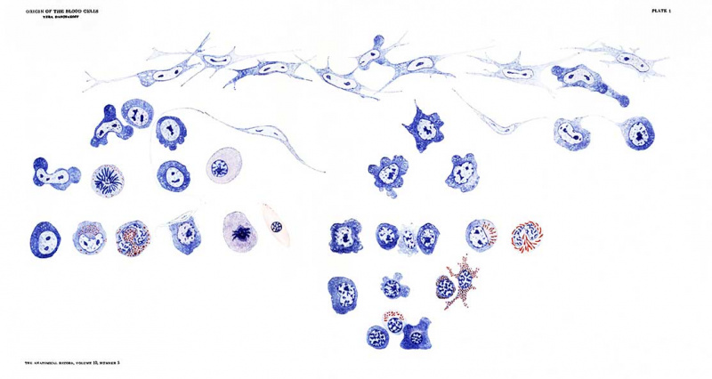
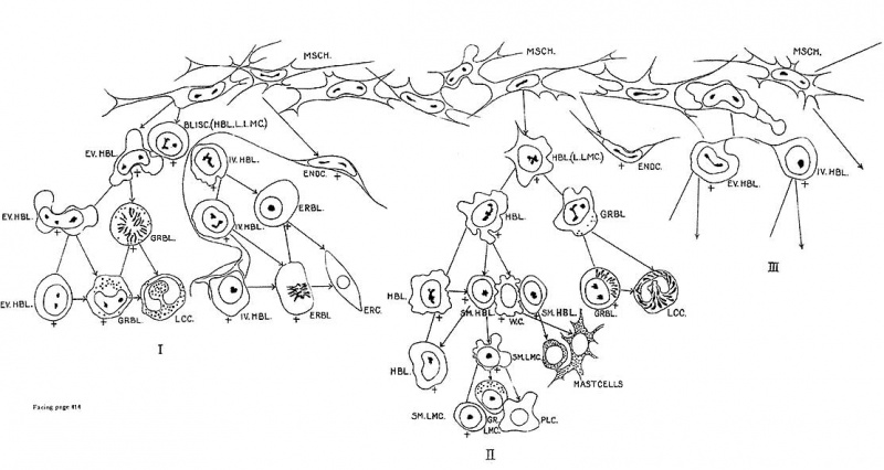
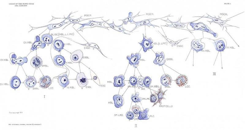
Through the kindness of the Rockefeller Institute for Medical Research I have been enabled to include a colored plate, illustrating the course of the gradual differentiation of various cells in the haematopoetic organs.
Plate 1
Explanation of Figures
The plate shows the development and the gradual differentiation of the blood cells.
| I Erythro- and granulopoesis in the yolk—sac of birds. | II Lympho- and granulopoesis in the special lymphatic glands and in the connective tissue. | III Starting point of the development of the erythro-granulopoesis in the bone-marrow, which proceeds as is shown in I. |
Arrow denotes differentiation. Simple line — denotes multiplication. Cross — indicates cells,. capable of mitosis.
Abbreviations
- BLISC. (HBL.L.LMC.), blood island.
- HBL., haemoblast cell (haemoblast) large lymphocyte
- I V.HBL., intravascular haemoblast
- ENDC. endothelial cell
- LCC., leucocyte
- ERBL., erythroblast
- M SCH ., mesenchyme
- ERC., erythrocyte
- PLC., plasma cells
- EV.HBL., extravascular haemoblast
- SM .HBL., small haemoblast
- GRBL., granuloblast
- SM .LM C ., small lymphocyte
- GR.LMC., granular lymphocyte
References
- ↑ Stockard CR. The origin of blood and vascular endothelium in embryos Without a circulation of the blood and in the normal embryo. Amer. Jour. Anat., vol. 18, no. 2, Sept, 1915.
- ↑ Analogous results of disintegration of spermatogenetic cells are offered by the interesting studies of B. D. Myers. The disintegration of the undifferentiated spermatogenetic cells is caused in his experiments also by a lack of withdrawing of the differentiated cells. See: Histological changes in testes following vasectomy. Proceedings of the Am. Assoc. of Anat., 1916.
Cite this page: Hill, M.A. (2024, April 30) Embryology Paper - The origin of blood cells (1916). Retrieved from https://embryology.med.unsw.edu.au/embryology/index.php/Paper_-_The_origin_of_blood_cells_(1916)
- © Dr Mark Hill 2024, UNSW Embryology ISBN: 978 0 7334 2609 4 - UNSW CRICOS Provider Code No. 00098G


