Paper - Significant superficial anastomoses in the arterial blood supply to the human brain (1959)
| Embryology - 27 Apr 2024 |
|---|
| Google Translate - select your language from the list shown below (this will open a new external page) |
|
العربية | català | 中文 | 中國傳統的 | français | Deutsche | עִברִית | हिंदी | bahasa Indonesia | italiano | 日本語 | 한국어 | မြန်မာ | Pilipino | Polskie | português | ਪੰਜਾਬੀ ਦੇ | Română | русский | Español | Swahili | Svensk | ไทย | Türkçe | اردو | ייִדיש | Tiếng Việt These external translations are automated and may not be accurate. (More? About Translations) |
Gillilan LA. Significant superficial anastomoses in the arterial blood supply to the human brain. (1959) J Comp. Neurol. 112: 55-74. PMID 13850118
| Historic Disclaimer - information about historic embryology pages |
|---|
| Pages where the terms "Historic" (textbooks, papers, people, recommendations) appear on this site, and sections within pages where this disclaimer appears, indicate that the content and scientific understanding are specific to the time of publication. This means that while some scientific descriptions are still accurate, the terminology and interpretation of the developmental mechanisms reflect the understanding at the time of original publication and those of the preceding periods, these terms, interpretations and recommendations may not reflect our current scientific understanding. (More? Embryology History | Historic Embryology Papers) |
Significant Superficial Anastomoses In The Arterial Blood Supply To The Human Brain
Lois A. Gillilan
Department of Anatomy, Graduate School of Medicine,
University of Pennsylvania, Philadelphia
Three figures
Introduction
Throughout the phylogenetic scale the conducting arteries to the mammalian brain are not uniform in pattern. In the cat, sheep, and goat the internal maxillary artery is the chief vessel of supply (Daniel, Dawes and Prichard, ’53). The ex brain receives its major blood supply from the internal maxillary artery, with a substantial contribution from the occipital artery. In the pig the ascending pharyngeal artery is the principal artery to the brain. The pattern in the dog and rabbit is more nearly like that in man. Subhuman primates have patterns very similar to those of man, but there are some interesting differences (Shellshear, ’27b; Watts, ’34).
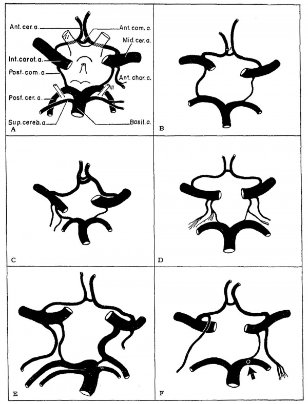
The human brain is supplied by 4 arteries: two internal carotid arteries and two vertebral arteries. The internal carotid arteries are usually larger than the Vertebral arteries. One or more of the 4 may be significantly smaller or even absent. Within the cranial cavity the internal carotid and vertebral arteries form an anatomically anastomosing system of vessels, the circle of Willis, which has heretofore been considered a more or less constant and uniform pattern. It has been so pictured in textbooks and in the minds of most students. Duret (187 4) pointed out that there was great individual Variation in the “hexagon” of Willis. Careful studies of large numbers of brains show that there are indeed many variations of the so-called “normal pattern” (DeVriese, ’05; Fawcett and Blachford, ’06; Blackburn, ’O7 ; Adachi and Hasehe, ’28; Fetterman and Moran, ’41; Padget, ’44; and Riggs [in Hodes et al.] ’53).
With present-day diagnostic techniques for visualizing the cranial arteries, modern surgical procedures for correcting vascular abnormalities (whether congenital or acquired), and with many studies being carried out to clarify the functions of the brain, a more detailed knowledge of the blood supply to the brain is essential. In this and later papers, various aspects of cerebral blood supply will be discussed.
Material and Methods
Observations reported in this paper are based on studies of approximately 300 human brains, both adult and fetal, prepared by several methods. The arteries of most of the adult brains were injected with latex, a viscous mass which fills only the larger Vessels. Some uninjected brains were also included. The brains of fetuses and newborn infants were prepared by injecting a roentgen-opaque mass into the intact head. The brains were later removed and cleared, using the Spalteholz, (’11 and ’l4) technique. A few adult brains were prepared in this way, but because of the difficulty in procuring intact whole heads and the relatively poor results obtained, the method was discontinued. These cleared preparations are most useful for studying the large surface Vessels, grossly or by stereo-roentgenograms.
The largest series of specimens was prepared by the plastic corrosion method of Batson (’55) using infant brains. A sufficient number of adult brains were prepared by this technique to determine that there is no difference between the blood vessel patterns of the infant and the adult. The size of the blood vessels is proportional to the size of the specimen. Although this series was most useful for demonstrating the rich vascularity of the brain and for studying the finer Vessels, they were also included in the study of the surface arteries.
Fig. 1.
Fig. 1 Diagrams showing different patterns of the circle of Willis, made from specimens of human brains. A—D, variations in the configuration of the anterior communicating artery are seen. The posterior communicating artery is the most Variable vessel (B—F), being asymmetric in its origin, diameters, and branches. Sometimes a branch from the posterior communicating artery (C, D) parallels the anterior chorioidal artery, giving olf rami usually ascribed to the anterior chorioidal artery. E, the left posterior cerebral artery is a major branch of the internal carotid artery. F, the right posterior communicating artery is absent; the right anterior chorioidal artery branches from the right anterior cerebral artery; the arrow indicates a small aneurysm.
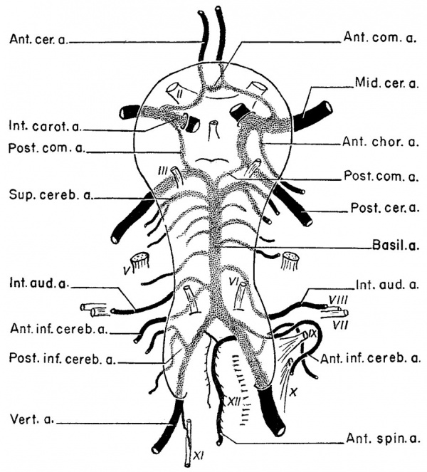
Fig. 2.
Fig. 2 A diagram illustrating the complex of arteries seen on the ventral surface of the brain. The arteries drawn in solid black represent the 4 large arteries supplying the brain and the constant group of small arteries supplying specific areas of the brain. The arteries lying inside the elliptoid line and which are stippled represent the highly variable, medium-sized group. Eighty-five % of cerebral aneurysms are located inside the elliptoid line, occurring on the variable group of arteries.
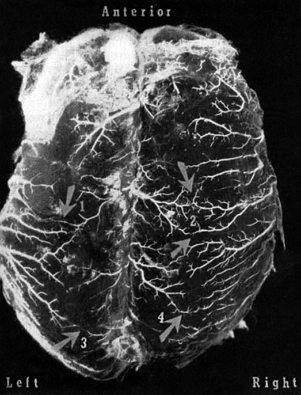
Fig. 3.
Fig. 3 A photograph of a 6-months old fetal brain, with the large superficial arteries injected, and the brain cleared by the Spalteholz technique.
Arrows 1 and 2 indicate anastomoses between large branches of the anterior cerebral arteries and the middle cerebral arteries.
Arrows 3 and 4 point out anastomoses between the posterior cerebral and the middle cerebral arteries. These large superficial anastomoses can be seen clearly only before the development of the gyri and sulci.
Cite this page: Hill, M.A. (2024, April 27) Embryology Paper - Significant superficial anastomoses in the arterial blood supply to the human brain (1959). Retrieved from https://embryology.med.unsw.edu.au/embryology/index.php/Paper_-_Significant_superficial_anastomoses_in_the_arterial_blood_supply_to_the_human_brain_(1959)
- © Dr Mark Hill 2024, UNSW Embryology ISBN: 978 0 7334 2609 4 - UNSW CRICOS Provider Code No. 00098G


