Paper - On the cervical veins and lymphatics in four human embryos, with an interpretation of anomalies on the subclavian and jugular veins in the adult (1909)
| Embryology - 30 Apr 2024 |
|---|
| Google Translate - select your language from the list shown below (this will open a new external page) |
|
العربية | català | 中文 | 中國傳統的 | français | Deutsche | עִברִית | हिंदी | bahasa Indonesia | italiano | 日本語 | 한국어 | မြန်မာ | Pilipino | Polskie | português | ਪੰਜਾਬੀ ਦੇ | Română | русский | Español | Swahili | Svensk | ไทย | Türkçe | اردو | ייִדיש | Tiếng Việt These external translations are automated and may not be accurate. (More? About Translations) |
Lewis FT. On the cervical veins and lymphatics in four human embryos, with an interpretation of anomalies on the subclavian and jugular veins in the adult. (1909)
| Historic Disclaimer - information about historic embryology pages |
|---|
| Pages where the terms "Historic" (textbooks, papers, people, recommendations) appear on this site, and sections within pages where this disclaimer appears, indicate that the content and scientific understanding are specific to the time of publication. This means that while some scientific descriptions are still accurate, the terminology and interpretation of the developmental mechanisms reflect the understanding at the time of original publication and those of the preceding periods, these terms, interpretations and recommendations may not reflect our current scientific understanding. (More? Embryology History | Historic Embryology Papers) |
On the Cervical Veins and Lymphatics in Four Human Embryos, with an Interpretation of Anomalies on the Subclavian and Jugular Veins in the Adult
By
From the Department of Anatomy, Harvard Medical School.
Introduction
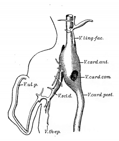
In order to explain an anomaly of the subclavian vein occurring in a man 68 years old, reconstructions of the cervical veins and lymphatics in four human embryos were made, with the following results. In an embryo having a maximum measurement of 10 mm., the right arm is drained by a primitive ulnar vein which unites with a thoracoepigastric vein to form the dorsal subclavian vein. These vessels are shown in Fig. 1. The primitive ulnar vein is seen to receive a branch near the elbow, and distally it has many smaller tributaries which have been omitted from the drawing. There are no veins larger than capillaries along the radial border of the arm.
The thoraco—epigastric vein, which is found in the lateral body wall, is represented in fishes and in all the higher classes of vertebrates. Unfortunately it has been given a great variety of names. In the rabbit it is called the external mammary vein (Krause[1] Lewis[2]) and this name has been applied to it in man (Poirier[3]). In man it has been called, in part at least, the long, lateral‘, or inferior thoracic vein, but the term thomco-epigastmb is preferable to any of these.
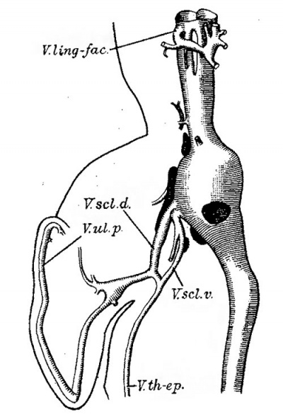
The primitive ulnar and thoraco-epigastric veins unite to form a subclavian vein which passes dorsal to the brachial plexus to enter the anterior cardinal vein. Small branches of the anterior cardinal vein are found Ventral to the brachial plexus. In a slightly older embryo (Fig. 2) these ventral branches are continuous with the thoraco-epigastric vein so that the brachial plexus is enclosed in a venous ring. This arrangement of the subclavian veins has been described in the rabbit and man by Hochstetter[4] and the corresponding stage in the rabbit has been figured.[5]
In addition to the subclavian vein, the anterior cardinal has another important branch. This begins as a median vessel in the lingual region (Fig. 1). It turns sharply to the right, receives tributaries from the lateral superficial tissues and descends in front of the vagus nerve. In the lower part of its course it is close to the dorsal portion of the pericardial cavity. It passes on the lateral side of the vagus nerve to enter the anterior cardinal vein. In the older embryo this linguo—facial branch (V. lingfac.) is easily recognized. The median vessel in the sublingual region is present, but it has not been drawn, since in this embryo it is drained by the linguo—facial branch of the left cardinal vein. As seen in the reconstruction, the linguo—facial vein has acquired a new outlet, which is anterior to the place Where the hypoglossal nerve cro-sses the cardinal vein. The hypoglossal nerve, which before was lateral to the cardinal vein, is now surrounded by it,[6] and the venous loop through which it passes is shown near the top of Fig. 2. That part of the linguo—facial vein which descended in front of the vagus nerve has apparently disappeared, and the vein is no longer in relation with the pericardial cavity.
It is perhaps worth while to call attention to the scattered data regarding this linguo—facial vein. It was discovered by Grosser in embryo bats and described as follows:[7]
The veins of the branchial arches consist in young embryos of Rhinolophus of a median longitudinal vein near the ventral surface of the mandibular and hyoid arches, which at its caudal end divides into two symmetrical parts. Each of these turns abruptly to one side, receives small tributaries from the lateral parts of the branchial arches, and empties, lateral to the aortic arches, into the anterior cardinal‘ vein. Later the vessel cannot be demonstrated. A part of it may perhaps be incorporated in the external jugular vein.”
|
|
| Fig. 1. Reconstruction, as seen from the ventral side, of the right arm and adjacent part of the body of a human embryo of 16 mm (Harvard Embryological Collection 1000), to show the veins and lymphatic vessels. The latter, in this and the three following figures, have been heavily shaded.x 20 diam. | Fig. 2. Similar reconstruction from a human embryo of 11.5 mm (H. E. C. Template:HEC189). X 20 diam. V. curd. ¢mt., Vena cardinalis anterior; V. card. com., V. cardinalis communis (duct of Cuvier) ; V. card. post., V. cardinalis posterior; V. ling-fac., V. linguo-facialis; V. sol. d.. V. subclavia dorsalis; V. sol. 12., V. subclavia ventralis; V. th—ep., V. thoraco-epigastrica; V. M. 1)., V. ulnar-is prima. |
This description is illustrated by an excellent reconstruction. Two years later these veins were recorded in pig embryos of 6, 12, 14 and 20 mm.[8] Because of their course across the throat they were called “transverse veins.” It was stated that the median vessel, instead of bifurcating, sometimes passes wholly to the right and sometimes to the left. The early appearance of corresponding veins in the rabbit was reported subsequently.[8] They are present in an embryo of 9 1/3 days (3 mm). Usually the linguo-facial vein arises from the anterior cardinal near its outlet, but sometimes it connects with the common cardinal vein (duct of Cuvier) and in one exceptional case — the left side only of an embryo of 12 days (5 mm.)—it emptied into the posterior cardinal vein. In a 7 mm. rabbit the median lingual portion of the vein has been seen to bifurcate symmetrically, agreeing with Grosser’s description for the bat. In rabbits from 9.5 to 29 mm. the main trunk of this vein has been shown in a series of six reconstructions.[5] Its terminal branches are the anterior and posterior facial veins, the latter receiving a posterior auricular branch; thus it corresponds with the external jugular vein of the adult rabbit as described by Krause, and it has been so labeled. This vein, however, seems to be homologous with the common facial vein in man, the external jugular vein of human anatomy arising independently as will be seen presently.
In a recent publication Grosser has shown that the linguo-facial vein is homologous with the inferior jugular vein of fishes, amphibia, and reptiles,— a paired ventral vessel draining the floor of the branchial region.[9] In the same paper he states that this vein is well developed in cat and guinea-pig embryos and in a human embryo of 6.5 mm. Grosser’s comparative studies demonstrate the fundamental importance of this vein. The name inferior jugular has, however, not been adopted in this paper since it is an unfortunate designation for a ventral vessel in fishes, which has nothing to do with the anterior (vi. e., ventral) jugular vein of man, but gives rise to veins which empty anterior or superior to the other jugulars. The term linguo—facial is justified by the embryonic distribution of the vessel,— in part to the lingual region (hyoid and mandibular arches) and in part to the superficial tissues of the mandibular region. Although there is a complex rearrangement and new formation of branches, a single large vein drains this territory from early embryonic stages to the adult. Thus in man, according to Sebileau and Demoulin,[10] “Faraboeuf has well shown that the facial vein, lingual vein, and superior thyreoid vein unite to form a common canal very happily termed the thyreo-linguowfacial venous trunk. This arrangement is in fact very frequent.” Except for the superior thyreoid vein, which has not yet developed, this description may be applied to the embryonic vessels shown in Fig. 2.
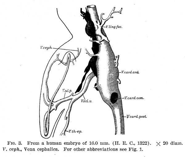
Fig. 3. From a human embryo of 16.0 mm (H. E. 0., 1322). X 20 diam. V. ceph., Vena cephalica. For other abbreviations see Fig. 1.
The study of the veins in the embryos just described led to the following observations upon the lymphatic vessels. In the 10 mm. embryo (Fig. 1), the jugular sac is represented by a single lymph space in close relation with the anterior cardinalvein. It appears to be lined with endothelium, which in some sections has shrunken away from the surrounding mesenchyma. It contains a few blood corpuscles, and appears to communicate with the vein by a slender oblique passage which is completely filled by a single file of blood corpuscles. This lymphatic space is larger and more irregular in outline than the neighboring small tributaries of the vein. No lymphatics could be found in a 9.2 mm. embryo, so that the jugular lymphatics probably arise in human embryos of about 10 mm. This accords with the observation that they first appear in rabbits of 9.5—10.0 mm., but does not agree with Ingalls’ opinion that in a 4.9 mm. human embryo certain Vessels represent “the first anlage, or earliest forerunners, perhaps, of the lymphatic system in man.[11] The vessels in question are clearly veins.
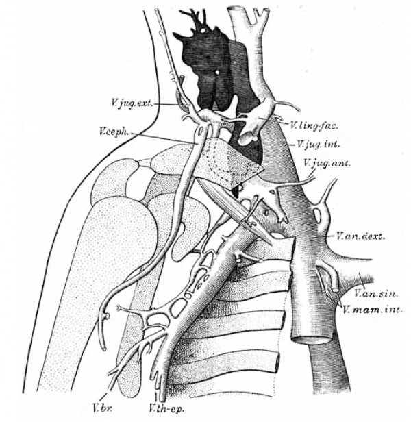
Fig. 4. From a human embryo of 22.8 mm (H. E. C., 871). X 20 diam. The ribs, clavicle, scapula, and humerus have been stippled, and the subclavius muscle has been drawn. In addition to the veins shown in Fig. 1, the following are included: V. cm. deml, Vena anonyma dextra; V. an. 31%., V. anonyma sinistra; V. bra, V. brachialis; V. 061%., V. cephalica; V. jug. ant., V. jug. ea2t., V. jug. ma, V. jugularis anterior, externa, interna; V. mam. int., V. mammaria interna. I
In the 11.5 mm. embryo (Fig. 2) there are four or perhaps five lymphatic spaces, apparently separate from each other. They contain some blood corpuscles and in two cases they seem to connect with the vein, but the apertures are very small. At 16 mm. (Fig. 3) the lymphatics have increased and extend along a considerable portion of the anterior cardinal vein. There is a separate space in relation with the linguo—facia1 vein. A lymphatic vessel extends from the ugular sac dorsal to the brachial plexus, but the dorsal subclavian vein which it accompanied in the earlier stage has disappeared. Similarly in the rabbit the dorsal subclavian vein is accompanied by a lymphatic vessel and later both vein and lymphatic disappear. No outlet from the jugular sac into the cardinal vein was found in the transverse sections studied, but, as Professor Sabin has recently demonstrated, frontal sections are more favorable for detecting the valvular orifice. In an embryo of 22.8 mm. (Fig. 4) there is a very large ugular sac which has grown around certain nerves. The upper small aperture transmits a branch of the third cervical nerve, and the lower one is for branches of the third and fourth. In a rabbit of 14.5 mm. the jugular sac showed similar openings for the third and fourth cervical nerves.
Returning to the veins in Fig. 3, it will be seen that the branch of the primitive ulnar vein near the elbow now joins the radial extremity of the ulnar vein, and from their junction a vessel can be followed along the radial border of the limb toward the shoulder. It is very probable that at this stage there is a capillary union between this cephalic vein and the lateral branch of the anterior cardinal which is shown in the figure. An obvious connection between them is seen in Fig. 4. Here the lateral branch of the anterior cardinal may be identified as the external jugular vein and it can be traced upward behind the car as the posterior auricular vein. It does not yet connect with the linguo-facial vein.
Fig. 4 shows the brachial vein (derived from the primitive ulnar) joining the larger thoraco-epigastric vein to make the (ventral) subclavian vein. Along the latter, several branches anastomose to make a small vena comitans for the subclavian artery. Higher up both the subclavian vein and the vena comitans are separated from the artery by the scalenus anterior muscle, and both are dorsal to the subclavius muscle, which has been drawn in the figure. Ventral to the subclavius muscle there is a small branch distributed beneath the pectoral muscles; this branch probably is the principal factor in the
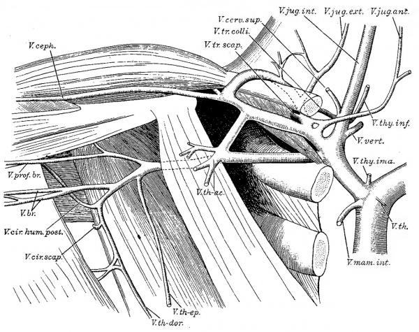
Fig. 5. Dissection of Veins in a man 68 years old. Two-thirds natural size. V. br., Venae brachiales; V. ceph., V. cephalica ; V. cerrv. sup, V. cervic-alis super.ficialis; V. Cir. hum. post, V. circumflexa humeri posterior; V. 0171‘. sCap., V. circumflexa scapulae; V. jug. ant, V. jug. ea:t., V. jug. int., V. jugularis anterior, externa, interna; V. mam. imfi, V. mammaria interna; V. prof. br., V. profunda brachii (with a branch, the V. circumflexa humeri anterior); V. th., V. thymica; V. th-ac., V. thoraco-acroinialis; V. th—dm'., V. thoracodorsalisg V. th-ep., V. thoraco—epigastrica; V. thy. ima, V. thy. inf., V. thyreoidea ima, inferior; V. tr. com, V. transversa colli (from vertebral border of scapula) ; V. tr. scap., V. transversa scapulae (from scapular notch) ; V. a;ert., V. vertebralis.
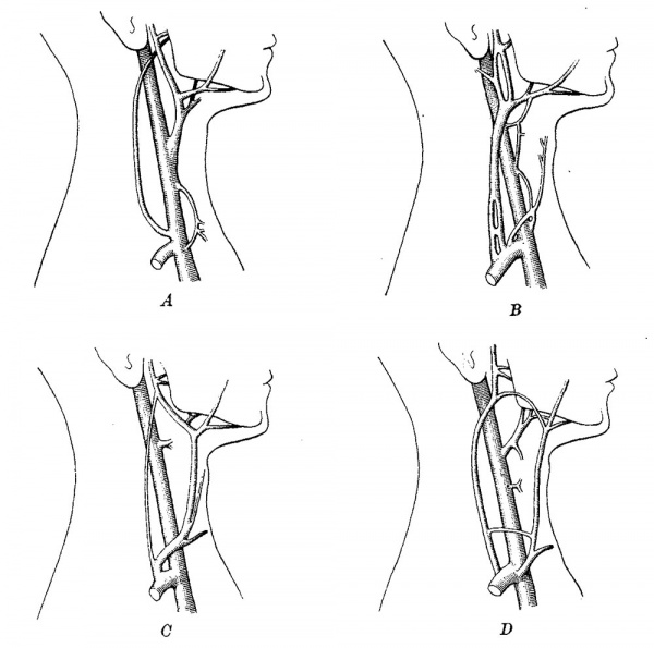
Fig. 6. The relation of the linguo—facial vein to the jugular veins in the adult. (From dissections.)
A. The primary relation. The linguo—facial vein is a branch of the internal jugular; its submental, anterior facial, lingual, and posterior facial branches are shown in the drawing. The 1inguo—facial vein has only small anastomoses with the external jugular vein, and none with the anterior jugular, the latter vessel being scarcely represented in this case. The similarity to the embryonic relations shown in Fig. 4 is apparent.
B. The linguo—facial vein has been tapped by the external jugular so that its branches appear to belong to the latter.
C. The linguo-facial vein is drained chiefly by the anterior jugular, to which its branches appear to belong.
D. The linguo~facial vein is subdivided, so that its posterior facial branch empties into the external jugular, its anterior facial branch empties into the anterior jugular, and the lingual branch remains as a tributary of the internal jugular anomaly to be described. At the outlet of the subclavian vein there are several branches, and among them the anterior jugular vein can be identified.
The anomaly which led to the examinatioh of the embryos is pictured in Fig. 5. If the small vein ventral to the subclavius muscle in Fig. 4 is considered to have enlarged and anastomosed both with the cephalic vein and the subclavian vein, the conditions found in the anomaly will be strikingly reproduced. Both the clavicle and the subclavius muscle will then be surrounded by a venous ring. In the anomaly the “jugulo—cephalic” vein, which is above the clavicle, is as large as the subclavian vein. The “accessory subclavian vein,” which is between the clavicle and the subclavius muscle, is somewhat larger. The subclavian vein occupies its normal position between the subclavius muscle above, the scalenus anterior behind, and the first rib below. '
Finally it may be noted that the formation of a venous ring around the scalenus anterior muscle, occasionally recorded in the adult, is suggested in the 22.8 mm embryo. Where the subclavian veiin empties into the jugular, these vessels are molded about the muscle, and since a vertebral branch joins the jugular at this point, nearly two-thirds of the circumference of the scalenus anterior are in relation with the veins. The completion of this ring by venous outgrowths would account for the anomaly. The conclusion may be drawn that although the jugulo—cephalic vein in man is a persistence of an important and normal embryonic vein, the accessory subclavian vein, whether situated behind the scalenus or above the subclavian, is an abnormal vessel.
Of wider interest is the conclusion that the linguo—facial vein is a morphological constant. In mammals it appears at an early stage, and although it often becomes resolved into a group of branches, it is present in the adult. Some of its transformations in man are shown in Fig. 6, on the preceding page. It may have large anastomoses with the external jugular vein or the anterior jugular vein or with both, but these can be readily described and understood on the basis of a primary linguo-facial vein.
| Historic Disclaimer - information about historic embryology pages |
|---|
| Pages where the terms "Historic" (textbooks, papers, people, recommendations) appear on this site, and sections within pages where this disclaimer appears, indicate that the content and scientific understanding are specific to the time of publication. This means that while some scientific descriptions are still accurate, the terminology and interpretation of the developmental mechanisms reflect the understanding at the time of original publication and those of the preceding periods, these terms, interpretations and recommendations may not reflect our current scientific understanding. (More? Embryology History | Historic Embryology Papers) |
References
- ↑ Krause, W. Die Anatomie des Kaninchens. Leipzig, 1868.
- ↑ Lewis FT. The development of the vena cava inferior. (1902) Amer. J Anat. 1(3): 229-244.
- ↑ Poirier, P. Traité d’anatomie humaine. Paris, 1896. Vol. 2, p. 911.
- ↑ Hochstetter, F. Ueber die Entwicklung der Extremitatsvenen bei den Amnioten. Morph. Jahrb., 1891, V01. 17, pp. 26-27 and 32-33.
- ↑ 5.0 5.1 Template:Ref-Lewis1905 Cite error: Invalid
<ref>tag; name 'Lewis1905' defined multiple times with different content - ↑ This accords with Tandler’s observation that the hypoglossal nerve is lateral to the internal jugular vein in human embryos of 8 and 9 min. and medial at 12.5 mm. Anat. Anz., 1907, Vol. 31, pp. 473-480.
- ↑ Grosser, O. Zur Anatomic und Entwickelungsgeschichte des Geféisssystenis der Chiropteren. Anat. Hefte, 1901, Heft 55, p. 131.
- ↑ 8.0 8.1 Lewis FT. The gross anatomy of a 12 mm pig. (1903) Amer. J Anat. 2: 221-225.
- ↑ Grosser, 0. Die Elemente des Kopfvenensystems der Wirbeitiere. Verh. d. anat. Gesellschaft, 1907, pp. 179-192. F. R. Lillie refers to this vein in the chick as the external jugular. Development of the Chick, Chicago, 1908.
- ↑ Sebileau, P., and Demoulin, A. Comment il faut comprendre le systeme des veines jugulaires antérieures. Bull. de la soc. anat. de Paris, 1892, année 67, pp. 120-132.
- ↑ lngalls. N. W. A contribution to the embryology of the liver and vascular system in man. Anat. Record. 1908, Vol. 2, p. 343.
Cite this page: Hill, M.A. (2024, April 30) Embryology Paper - On the cervical veins and lymphatics in four human embryos, with an interpretation of anomalies on the subclavian and jugular veins in the adult (1909). Retrieved from https://embryology.med.unsw.edu.au/embryology/index.php/Paper_-_On_the_cervical_veins_and_lymphatics_in_four_human_embryos,_with_an_interpretation_of_anomalies_on_the_subclavian_and_jugular_veins_in_the_adult_(1909)
- © Dr Mark Hill 2024, UNSW Embryology ISBN: 978 0 7334 2609 4 - UNSW CRICOS Provider Code No. 00098G


