Carnegie stage 6
| Embryology - 24 Jul 2026 |
|---|
| Google Translate - select your language from the list shown below (this will open a new external page) |
|
العربية | català | 中文 | 中國傳統的 | français | Deutsche | עִברִית | हिंदी | bahasa Indonesia | italiano | 日本語 | 한국어 | မြန်မာ | Pilipino | Polskie | português | ਪੰਜਾਬੀ ਦੇ | Română | русский | Español | Swahili | Svensk | ไทย | Türkçe | اردو | ייִדיש | Tiếng Việt These external translations are automated and may not be accurate. (More? About Translations) |
Introduction

Bilaminar embryonic disc |
The bilaminar (epiblast and hypoblast) embryo is now about 0.2 mm diameter in size. The three extra embryonic spaces (amniotic, primitive yolk sac and chorionic) are present.
The large chorionic cavity is surrounded by the cell layers extending to form chorionic villi. These villi are the beginning of the functional units of the placenta. Outside of these villi, and extending from their tips, trophoblasts invade the maternal decidua forming maternal blood-filled lacunae (lakes). Emptying into these maternal lacuna are both uterine glands and spiral arteries held open by trophoblast cells. The maternal uterine wall continues to undergo expanding decidualization in response to the implantation process. SummaryHuman embryonic stage 6 occurs towards the end of week 2 at approximately 13-14 days.
|
| Stage 6 Links: Week 2 | Implantation | Lecture | Practical | Carnegie Embryos | Category:Carnegie Stage 6 | Next Stage 7 |
| Historic Papers: 1909 | 1925 | 1937 |
| Week: | 1 | 2 | 3 | 4 | 5 | 6 | 7 | 8 |
| Carnegie stage: | 1 2 3 4 | 5 6 | 7 8 9 | 10 11 12 13 | 14 15 | 16 17 | 18 19 | 20 21 22 23 |
- Carnegie Stages: 1 | 2 | 3 | 4 | 5 | 6 | 7 | 8 | 9 | 10 | 11 | 12 | 13 | 14 | 15 | 16 | 17 | 18 | 19 | 20 | 21 | 22 | 23 | About Stages | Timeline
Carnegie Collection

|

|
| Embryonic Disc | Primitive streak region (gastrulation) |
| iBook - Carnegie Embryos | |
|---|---|

|
|
Kyoto Collection

Other Embryos
This stage was previously subdivided into 6a and 6b.[2]
Stage 6a
- Carnegie No. 8905, Merrill. Unbranched villi. Although an abnormal leucocytic reaction is present, this specimen “represents the best example in the author’s collection of formation of early primordial villi, active mesogenesis and angiogenesis, completion of the amnion and the transitional phase between the primary and definitive [secondary] yolk sac formation” (Hertig, 1958[3], who reproduced a photomicrograph as fig. 55). Presumed age, 12-13 days.
- Carnegie No. 6800, Stöckel. Described by Linzenmeier (1914). Hysterectomy. Angiogenesis in chorion described by Hertig (1935). Photomicrographs reproduced by Hertig (1958)[3], Figs. 56-58). Important as one of the youngest specimens having “true villi” (Hertig and Rock, 1941). Chorionic villi show “an occasional tendency to dichotomous branching” (Krafka, 1941). Indication of blood vessel formation in villi. Chorion, 2.75 x 1.05 x 0.9 mm. Chorionic cavity, 0.75 x 0.61 x 0.52 mm; capacity, 0.13 mm3 (Odgers, 1937).[4] Embryonic disc, 0.21 x 0.105 mm (Krafka, 1941). Allantoic diverticulum doubtful. Presumed age, 13 days.
- Carnegie No. 8672. Photomicrograph illustrated by Hertig, Rock, and Adams (1956, fig. 40). Chorion, 1.14 x 1.08 mm. Chorionic cavity, 0.8 x 0.79 mm. Embryonic disc, 0.203 x 0.07 mm. Presumed age, 13 days.
- Harvard No. 55. Studied histochemically by Hertig et al. (1958). Hysterectomy. Chorion, 1.77 x 1.33 x 0.598 mm.Chorionic cavity, 0.73 x 0.68 x 0.221 mm. Embryonic disc,0.296 x 0.196 x 0.044 mm. Chorionic villi essentially solid,with earliest suggestion of mesoblastic core formation. “Apparently without axial differentiation.” Possesses “a very recently formed definitive [secondary] yolk sac.” Possible primordial germ cells (“stuffed with glycogen”) within endoderm near edge of disc. Presumed age, 13 days. For histochemical details, the original paper should be consulted.
- Carnegie No. 8360. Photomicrographs illustrated by Hertig, Rock, and Adams (1956, figs. 42 and 46)[5]. Chorion, 1.466 x 1 mm. Chorionic cavity, 1 x 0.66 mm. Embryonic disc, 0.188 x 0.055 mm. Presumed age, 13 days.
- Peters. Described in a monograph by Peters (1899)[6]. Autopsy. A famous embryo, for long the youngest known and the first to be described in detail. Photomicrographs have since been published (Rossenbeck, 1923, plate 42; Odgers, 1937[4], plate 2, fig. 2). The chorionic villi, some of which display a mesenchymal core, send cellular columns externally and these latter are beginning to form a cytotrophoblastic shell. Slight branching of villi (Krafka, 1941). Chorionic cavity contains magma réticulé of Velpeau (Mall, 1916). Blood islands on umbilical vesicle. Chorion, 1.5 x 2 mm. Chorionic cavity, 1.6 x 0.9 x 0.8 mm; capacity, 0.7 mm3 (Odgers, 1937)[4]. Embryonic disc, 0.18 x 0.24 mm (Krafka, 1941). The basement membrane (Hensen’s membrane prima) of the epiblast was noted by Graf Spee. Allantoic diverticulum and primitive streak uncertain. Presumed age, 13 days (Krafka,1941). A tabulation of normal human embryos compiled from the literature prior to 1900 and from Mall’s own collection was published by Mall (1900, pp. 38-46). The least advanced specimen was the Peters embryo, and included in the list were 92 embryos of 0.19-32 mm, as well as 17 fetuses of 33-210 mm.
- E.B. (E. Béla v. Entz). Described by Faber (1940). Curettage. Incomplete. Primitive villi. Chorionic cavity, 0.935 x 0.697 mm. Embryonic disc, 0.231 mm. No primitive streak, node, or groove. Secondary umbilical vesicle. No allantoic diverticulum. Said to resemble the Peters specimen closely.
- Carnegie No. 7634, Torpin. Described in detail by Krafka (1941), who provided also an extensive discussion of the decidua. Hysterectomy. Posterior wall of uterus. Chorionic villi with mesoblastic cores, 0.1-0.2 mm in length. Villi “generally single, but two or more may arise from a common base,” although no branching was recorded. Chorion (possessed 85 villi), 1.76 x 1.7 x 1.5 mm. Chorionic cavity, 1.3 x 1.1 x 1 mm. Embryonic disc, 0.216 x 0.21 mm. Amniotic duct. Neither primitive node nor primitive streak. Cloacal cord (rather than membrane) claimed, but doubted by Mazanec (1959). No allantoic diverticulum. Diverticulum of umbilical vesicle. Presumed age, 13 days. Dorsal and transverse projections published (Krafka, 1941, figs. 1 and 3, and plate 2).
- VMA-I. Specimen of Knorre, summarized by Mazanec (1959). Chorion, 3.24 x 2.04mm. Chorionic cavity, 1.53 x 1.02 mm. Embryonic disc, 0.23 x 0.2 mm. Development thought to be between Torpin and Yale specimens.
- Carnegie No. 6734, Yale. Described in detail by Ramsey (1938). Necropsy. Left lateral uterine wall. Chorion, 2.75 x 1.9 x 0.76 mm. Chorionic cavity, 1.3 x 1.1 x 1 mm. Some of the chorionic villi “show dichotomous division, but no more complicated branching has occurred.” Some angioblastic strands in villi. Embryonic disc (damaged and distorted), 0.15 mm. Allantoic diverticulum stated to be present but denied by Krafka (1941). Presumed age, 13-14 days. Drawings of model published (Ramsey, 1938[7], fig. 1).
- Noback, Paff, and Poppiti (1968) described an autopsy specimen that possessed a chorion of 2.25 x 1.25 x 2 mm. Chorionic villi avascular. Embryonic disc, 0.22 x 0.2 mm. No primitive node, notochordal process, cloacal membrane, or allantoic diverticulum. Axial differentiation, however, was suggested by the possible primordia of the prechordal plate and the primitive streak. Hence this specimen may be regarded as transitional between 6a and 6b.
Stage 6b
(listed in order of length of primitive streak)
- Liverpool I. Described by Harrison and Jeffcoate (1953). Curettage. Chorionic villi “are only beginning to show evidence of branching” (ibid., Plate 1, fig. 1). Chorion, 1.86 x 1.47 mm. Chorionic cavity, 1.5 x 0.84 mm. Embryonic disc, 0.161 x 0.199 x 0.033 mm. Primitive streak, 0.021 mm. Allanto-enteric diverticulum claimed. Median projection published (ibid., fig. 1; Mazanec, 1959, fig. 31).
- Liverpool II. Described by Lewis and Harrison (1966), who, in view of “the dimensions, degree of differentiation and decidual appearances,” assigned the specimen to horizon VII. Hysterectomy. The chorionic villi are localized to the embryonic pole, and their mesoblastic cores contain “isolated vascular primordia formed by coalescence of angioblasts.” “The villi are branched; lacunae and intervillous spaces have formed.” Chorion, 2.72 x 2.35 x 1.54 mm. Embryonic disc, 0.264 x 0.22 mm. Primitive streak, 0.024 mm. Amniotic duct and long duct of umbilical vesicle. Resembles Teacher-Bryce II embryo. Median projection published (ibid., fig. 6).
- Carnegie No. 7801 (figs. 6-5 to 6-10). Described in detail and illustrated by Heuser, Rock, and Hertig (1945). Hysterectomy Posterior wall of uterus. “The primitive villi are short and stubby; a few reach a length of about 0.25 mm.” Chorion, 2.6 x 1.9 x 1.4 mm. Chorionic cavity, 1.3 x 1.1 x 0.8 mm. Embryonic disc, 0.04 x 0.22 x 0.253 mm. Primitive streak, 0.04 mm. “Axial differentiation is just appearing.” “In this embryo the site of the future [cloacal] membrane seems indicated, but not the structure itself.” Probably no allantoic duct. Presumed age, 13-13½ days. Median projection published (ibid., plate 3).
- Carnegie No. 8819, Edwards-Jones-Brewer. Described in detail by Brewer (1937, 1938). Hysterectomy. “There is no branching of a mesodermal villus.” Chorion, 3.6 x 3 x 1.9 mm. Chorionic cavity, 1.85 x 1.71 x 1.01 mm; capacity, 13.38 mm3. Embryonic disc, 0.209 x 0.177 mm; volume, 0.0814 mm3. Primitive streak, 0.04 mm, claimed, but its presence was denied by Krafka (1941). No allantoic diverticulum. Dorsal and median projections published (Brewer, 1938, plate 1, figs. 2 and 3; Mazanec, 1959, fig. 33). Some authors have attempted to identify a prechordal plate from the median projection.
- Carnegie No. 7762, Rochester. Described by Wilson (1945). Curettage. Chorion, 2.3 x 2.2 x 2 mm. Larger villi have a mesoblastic core “and some show a tendency toward branching.” Moreover, “no evidence of actual blood vessels is seen in the villi, but in many of them groupings of angioblasts are observed.” Chorionic cavity, 1.75 x 1.3 x 1 mm. Embryonic disc, 0.313 x 0.22 mm. Primitive streak, 0.04 mm. No definite allantoic diverticulum. Amniotic duct. Median reconstruction published (ibid., plate 3; Mazanec, 1959, fig. 36).
- Op (Opitz). Described by von Möllendorff (1921b). Hysterectomy. Chorionic villi show first branching in many places. Chorionic cavity, 1.5 x 1.15 x 1 mm. Embryonic disc, 0.19 mm. Primitive streak, 0.045 mm. Allantoic diverticulum denied by Florian (1930a). Disintegrating epithelial proliferation of amnion behind caudal end of embryonic plate (ibid.). Median reconstruction published (Mazanec, 1959, fig. 34).
- Fetzer. Described by Fetzer (1910) and by Fetzer and Florian (1929, 1930). Curettage. Chorionic villi “show a beginning tendency to branch” (Streeter, 1920). Chorion, 2.2 x 1.8 mm. Chorionic cavity, 1.6 x 0.9 mm. Embryonic disc, 0.26 x 0.215 mm, Primitive streak (denied by Rossenbeck, 1923), 0.05 mm. Cloacal membrane (Florian, 1933) but no allantoic diverticulum. Area of mesoblastic proliferation from adjacent disc and amniotic ectoderm, “caudal” to cloacal membrane (Hill, 1932; Florian, 1933). Stated to lie between Wo and Bi I in development. Dorsal and median projections published (Fetzer and Florian, 1930, figs. 1a, 1b, 2 and 53; Florian, 1945, plate 4, fig. 40; Mazanec, 1959, fig. 37).
- H.R. 1 (Hesketh Roberts). Described by Johnston (1940) who included Florian’s divergent interpretation of the specimen Hysterectomy. Chorion and endometrium described by Johnston (1941). According to Florian, the embryonic disc is 0.048 mm and the primitive streak is 0.06 mm in length. Primitive node, notochordal process, and prechordal plate all absent (but described as present by Johnston). Embryo abnormal in shape, the result of an abnormal growth process. Median projection published (Johnston, 1940, fig. 35).
- Wo (Wolfring). Described by von Möllendorff (1925). Chorionic cavity, 2.52 x 2.16 x 2.06 mm. Embryonic disc, 0.25 x 0.22 mm. Primitive streak, 0.065 mm. Cloacal membrane rather than solid allantois (Florian, 1933). Median projection published (von Möllendorff, 1925, fig. 4; Florian, 1928a, fig. 40; Mazanec, 1959, fig. 38).
- Beneke (Strahl-Beneke). Described originally by Strahl and Beneke in 1916 in a monograph and later by Florian and Beneke (1931). Chorionic cavity, 3.8 x 2.2 x 1.2 mm. Embryonic disc (narrow type), 0.375 mm (Florian, 1934a). Primitive streak (doubted by Rossenbeck, 1923, and denied by Fahrenholz, 1927, but acknowledged by Florian, 1928a), 0.1 mm. No notochordal process (Hill and Florian, 1931b). Prechordal plate, 0.066 mm. Dorsal and median projections published (Florian and Beneke, 1931, figs. 2 and 1; Florian, 1928a, fig. 42; Florian, 1945, plate 4, fig. 41; Mazanec, 1959, fig. 40).
- Am. 10. Described by Krause (1952). Hysterectomy. Chorionic cavity, 3.6 x 2.5 x 2.5 mm. Embryonic disc (broad type), 0.32 x 0.3 x 0.06 mm. Primitive streak, 0.135 mm. No notochordal process (but see Mazanec, 1959) although a small lumen was suggested as a possible Anlage of “Lieberkühn’s canal,” Dorsal and median projections published (Krause, 1952, figs. 13 and 15; Mazanec, 1959, fig. 42). Could be stage 7.
- Bi I (Bittman). Described by Florian (1927) and in 1928 in a Czech publication, (For general appearance, see Mazanec, 1959, figs. 95 and 112.) Chorionic cavity, 2.13 x 2.13 x 2.12 mm. Embryonic disc (broad type), 0.35 x 0.34 mm. Primitive streak, which appears as an “indifferent cellular knot” (Florian, 1928b, 0.135 mm). Possible primordial germ cell in ventral wall of umbilical vesicle (Politzer, 1933). Median projection published (Florian, 1928a, fig. 41; Florian, 1945, plate 5, fig. 42; Mazanec, 1959, fig. 41).
- Lbg (Lönnberg). Described by Holmdahl (1939). Chorion, 16 x 15 mm. Embryonic disc, 0.285 x 0.236 x 0.032 mm. Primitive streak, 0.144 mm. No allantoic duct. T.F. Described by Florian (1927, 1928a). Autopsy. Chorionic cavity, 4.578 x 3.078 x 1.76 mm. Embryonic disc, 0.468 x 0.397 x 0.485 mm. Primitive streak (Mazanec, 1959, fig. 100) 0.162 mm. No notochordal process. Median projection published (Florian, 1928a, figs. 27 and 43; Mazanec, 1959, fig. 43).
- HEB-28. Described by Mazanec (1960). Abortion. Abnormal features, Chorionic cavity, 4.29 x 4 x 3.55 mm. Embryonic disc (broad type), 0.44 X 0.47 mm. Primitive streak, 0.187-0.22 mm, and node, 0.071 mm. No notochordal process. Dorsal and median projections published (ibid., figs. 1 and 2). Regarded as transitional between stages 6 and 7.
Additional Specimens
- Precise measurements of the primitive streak have not been provided in accounts of the following embryos. The specimens are listed in order of year of publication.
- Minot. Described by Lewis in Keibel and Mall (1912). Primitive streak present. Median projection published (ibid., vol. 2, fig. 229).
- Schlagenhaufer and Verocay (1916) described an autopsy specimen that possessed an embryonic disc of 0.24 X 0.28 mm. Although a primitive streak was not found, the development of the specimen is such that it was probably present (Mazanec, 1959).
- Teacher-Bryce II[8]. Described by Bryce (1924)[9] and Mc’Intyre (1926)[10]. Autopsy. Chorionic villi are “well developed but are still simple and little branched.” Chorion, 4.5 X 4 X 3.5 mm. Chorionic cavity, 2.8 X 2.6 X 2.25 mm. Embryonic disc, 0.2 X 0.1 X 0.15 mm. Primitive streak present (Mazanec, 1959). Long stalk from umbilical vesicle. Blood vessel primordia in connecting stalk.
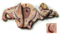
Uterus laid open, embryo imbedded in posterior wall
lobule containing embryo

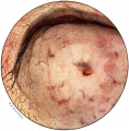
whole ovum drawing
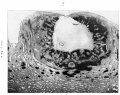
Photomicrograph of Section 28.2.2


- H381. Described by Stump (1929). Chorionic villi branched. Chorion, 4.38 x 4.2 x 1.4 mm. Chorionic cavity, 3.48 X 3.44 X 0.81 mm. Embryonic disc, 0.58 X 0.3 mm. Primitive groove and streak or node believed to be present. Said to resemble Hugo and Debeyre specimens.
- Andô Described by Hiramatsu (1936). Hysterectomy. Chorion, 4.2 X 3.25 X 1.9 mm. Chorionic cavity, 4.2 X 2.4 X 1 mm. Embryonic disc, 0.24 X 0.26 x 0.04 mm. Some villi branched. Stated to resemble the Peters specimen. Described as possessing no primitive streak but Mazanec (1959) detected a very early Anlage of the primitive streak in one of the illustrations. Presumed age, 14-15 days. A median interpretation has been published (Mazanec, 1959, fig. 35).
- Carnegie No. 6026, Lockyer. A pathological specimen described by Ramsey (1937). Necropsy. Branching villi. Chorionic cavity, 2.12 X 1.48 x 1.6 mm. Embryonic disc degenerated. Primitive groove and embryonic mesoblast probably present. Formerly classified under horizon VIII.
- Thomson. Described by Odgers (1937)[4]. Chorionic cavity, 2.1 x 1.51 x 0.7 mm; capacity, 1.55 mm3. Embryonic disc (which shows “a good deal of disorganization”), 0.26 X 0.31 (?) X 0.16 (?) mm. Compared by author to various embryos of stage 6.
- Fife-Richter. Described briefly in an abstract by Richter (1952). Hysterectomy. Branching villi. Chorion, 3.44 mm. Chorionic cavity, 2.24 mm. Embryonic disc, 0.29 X 0.4 mm. “A poorly defined primitive streak with groove is present.”
- Kistner (1953) had only one slide through the embryonic disc. Curettage. Primitive streak thought to be present. Presumed age, 13 days.
- Jahnke and Stegner (1964) described a specimen that possessed a chorionic cavity of 2.5 x 2.3 x 1.5 mm. Embryonic disc, 0.31 X 0.29 mm. Primitive streak not precisely ascertainable but thought to be present. Presumed age, 15-16 days. Median projection published (ibid., fig. 2).
- Hamilton, Boyd, and Misch (1967) described twins. Hysterectomy. Chorion 2.37 X 2 x 1.4 mm. Early villi. Embryonic disc with primitive node. Twin represented by (1) a vesicle interpreted as “a poorly developed embryonic disc and amnion,” and (2) a detached umbilical vesicle. Monozygotic twinning here probably caused by unequal division of inner cell mass of blastocyst.
- Carnegie No. 8290. Abnormal specimen illustrated by Hertig (1968, fig. 129). Polypoid implantation site, deficient polar trophoblast, and buckled embryonic disc. Presumed age, 13 days.
- Carnegie No. 7800. Abnormal specimen illustrated by Hertig (1968, fig. 126). Trophoblastic hypoplasia with virtual absence of chorionic villi. Presumed age, 13 days. Thought to belong to either stage 6 or stage 7.
- Liverpool III. Described by Rewell and Harrison (1976). Some chorionic villi branched. Embryonic disc, 0.238 mm. Primitive streak.
Several other unsatisfactory specimens (in poor condition or inadequately described or both) have been published but will not be referred to here. These include Bayer (Keibel, 1890) and von Herff (von Spee, 1896), and the specimens of Giacomini (1898), van Heukelom (1898), Jung (1908) Herzog (1909), Heine and Hofbauer (1911) Johnstone (1914), Greenhill (1927), and Thomas and van Campenhout (1953).[11] These probably belong to stage 6, and further examples may be found in Mazanec (1959). Moreover, frankly pathological specimens have also been recorded, e.g., by Harrison, Jones, and Jones (1966).

|

|
| Fully implanted conceptus showing embryonic disc extra-embryonic cavities (amnion, yolk sac and chorion) then chorionic villi and maternal decidua | Detail of embryonic disc (gastrulation) showing extra-embryonic cavities (amnion and yolk sac) |
Events
References
- ↑ Heuser CH. Rock J. and Hertig AT. Two human embryos showing early stages of the definitive yolk sac. (1945) Contrib. Embryol., Carnegie Inst. Wash. Publ. 557, 31: 85-99.
- ↑ O'Rahilly R. and Müller F. Developmental Stages in Human Embryos. Contrib. Embryol., Carnegie Inst. Wash. 637 (1987).
- ↑ 3.0 3.1 Hertig AT. Adams EC. Mckay DG. Rock J. Mulligan WJ. and Menkin MF. A thirteen-day human ovum studied histochemically. (1958) Am. J. Obstet. Gynecol., 76(5): 1025-40. PMID 13583048
- ↑ 4.0 4.1 4.2 4.3 Odgers PN. An early human ovum (Thomson) in situ. (1937) J Anat. 71(2): 161-168.3. PMID 17104634
- ↑ Hertig AT. Rock J. and Adams EC. A description of 34 human ova within the first 17 days of development. (1956) Amer. J Anat., 98:435-493.
- ↑ Peters H. Ueber die Einbettung des menschlichen Eies und das früheste, bisher bekannte, menschliche Placentationsstadium (About the embedding of the human egg and the earliest known human placentation stage). (1899) Deuticke, Leipzig.
- ↑ Ramsey EM. The Yale embryo. (1938) Contrib. Embryol., Carnegie Inst. Wash. Publ. 496, 27: 67-84.
- ↑ Teacher JH. On the implantation of the human ovum and the early development of the trophoblast. (1925) J Obst. Gynaecol. 31(2); 166-217.
- ↑ Bryce TH. Observations on the early development of the human embryo. (1924) Trans. Roy. Soc. Edinburgh, 53: 533-567.
- ↑ M'Intyre D. The development of the vascular system in the human embryo prior to the establishment of the heart. (1926) Trans. Roy Soc. 40(1): 12-20.
- ↑ THOMAS F & VAN CAMPENHOUT E. (1953). [A human egg of approximately 17 days found in medicolegal autopsy]. Ann Med Leg Criminol Police Sci Toxicol , 33, 193-9. PMID: 13125026
Additional Images
- Carnegie Stages: 1 | 2 | 3 | 4 | 5 | 6 | 7 | 8 | 9 | 10 | 11 | 12 | 13 | 14 | 15 | 16 | 17 | 18 | 19 | 20 | 21 | 22 | 23 | About Stages | Timeline
Glossary Links
- Glossary: A | B | C | D | E | F | G | H | I | J | K | L | M | N | O | P | Q | R | S | T | U | V | W | X | Y | Z | Numbers | Symbols | Term Link
Cite this page: Hill, M.A. (2026, July 24) Embryology Carnegie stage 6. Retrieved from https://embryology.med.unsw.edu.au/embryology/index.php/Carnegie_stage_6
- © Dr Mark Hill 2026, UNSW Embryology ISBN: 978 0 7334 2609 4 - UNSW CRICOS Provider Code No. 00098G