Cardiovascular System - Double Outlet Right Ventricle

From Embryology
Embryology - 19 Mar 2024    Facebook link Pinterest link Twitter link  Expand to Translate  
Google Translate - select your language from the list shown below (this will open a new external page)

العربية | català | 中文 | 中國傳統的 | français | Deutsche | עִברִית | हिंदी | bahasa Indonesia | italiano | 日本語 | 한국어 | မြန်မာ | Pilipino | Polskie | português | ਪੰਜਾਬੀ ਦੇ | Română | русский | Español | Swahili | Svensk | ไทย | Türkçe | اردو | ייִדיש | Tiếng Việt    These external translations are automated and may not be accurate. (More? About Translations)

LA85.2 Double Outlet Right Ventricle

 ICD-11
LA85.2 Double outlet right ventricle
A congenital cardiovascular malformation in which both great arteries arising entirely or predominantly from the morphologically right ventricle.

Introduction

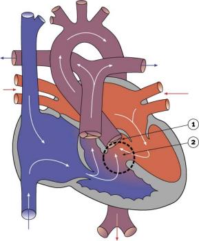
Double Outlet Right Ventricle cartoon
Double Outlet Right Ventricle
  • 1-1.5% of Congenital Heart Disease
  • Both large arteries arise wholly or mainly from the right ventricle.
  • Arrangement of the atrioventricular valves and the ventriculoarterial connections are variable.
  • Clinical manifestations variable.


If the pulmonary trunk retains its location above the morphologically right ventricle, then the end result will be yet another variant of double outlet right ventricle, specifically with a subpulmonary interventricular communication. This variation is known as the Taussig-Bing malformation.


Heart Abnormal: Tutorial Abnormalities | atrial septal defects | double outlet right ventricle | hypoplastic left heart | patent ductus arteriosus‎ | transposition of the great vessels | Tetralogy of Fallot | ventricular septal defects | coarctation of the aorta | Category ASD | Category PDA | Category ToF | Category VSD | ICD10 - Cardiovascular | ICD11


Cardiovascular Links: cardiovascular | Heart Tutorial | Lecture - Early Vascular | Lecture - Heart | Movies | 2016 Cardiac Review | heart | coronary circulation | heart valve | heart rate | Circulation | blood | blood vessel | blood vessel histology | heart histology | Lymphatic | ductus venosus | spleen | Stage 22 | cardiovascular abnormalities | OMIM | 2012 ECHO Meeting | Category:Cardiovascular
Historic Embryology - Cardiovascular 
1902 Vena cava inferior | 1905 Brain Blood Vessels | 1909 Cervical Veins | 1909 Dorsal aorta and umbilical veins | 1912 Heart | 1912 Human Heart | 1914 Earliest Blood-Vessels | 1915 Congenital Cardiac Disease | 1915 Dura Venous Sinuses | 1916 Blood cell origin | 1916 Pars Membranacea Septi | 1919 Lower Limb Arteries | 1921 Human Brain Vascular | 1921 Spleen | 1922 Aortic-Arch System | 1922 Pig Forelimb Arteries | 1922 Chicken Pulmonary | 1923 Head Subcutaneous Plexus | 1923 Ductus Venosus | 1925 Venous Development | 1927 Stage 11 Heart | 1928 Heart Blood Flow | 1935 Aorta | 1935 Venous valves | 1938 Pars Membranacea Septi | 1938 Foramen Ovale | 1939 Atrio-Ventricular Valves | 1940 Vena cava inferior | 1940 Early Hematopoiesis | 1941 Blood Formation | 1942 Truncus and Conus Partitioning | Ziegler Heart Models | 1951 Heart Movie | 1954 Week 9 Heart | 1957 Cranial venous system | 1959 Brain Arterial Anastomoses | Historic Embryology Papers | 2012 ECHO Meeting | 2016 Cardiac Review | Historic Disclaimer

Some Recent Findings

  • Double-outlet right ventricle is not hypoplastic left heart syndrome[1] "We write to express our concerns that the report ‘The complex genetics of hypoplastic left heart syndrome’ by Liu et al.[2] is misleading, and the data presented in the figures and in the Mouse Genome Informatics repository do not convincingly demonstrate hypoplastic left heart syndrome (HLHS)."
  • Imaging Spectrum of Double-Outlet Right Ventricle on Multislice Computed Tomography[3] "Double-outlet right ventricle is a complex congenital heart disease that encompasses various common and rare subtypes. Surgical management of these patients needs to be individualized owing to extremely variable morphology and hemodynamics. Imaging plays a crucial role in determination and characterization of outflow tract morphology. The assessment of ventricular septal defect routability with identification of associated anomalies has therapeutic implications in these patients. Multislice computed tomography with advanced 3-dimensional post-processing techniques and dose-reduction strategies is invaluable in defining the anatomy and morphology of double-outlet right ventricle with simultaneous assessment of associated anomalies."
  • HAND1 loss-of-function mutation contributes to congenital double outlet right ventricle[4] "In the present study, the coding exons and flanking introns of the heart and neural crest derivatives expressed transcript 1 (HAND1) gene, which encodes a basic helix‑loop‑helix transcription factor crucial for cardiovascular development, were sequenced in 158 unrelated patients with CHDs, and a de novo heterozygous mutation, p.K132X, was identified in a patient with double outlet right ventricle (DORV), as well as ventricular septal defect. The nonsense mutation, which was predicted to produce a truncated HAND1 protein lacking 84 carboxyl‑terminal amino acids, was absent in 600 control chromosomes. Functional analyses revealed that the HAND1 K132X mutant had no transcriptional activity. Furthermore, the mutation disrupted the synergistic activation between HAND1 and GATA binding protein 4 (GATA4), another cardiac core transcription factor causally linked to CHDs. To the best of our knowledge, this is the first report on the association of HAND1 loss‑of‑function mutation with an enhanced susceptibility to DORV in humans. These findings expand the phenotypic spectrum linked to HAND1 mutations, suggesting potential implications for the development of novelo prophylactic and therapeutic strategies for DORV."
More recent papers  
Mark Hill.jpg
PubMed logo.gif

This table allows an automated computer search of the external PubMed database using the listed "Search term" text link.

  • This search now requires a manual link as the original PubMed extension has been disabled.
  • The displayed list of references do not reflect any editorial selection of material based on content or relevance.
  • References also appear on this list based upon the date of the actual page viewing.


References listed on the rest of the content page and the associated discussion page (listed under the publication year sub-headings) do include some editorial selection based upon both relevance and availability.

More? References | Discussion Page | Journal Searches | 2019 References | 2020 References

Search term: Double Outlet Right Ventricle | Taussig-Bing malformation

Anatomy

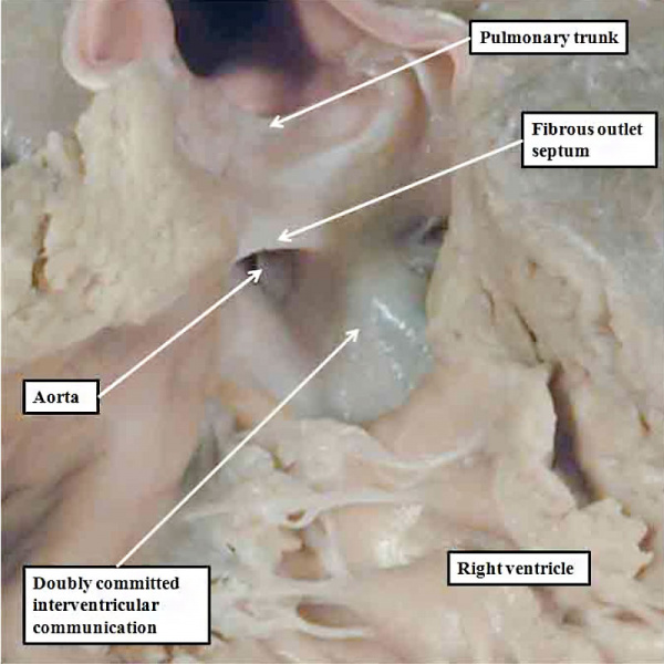
A human heart with double outlet from the right ventricle, but with a doubly committed interventricular communication. The proximal outflow cushions have fused, but have failed to muscularise. Because of this, the outlet septum, derived from the fused cushions, is fibrous and hypoplastic.

Anderson2016-fig47.jpg

Double Outlet from Right Ventricle

Genetics

HAND1

Heart-And Neural Crest Derivatives-Expressed 1 (HAND1) gene located at 5q32, is a 215-amino acid protein transcription factor that contains a bHLH region and several potential phosphorylation sites. The mouse and human sequences share 93% overall sequence identity.


Links: OMIM - HAND1

References

  1. Chaudhry B, Henderson D & Anderson R. (2019). Double-outlet right ventricle is not hypoplastic left heart syndrome. Nat. Genet. , 51, 198. PMID: 30617254 DOI.
  2. Liu X, Yagi H, Saeed S, Bais AS, Gabriel GC, Chen Z, Peterson KA, Li Y, Schwartz MC, Reynolds WT, Saydmohammed M, Gibbs B, Wu Y, Devine W, Chatterjee B, Klena NT, Kostka D, de Mesy Bentley KL, Ganapathiraju MK, Dexheimer P, Leatherbury L, Khalifa O, Bhagat A, Zahid M, Pu W, Watkins S, Grossfeld P, Murray SA, Porter GA, Tsang M, Martin LJ, Benson DW, Aronow BJ & Lo CW. (2017). The complex genetics of hypoplastic left heart syndrome. Nat. Genet. , 49, 1152-1159. PMID: 28530678 DOI.
  3. Priya S, Nagpal P, Sharma A, Pandey NN & Jagia P. (2019). Imaging Spectrum of Double-Outlet Right Ventricle on Multislice Computed Tomography. J Thorac Imaging , , . PMID: 30801451 DOI.
  4. Li L, Wang J, Liu XY, Liu H, Shi HY, Yang XX, Li N, Li YJ, Huang RT, Xue S, Qiu XB & Yang YQ. (2017). HAND1 loss-of-function mutation contributes to congenital double outlet right ventricle. Int. J. Mol. Med. , 39, 711-718. PMID: 28112363 DOI.

Reviews

Fraser CD. (2017). The Neonatal Arterial Switch Operation: Technical Pearls. Semin Thorac Cardiovasc Surg Pediatr Card Surg Annu , 20, 38-42. PMID: 28007063 DOI.

Spicer DE, Hsu HH, Co-Vu J, Anderson RH & Fricker FJ. (2014). Ventricular septal defect. Orphanet J Rare Dis , 9, 144. PMID: 25523232 DOI.

Articles

Aherrera JA, Magno JD, Uy CC, Abrahan LL, Maria HF, Buitizon RR & Jara RD. (2015). The Triply Twisted Heart: Cyanosis in an Adult With Situs Inversus, Levocardia, Double Outlet Right Ventricle, and Malposition of the Great Arteries. Cardiol Res , 6, 362-366. PMID: 28197259 DOI.

Search Pubmed

Search Pubmed: Search PubMed

External Links

External Links Notice - The dynamic nature of the internet may mean that some of these listed links may no longer function. If the link no longer works search the web with the link text or name. Links to any external commercial sites are provided for information purposes only and should never be considered an endorsement. UNSW Embryology is provided as an educational resource with no clinical information or commercial affiliation.