ANAT2241 Nervous Tissue

| ANAT2241 This practical support page content is not part of the virtual science practical class and provides additional information for student self-directed learning purposes. All practical class pages are located on Moodle - ANAT2241 |
General Objective
To understand the main histological features of the peripheral and central parts of the nervous system (PNS and CNS).
Specific Objectives
- To know the histological features of peripheral nerves in cross, oblique and longitudinal section and the LM and EM features of the myelin sheath.
- To identify peripheral nerves in routinely stained sections of various organs.
- To know the structure of muscle spindles and Pacinian and Meissner’s corpuscles.
- To know the ultrastructure of a CNS synapse and the morphology of the neuromuscular junction.
- To distinguish between the components and location of grey and white matter, using the spinal cord and cerebellum as examples.
- To describe the parts of the neurone: perikaryon (soma), dendrites, axon, axon hillock, axon collateral, telodendria (terminal arborizations).
- To identify large multipolar neurones in the ventral horn of the spinal cord, Purkinje neurones in cortex of cerebellum, multipolar neurones in peripheral autonomic ganglia and pseudounipolar neurones in sensory ganglia.
- To know the basic histological features of astrocytes, oligodendrocytes, microglia and ependymal cells. To identify the nuclei of astrocytes and oligodendrocytes.
Learning Activities
- Examine the following virtual slides and in your course manual draw and label the features listed and state their function(s):
Virtual Slides: Nervous Tissue


This page provides histology support information for central nervous system structure.

Key concepts

The brain and spinal cord comprise the central nervous system (CNS). The nerves that emerge from the spinal cord and brain to pass to parts of the body are the peripheral nervous tissue (PNS). Nervous tissue, with many interconnections, forms a complex system of neuronal communication within the body and is specialized for detecting stimuli, integrating functions, controlling effectors and higher functions. Nervous tissue consists of cell bodies, cell processes (nerves), and neuroglia (supporting cells).
Neurons
- Structural and Functional units of the nervous system.
These cells (around 12 billion) are responsible for the receptive, integrative, and motor functions of the nervous system. They can generate nerve impulses (irritability), and can transmit these impulses along their processes (conductivity). They range in diameter from 5 to 150 μm and contain 3 parts: a cell body, multiple dendrites and a single axon.
- Cell body (soma, perikaryon) is the region of the neuron containing a large pale-staining spherical, nucleus with a conspicuous nucleolus and perinuclear cytoplasm.
- Dendrites project from the cell body and are specialized for receiving (afferent) stimuli from sensory cells, axons and other neurons which are then transmitted towards the soma.
- Axons arise as a single thin process extending longer distances from the cell body than the dendrite. As with dendrites, the terminals of the axon are branching and terminate in end bulbs (terminal boutons), which come close to another cell and form a synapse.
Glia
Glia are the support cells of both the central and peripheral nervous system. There are several different types of glial cells each with specific functions.
Central Glia
- Astrocytes - (astroglia) are "star-shaped" cells with processes often in contact with a blood vessel (perivascular foot processes).
- provide mechanical and metabolic support to neurons
- maintenance of the composition of the extracellular fluid.
- removal of transmitters and their metabolism at synapses
- scar-forming cells of the CNS.
- Oligodendrocytes - (oligoglia) cells have fewer and shorter processes
- form central myelin sheaths around axons
- can form parts of the myelin sheath around several axons
- Microglia - are small cells with complex shapes, embryonically mesoderm origin.
- same cells form monocytes (tissue macrophage precursor)
- tissue damage causes microglia to proliferate and differentiate into phagocytotic cells.
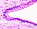
- Ependymal cells - cilated simple cuboidal or low columnar epithelium.
- line ventricles of the brain and spinal cord central canal
- lack of tight junctions, allows exchange between cerebrospinal fluid and nervous tissue.
Peripheral Nervous System
Peripheral Nerve Fibers
Peripheral nerves are bundles (fascicles) of nerve fibers (axons) surrounded by several CT sheaths. Each bundle contains sensory and motor components.
Myelinated Fibers (1-20μm diameter)
Myelin (rich in lipid) is the membrane of the Schwann cell organized into a spiral sheath that is wrapped several times around the axon. Schwann cells are cells whose cytoplasm contains a flattened nucleus, a small Golgi apparatus, and a few mitochondria. Myelinated fibers are capable of rapid transfer of impulses (touch sensory pathways).
Unmyelinated Fibers (less than 2μm in diameter)
Some axons in the PNS are surrounded by Schwann cells but not wrapped with layers of myelin. They are found in pain and temperature sensory pathways and motor paths to the viscera.

|
(text modified from original figure legend) |
Virtual Slide Box (Peripheral Nerve) and Zurich Virtual Slide database (Nerve; Goldner and Nerve; Haematoxylin and Eosin)
|

Simplified animation shown how an axon develops a myelin sheath consisting of layers of either oligodendrocyte (CNS) or Schwann (PNS) cell membrane. |

neurovascular bundle (Stain - Osmium) |

overview (Stain - Osmium) |

neural connective tissue (Stain - Haematoxylin Eosin) |

nerve longitudinal (Stain - Haematoxylin Eosin) |

myelin sheath (Stain - Haematoxylin Eosin) |

Schmidt-Lanterman cleft (diagram) |
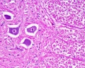
Sensory and Autonomic Ganglia

|

|
Virtual Slide Box (Sensory Trigeminal Ganglion, cat., Luxol Fast Blue and H&E).
- Identify the pseudounipolar somata, with central nuclei and satellite cells.
- Identify the axons, axon hillock, neurokeratin and Schmidt-Lanterman cleft.
- What is the function of the sensory ganglion?
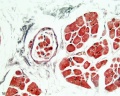
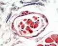
Pacinian Corpuscle


Meissner Corpuscle

|

|
Muscle Spindle
Muscle spindle HE x20
Muscle spindle HE x40
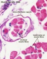
Muscle Spindle HE x40


Neuromuscular Junction

|

|
| Neuromuscular junction diagram | Neuromuscular junction |
Central Nervous System
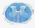
Spinal Cord


Virtual Slide Box (Spinal cord and Spinal cord smear) and Zurich Virtual Slide database (Spinal cord-Thoracic Segment-Luxol Fast Blue, Neutral Red and Spinal cord-Lumbar Segment-Azan and Meninges-Azan).
Identify gray and white matter, central canal (surrounded by ependymal cells), dorsal and ventral horns, meninges (pia, arachnoid and dura mater), subarachnoid space with dorsal and ventral rootlets, blood vessels, a motor neurone with a cell body (soma), nucleus, nucleolus, Nissl granules, an axon with axon hillock area, dendrites, glial cells (oligodendrocytes, astrocytes).
| Spinal cord (Luxol Fast Blue) | |
|---|---|

|

|
- What is the difference between white and gray matter?
| Spinal cord - Grey and white matter | |
|---|---|

|

|
| Spinal cord - Grey matter | |
|---|---|

Grey matter (Stain - Haematoxylin Eosin) |

Grey matter (silver) |
- What do Nissl granules represent?
- What is the functional difference between an axon and a dendrite?

Spinal Cord ependymal cells
- What is the function of ependymal cells?
- What function do the meninges serve and what type of tissue are the meninges made up of?
- What is the function of an oligodendrocyte and of an astrocyte?
Histology Images
Overview
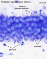
Grey matter
Grey matter
White matter
Ependymal cells


- Spinal Cord: Overview 1 | Overview 2 | Overview animation | Grey matter | Grey matter | Grey matter | White matter | Overview unlabeled | Grey matter unlabeled 1 | Grey matter unlabeled 2 | White matter unlabeled 1 | Ependymal cells unlabeled
Cerebellum
|
Virtual Slide Box (Brain/Cerebellum and Cerebellum silver stain) and Zurich Virtual Slide database (Cerebellum silver stain).
|

Cerebellum - historic drawing |
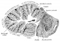
Sagittal section of the cerebellum
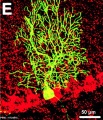
Mouse Purkinge neuron


Cortex
Overview Cortex (mouse)
Cortex (mouse)


Development
The following information is not part of the current class, but for students interested in issues of normal and abnormal neural development.
Developing Cortex
|
Human Embryo - developing cortex
|

|
Terms
- arachnoid mater - (Greek, arachne = spider + -oeides = form) A meshwork (spider web-like) connective tissue covering of the central nervous system, forms part of the meningial layers. Lies between tough outer dura mater layer and the inner fine pia mater layer. These three connective tissue layers have different embryonic origins: dura is from mesoderm, pia and arachnoid layers are neural crest in origin. The space underlying the arachnoid mater (subarachnoid space) is filled with cerebrospinal fluid.
- artifact - changes and distortions introduced to the normal tissue structure by the histological processing. Common artifacts include: folds (gives the tissue a darker appearance), tears (rips in the tissue can be seen in epithelia), shrinkage when tissues loose mainly liquid through histological processing, and cuts often used in tissue preparation.
- axon hillock - point of axon beginning or initiation from the neuron cell body (soma). (hillock, is a small hill)
- blood-brain barrier - (BBB) formed by endothelial cells of brain capillaries, differing in junctions, transport and glial association from those found in peripheral capillaries. Damage to this layer can occur following injuries involving ischemia and reperfusion. Note a similar vascular "barrier" can be found in the testis.
- cell body - (soma, perikaryon) is the region of the neuron containing a large pale-staining spherical, nucleus with a conspicuous nucleolus and perinuclear cytoplasm.
- dura mater - (Latin, dura mater = hard mother) The outer tough connective tissue meningial coat of the 3 layers that cover the central nervous system of 3 layers (overlays the arachnoid mater middle layer and pia mater inner layer). These three connective tissue layers have different embryonic origins: dura is from mesoderm, pia mater and arachnoid layers are neural crest in origin.Duramater at the level of the spinal cord is separated from the periosteum of the vertebral canal by an epidural space.
- leptomeninges - (thin meninges) Term referring to just the pia mater and arachnoid mater layers of the meninges.
- Luxol fast blue - stain for myelin sheath of nerve fibres. (More? Histology Stains)
- meninges - (singular meninx; Greek, meninx = membrane) Anatomical term describing the three connective tissue layers that surround the entire central nervous system (brain and spinal cord). The 3 layers from the central nervous outward are: pia mater, arachnoid mater, and the dura mater. These layers also have differing embryonic origins; dura mater is mesoderm, pia mater and arachnoid are neural crest. The space under the arachnoid layer (subarachnoid space) is filled with cerebrospinal fluid. (More? Image - Meninges cartoon)
- meningococcal disease - (meningitis) Term describing the bacterial infection of cerebrospinal fluid of the spinal cord and brain. Note meningitis can also be caused by a viral or other organism infection. Treatment and outcomes differ for either viral (less severe, resolves without specific treatment) or bacterial (severe, may result in brain damage, hearing loss, or learning disability) infections. (More? Bacterial Infection | Postnatal Development | CDC - meningococcal disease | Medline Plus - Meningitis)
- myelin - (myelin sheath) is the membrane of a glial cell (brain, oligodendrocyte; peripheral nerve, Schwann cell) organized into a spiral sheath that is wrapped many times around the axon.
- neuropil - (neuropile) is the region of nerve fibres (axons and dendrites) with numerous synapses and also glia, with few neural cell bodies. (Historic term, located in grey matter and does not refer to white matter.)
- neurokeratin - Term describing the protein network remaining after the myelin has been removed from the myelin sheath of nerves and seen in the histological sections. (Historically used by Ewald and Kuhnefirst in 1877 to describe "the substance contained in medullated nerves and in the central nerve organs which is insoluble in alcohol and ether".)
- Nissl body - Term describing the chromatophilic granules in the neuronal soma formed by the rough endoplasmic reticulum, very apparent in spinal cord motor neuron histology. (Named after Franz Nissl (1860 - 1919) a German neurologist.)
- node of Ranvier - Distinct myelinated axon region where two adjacent segments of myelin are separated an short unmyelinated region. Physiological site for action potential saltatory conduction jumping. (Named after their discoverer Louis Antoine Ranvier (1835 – 1922), a German anatomist.)
- perineurium - A lamellated sheath of connective tissue and cells around a nerve fascicle. (3 CT layers: epineurium-perineurium-endoneurium)
- pia mater - A fine connective tissue covering of the central nervous system, forms innermost part of the meningial layers. Lies beneath the arachnoid mater and then tough outer dura mater layer. These three connective tissue layers have different embryonic origins: pia mater and arachnoid layers are neural crest in origin, dura is from mesoderm. The space overlying the pia mater (subarachnoid space) is filled with cerebrospinal fluid. The pia mater has close contact with the spinal cord and brain, in the brain it follows down into the sulci and fissures of the cortex. This layer also fuses with the membranous lining of the ventricles (ependyma) forming the choroid plexus.
- Purkinje (Purkyne), Johannes Evangelista, Ritter von. (1787 - 1869) Breslau pathologist, Prague physiologist; famous microscopist; early use of microtome; P. cells = largest cerebellar neurones with extensive dendrites (1837); P. cells = conducting heart cells (1845); P. phenomenon = casting shadows of retinal blood vessels.
- satellite cells - (satellite glial cells) Glial cells that surround each neuron in peripheral ganglia (sensory, sympathetic). (Note these should not be confused with entirely different "satellite cells" found in skeletal muscle.)
- Schmidt-Lanterman cleft - (Schmidt-Lanterman incisure) remnants of Schwann cell cytoplasm forming funnel-shaped circumferential discontinuities distributed irregularly (10 to 100 micron intervals) throughout the internodal myelin. May have a role in peripheral nerve stretch, these incisures are not usually present in the CNS. (More? Schmidt-Lanterman cleft cartoon | Figure 4-9)
- soma - see cell body.
- substantia gelatinosa - (substantia gelatinosa of Rolando) Distinct region of spinal cord grey matter in dorsal horn containing both small neurons and neuroglia, appears pale due to presence of very few myelinated fibers.
- vasa nervorum - the small arteries providing the blood supply to peripheral nerves.
References
Textbook
Neuroscience 2nd edition. Purves D, Augustine GJ, Fitzpatrick D, et al., editors. Sunderland (MA): Sinauer Associates; 2001.
- The Cellular Components of the Nervous System
- The Internal Anatomy of the Spinal Cord
- The Internal Anatomy of the Cerebral Hemispheres and Diencephalon
External Links
External Links Notice - The dynamic nature of the internet may mean that some of these listed links may no longer function. If the link no longer works search the web with the link text or name. Links to any external commercial sites are provided for information purposes only and should never be considered an endorsement. UNSW Embryology is provided as an educational resource with no clinical information or commercial affiliation.
- The Cerebral Circulation. Cipolla MJ. San Rafael (CA): Morgan & Claypool Life Sciences; 2009. Chapter 2 Anatomy and Ultrastructure
- Clinical Methods: The History, Physical, and Laboratory Examinations. 3rd edition. Walker HK, Hall WD, Hurst JW, editors. Boston: Butterworths; 1990. Chapter 55 Cerebrovascular Disease
- Medical Microbiology. 4th edition. Baron S, editor. Galveston (TX): University of Texas Medical Branch at Galveston; 1996. Chapter 96 Microbiology of the Nervous System
- Meningitis CDC - meningococcal disease | Medline Plus - Meningitis
- Neuroscience. 2nd edition. Purves D, Augustine GJ, Fitzpatrick D, et al., editors. Sunderland (MA): Sinauer Associates; 2001. Chapter 1 The Cellular Components of the Nervous System
Course Links
- Histology Glossary: A | B | C | D | E | F | G | H | I | J | K | L | M | N | O | P | Q | R | S | T | U | V | W | X | Y | Z | ANAT2241 Support | Histology | Histology Stains | Embryology Glossary
| Common Histology Stains | ||||||||||||||||||||||||||||||||||||||||||||||||||||||||||||||||||||||||||||||||||||||||||||||||||||||||||||||||||||||||||||||||||||||||||||||||
|---|---|---|---|---|---|---|---|---|---|---|---|---|---|---|---|---|---|---|---|---|---|---|---|---|---|---|---|---|---|---|---|---|---|---|---|---|---|---|---|---|---|---|---|---|---|---|---|---|---|---|---|---|---|---|---|---|---|---|---|---|---|---|---|---|---|---|---|---|---|---|---|---|---|---|---|---|---|---|---|---|---|---|---|---|---|---|---|---|---|---|---|---|---|---|---|---|---|---|---|---|---|---|---|---|---|---|---|---|---|---|---|---|---|---|---|---|---|---|---|---|---|---|---|---|---|---|---|---|---|---|---|---|---|---|---|---|---|---|---|---|---|---|---|---|
| ||||||||||||||||||||||||||||||||||||||||||||||||||||||||||||||||||||||||||||||||||||||||||||||||||||||||||||||||||||||||||||||||||||||||||||||||
| ||||||||||||||||||||||||||||||||||||||||||||||||||||||||||||||||||||||||||||||||||||||||||||||||||||||||||||||||||||||||||||||||||||||||||||||||
Practical Support
- Pages can be accessed from any internet connected computer.
ANAT2241 Support Links: The Virtual Microscope | Covering and Lining Epithelia | Glandular Epithelia | CT Components | CT Types | Bone, Bone Formation and Joints | Muscle | Nervous | Blood | Eye | Cardiovascular | Respiratory | Integumentary | Gastrointestinal | Gastrointestinal Organs | Lymphatic and Immune | Endocrine | Urinary | Female Reproductive | Male Reproductive | Histology Stains | Histology Drawings | Practicals Health and Safety 2013 | Moodle - 2019
ANAT2241 This practical support page content is not part of the science practical class and provides only background information for student self-directed learning purposes.
Cite this page: Hill, M.A. (2026, July 30) Embryology ANAT2241 Nervous Tissue. Retrieved from https://embryology.med.unsw.edu.au/embryology/index.php/ANAT2241_Nervous_Tissue
- © Dr Mark Hill 2026, UNSW Embryology ISBN: 978 0 7334 2609 4 - UNSW CRICOS Provider Code No. 00098G