2010 Lab 2
Week 1 to 3
Introduction
This laboratory will look at the first 3 weeks of human development using online resources to study events that lead up to formation of the trilaminar embryo and early placental development. The laboratory will also begin work on the group online project.
Objectives
- Understand development of the blastocyst.
- Understand normal and abnormal implantation.
- Understand the events of early placentation.
- Understand gastrulation and early germ layer development.
From Oocyte to Blastocyst

First cell divisions of the zygote forming initially 2 blastomeres and continuing to divide to form the morula.
This early mitosis is a unique embryonic cell cycle (M, S, M phases) compared to adult (M, G1, S, G2, M phase). With virtually no G1 or G2 phases this results in a reduction in cytoplasmic volume of each daughter cell with each cell division. See also Human oocyte to blastocyst
Facts: Week 1, 2-3 days, size 0.1-0.2 mm Features: zona pellucida, blastomeres

|

|

|
| Human Embryo (day 2) | Human Embryo (day 3) | Human Embryo (day 5) |
![]()
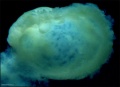
Blastocyst Hatching
By the end of the first week the blastocyst now consists of a ball of cells containing a large hollow fluid-filled (blastoceol) space.
Trophoblast Layer - Continued expansion of the blastoceol and cell division has led to the single layer of cells located at the periphery now being pressed against the inflexible zona pellucida wall and becoming flattened (squamous). This peripheral layer of cells is now called the trophoblast layer. Later in development this later will be involved in implantation and form part of the placenta.
Inner Cell Mass - On one wall of the blastoceol there is present a second layer of non-flattened cells. These inner cells are called the inner cell mass. Later in development from this layer the embryo will be formed.
Hatching - a combination of lysins (from the blastocyst or the uterus) and physical expansion, reduces the thickness and weakens the zona pelludica wall in preparation for hatching. Typically the blastocyst will hatch through a small opening (potentially at the site of fertilization) in the zona pellucida. The blastocyst is now ready to begin implantation.
Assisted Hatching - in vitro fertilization techniques use zona thinning, zona drilling, zona slitting or laser-assisted hatching to help blastocyst hatching and increase the probability of implantation occurring.
 Zona pellucida (left) with Blastocyst (right) hatching through a small opening in the wall. (More? Carnegie stage 3)
Links: MBoC - Mouse Blastocyst Development
Overview of development of the 1-chambered conceptus (blastocyst)

Ovulation is the initial release of oocyte.
Follicular fluid and fimbriae together aid oocyte movement into infundibulum then the ampulla of the uterine tube.
Sperm deposited in the vagina then enter the uterus, mature (capacitation), then actively migrate along the uterine tube.
Fertilization generally occurs in the ampulla region of the tube.
Following fertilization, repeated rounds of cell division occur without growth forming initially a solid ball of cells (morula), which cavitates to form the 1-chambered conceptus (blastocyst). Liberation of the blastocyst from the zona pellucida the allows attachment (adplantation) to the uterine wall.
Following ovulation the empty follicle within the ovary now forms the corpus luteum.
Note - the day timings shown above for the first week are approximate and may vary by several days for events following fertilization.
Blastocyst (right) hatching from zona pellucida (left)
Sex Determination
Mammalian sex determination is regulated by chromosomes.
- Females have two X chromosomes. (XX)
- Males have a single X and a small Y. (XY)
- The X and Y chromosome are morphologically and functionally different from each other.
- Evolutionary studies have shown that the Y was once the homologous pair for X.
- It is only in the last 5 years that we have some idea about how these two types of chromosomes may be regulated and genes of inportance located upon them.
- In females the main scientific problem was that of gene dosage, only one copy of X chromosome is needed to be active.
- In males the main scientific problem was what on the Y chromosome determined "maleness", and how did it do it.

The conceptus is now fully implanted witin the endometrial wall and the uterine epithelium has reformed over the site of implanation.
Note that all future development will now occur within the wall of the uterus, not within the cavity of the uterine body.
As the conceptus continues to implant and grow a series of fluid-filled cavities form both outside and inside the implanting conceptus.
- Outside the Conceptus - maternal blood-filled lacunae
- Inside the Conceptus - 3 separate spaces - amniotic sac, yolk sac, chorionic sac
Conceptus Cavities Week 2 and Week 3
Stage 7 showing the chorion and yolk sac
Stage 7 higher power of embryonic disc and yolk sac
Stage 9


Outside
Continued expansion of syncitiotrophoblasts within the endometrial wall opens both uterine glands and uterine blood vessels. These spaces outside the conceptus fill with uterine gland secretions and maternal blood forming maternal blood-filled lacunae. These lacunae provide the initial nutrition to the growing conceptus, which will later be provided through the placenta.
Inside
A cavity forms now between the inner cell mass and the trophoblast (cytotrophoblasts) wall. This cavity is the amniotic cavity.
A new cell layer forms from cells proliferating and lining the inside of the blastoceol cytotrophoblastic layer (extraembryoic mesoderm). This extraembryoic mesoderm layer continues to proliferate and then vacuolates, splitting into two separate cavities.
Overview of blastocyst implantation in uterine wall during the second week of development. (Image: Moore & Persaud, 1998)
Beginning of Carnegie Stage 6, blastocyst fully implanted in endometrial wall.
Continued expansion of the cavities in the extraembryonic mesoderm, allow the primary yolk sac to collapse. The collapsed space later becomes lined with endodermal cells and forms the yolk sac.
The space formed outside is lined with extraembryonic mesoderm and forms the third cavity the chorionic cavity.
A section of extraembryonic mesoderm links the now bilaminar embryonic disc to surrounding shell and is called the connecting stalk.
Human Conceptus Implantation 2nd and 3rd Week of Development
| Identify chorionic sac, yolk sac and amniotic sac, all containing different fluids.
Identify secondary chorionic villi, maternal blood "spaces" (empty) and uterine tissue (maternal decidua). The embryo lies between the yolk sac fluid and the amniotic sac fluid. Note that the ends ("bases") of some long secondary chorionic villi are attached to the maternal (decidual) tissue by dense clusters of trophoblast cells (the cytotrophoblastic column) - these are anchoring secondary villi. Other shorter, secondary villi are "free-floating" in the maternal intervillous spaces, from which the blood has drained into the large endometrial veins. (Image: Nishimura etal., 1977) |
 Carnegie Stage 6-7, human conceptus approximately 16-18 days. (about the time of the first missed menstrual period). Carnegie Stage 6-7, human conceptus approximately 16-18 days. (about the time of the first missed menstrual period).
|
--Mark Hill 15:34, 16 June 2010 (UTC) These 2 missing images Carnegie Stage 6-7 from the original practical class page have been added later and are not examinable.
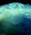
Human Conceptus (high power view)
| Identify the three fluid-filled sacs, the embryo, and the body (connecting) stalk indicating the caudal end of the embryo. Note the thin amnion and the thicker wall of the yolk sac and conversely, the thick ectoderm and thin endoderm. Vascular channels in connecting stalk. The broad elevation in the ectoderm is the expansion dome (primitive single brain bulge) and the cluster of cells at the caudal end of the embryonic disc is the primitive node region. Note the precipitated protein in the extra-embryonic coelom and in the yolk sac, and the start of the formation of the intra-embryonic coelom between ectoderm and entoderm, at the cranial (rostral) end of the expansion dome. | 
|
--Mark Hill 15:34, 16 June 2010 (UTC) These 2 missing Carnegie Stage 6-7 images from the original practical class page have been added later and are not examinable.
Human Conceptus (high power view)

|
Approximate cross-section of an 18 day human conceptus.
(Image: Nishimura etal., 1977)
|
|
|
Gastrulation means "gut forming" and converts the inner cell mass which then formed the bilaminar embryo (epiblast, hypoblast) into the trilaminar embryo (ectoderm, mesoderm, endoderm).
The process involves the migration of cells from the epiblast layer through the primitive streak to form first the endoderm layer and then a second intermediate layer the mesoderm layer. Once all cells have left the epiblast layer it now becomes the ectoderm layer. These three germ cell layers (ectoderm, mesoderm, endoderm) will form in a layer specific manner all the future tissues of the developing embryo.
Approximate cross-section of an 18 day human conceptus. Identify the 3 layers of the trilaminar embryo: ectoderm (columnar cells), intraembryonic mesoderm (mesenchymal cells, endodermal cells (cuboidal single layer). Identify primitive groove with dense cluster of primitive streak cells below it.
|
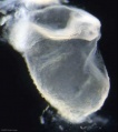
Stage 7

|

|
| Features: embryonic disc, primitive node, primative streak, primitive groove, yolk sac
Facts: Week 3, 15 - 17 days, 0.4 mm View 1: embryonic disc, showing the epiblast viewed from the amniotic (dorsal) side. Events: Gastrulation is continuing as cells migrate from the epiblast, continuing to form mesoderm. Mesoderm lies between the ectoderm and endoderm as a continuous sheet except at the buccopharyngeal and cloacal membranes. These membranes have ectoderm and endoderm only and will lie at the rostral (head) and caudal (tail) of the gastrointestinal tract.
|
Gastrulation: Through the primitive streak cells migrate continuously through week 3 into week 4. Initial cells replace hypoblast as an epithelial layer the endoderm. Later migrating cells spread between the two epithelial layers to form mesoderm. |

|

|
| Axes: embryonic disc is shown rostral (head) to top and caudal (tail) to bottom. Left and right are the lateral margins of the disc as shown. |
Gastrulation
Carnegie Stage 7 and 8, gastrulation, migration of cells through the primitive streak to form endoderm and mesodermal layers of embryo.

 Scanning electron micrograph showing the early forming 3 layers: ectoderm, mesoderm and endoderm.
![]()
Notochord
The embryonic structure which establishes body axes and patterns surrounding tissues is called the notochord.
The notochord is a midline column of cells running in a rostrocaudal direction (head-tail) within the mesoderm layer. It exists as a transient developmental patterning structure with a role in molecular signaling (patterning) and controlling the direction of embryonic disc folding (mechanical). These images are of the embryonic disc in week 3 (stage 7).


The notochordal process begins as a fold of ectoderm extending cranially toward the prechordal plate region. The sequence of differentiation: notochordal process -> notochordal plate -> notochord.
- Elongation of the notochordal process cranially from the primitive pit as a hollow tube (notochordal canal) in the midline of the embryonic disc underlying the ectoderm.
- The notochordal canal may appear to break down on the endodermal side forming a notochordal plate continuous with the endodermal layer.
- Notochordal plate folds to form notochord. The notochord (also called axial mesoderm) is an embryonic structure that regulates differentiation of surrounding structures including the overlying ectoderm (neural plate) and mesoderm (somites).
Disc Folding

Folding: all edges of the embryonic disc will fold ventrally, forming a rostro-caudal "C" shaped tube.
| Chorionic Cavity | Amniotic Cavity | Week 3 |
Stage 7
Facts
Human embryonic stage 7 occurs during week 3 between 15 to 17 days.
The embryo is now 0.4 mm diameter in size.
The initial images are displayed unlabeled to allow you to explore the embryo for yourself, linked labeled versions are also available for some images.
Events
Gastrulation is continuing as cells migrate from the epiblast, continuing to form mesoderm.
Mesoderm lies between the ectoderm and endoderm as a continuous sheet except at the buccopharyngeal and cloacal membranes. These membranes have ectoderm and endoderm only and will lie at the rostral (head) and caudal (tail) of the gastrointestinal tract.
From the primitive node a tube extends under the ectoderm in the opposite direction to the primitive streak. This tube forms first the axial process then notochordal process, then finally the notochord.
The notochord is a key to embryonic folding and regulation of ectoderm and mesoderm differentiation. It lies in the rostrocordal axis and the embryonic disc will fold either side ventrally, pinching off a portion of the yolk sac to form the lining of the gastrointestinal tract.
Carnegie Stage 8

Facts
Human embryonic stage 8 occurs during week 3 between 17 to 19 days.
The embryo is now 1.0 - 1.5 mm in size.
Events
Gastrulation is continuing as cells migrate from the epiblast, continuing to form mesoderm.
Mesoderm lies between the ectoderm and endoderm as a continuous sheet except at the buccopharyngeal and cloacal membranes. These membranes have ectoderm and endoderm only and will lie at the rostral (head) and caudal (tail) of the gastrointestinal tract.
From the primitive node a tube extends under the ectoderm in the opposite direction to the primitive streak. This tube forms first the axial process then notochordal process, then finally the notochord.
The notochord is a key to embryonic folding and regulation of ectoderm and mesoderm differentiation. It lies in the rostrocordal axis and the embryonic disc will fold either side ventrally, pinching off a portion of the yolk sac to form the lining of the gastrointestinal tract.
Identify
- embryonic disc
- primitive node, primative streak, primative groove
- connecting stalk
- cut amniotic membrane
Carnegie Stage 9

Facts
Human embryonic stage 9 occurs during week 3 between 19 to 21 days.
The embryo is now 1.5 to 2.5 mm in size and somites have begun to form and number between 1 to 3 somite pairs during this stage.
The initial images are displayed unlabeled to allow you to explore the embryo for yourself, linked labeled versions are also available for some images.
Events
Ectoderm - Neural plate brain region continues to expand, neural plate begins folding over the notochord. Gastrulation continues through the primitive streak region.
Mesoderm - Paraxial mesoderm segmentation into somites begins (1 - 3 somite pairs). Lateral plate mesoderm begins to vacuolate, dividing it into somatic and splanchnic mesoderm and to later form the intra-embryonic coelom. Prechordal splanchnic mesoderm begins to form the cardiogenic region, from which the primordial heart will develop.
Endoderm - Notochordal plate still visible which will form the notochord. Endoderm is still widely open to the yolk sac and germ cells form part of this layer. Extra-embryonic mesoderm on the yolk sac surface begins to form "blood islands".
Identify
- Neural groove and neural folds, the mesoderm showing first somite bulges, that segments beside the neural groove to form somites but extends laterally to margin of embryonic disc lateral plate mesoderm, where it merges with the covering extraembryonic mesoderm.
- The intra-embryonic coelom develops in the middle of the lateral plate mesoderm.
- Carnegie Stages: 1 | 2 | 3 | 4 | 5 | 6 | 7 | 8 | 9 | 10 | 11 | 12 | 13 | 14 | 15 | 16 | 17 | 18 | 19 | 20 | 21 | 22 | 23 | About Stages | Timeline
Human Development Timeline
The table below shows human development features and approximate timing during the menstrual cycle to fertilization and the first 3 weeks of development.
The timing assumes fertilization the day after ovulation and the "weeks" refer to embryonic development and differ from clinical weeks (shown in brackets, from last menstrual period) and "stages" refer to Carnegie stages of development.
Week -2
(Clinical Week 1)
| Event | ||
| Menstrual Phase |  Menstrual Cycle changes: Uterine endometrium (loss), Ovary (Follicle Development) | |

| ||
| Proliferative Phase |   Menstrual Cycle changes: Uterine endometrium (proliferation), Ovary (Follicle Development) Menstrual Cycle changes: Uterine endometrium (proliferation), Ovary (Follicle Development)
| |
Week -1
(Clinical Week 2)
| Menstrual cycle | Event | |
| Proliferative Phase | ||
    Menstrual Cycle - Mid proliferative Menstrual Cycle - Mid proliferative
| ||
  Menstrual Cycle - Late Proliferative Menstrual Cycle - Late Proliferative
| ||
| Ovulation
Capacitation |
 |
Week 1
Week 1 (Clinical Week 3)
| Event | ||
| Secretory PhaseStage 1 |    Fertilization, Secretory Phase Fertilization, Secretory Phase
| |
| Stage 2 | | |
| Stage 3 | Blastocyst Hatching (zona pellucida lost)
| |
  Late Secretory, Blastocyst (free floating) Late Secretory, Blastocyst (free floating)
| ||
| Stage 4 | Adplantation | |
| Stage 5 |
Week 2
Week 2 (Clinical Week 4)
| Event | ||
| Stage 6 | ||
Week 3
Week 3 (Clinical Week 5)
| Event | ||
| Stage 7 | 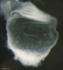 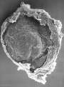 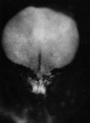
| |
| Stage 8 | 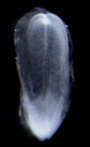 | |
| ||
| Stage 9 | 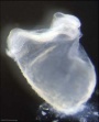  Musculoskeletal somitogenesis, first somites form and continue to be added in sequence caudally Musculoskeletal somitogenesis, first somites form and continue to be added in sequence caudally
Neural the three main divisions of the brain, which are not cerebral vesicles, can be distinguished while the neural groove is still completely open Neural Crest mesencephalic neural crest is visible PMID: 17848161 | |
| Heart cardiogenesis, week 3 begins as paired heart tubes. |
Terms
- bilaminar- having 2 layers
- blastocyst- the developmental stage following morula, as this stage matures, the zona pellucia is lost allowing the conceptus to adplant and then implant into the uterine wall.
- corpus albicans - (Latin, corpus = body, albicans = whitish) The histological structure formed by luteolysis of the corpus luteum in the ovary. If implantation does not occur and the hormone hCG is not released the corpus luteum degenerates and the structure is white, not yellow, because of the absence of steroid hormone synthesis/accumulation.
- corpus luteum - (Latin, corpus = body, luteum = yellow) The remains of ovarian follicle formed after ovulation that acts as an endocrine organ (produce progesterone and oestrogens) supporting pregnancy and preventing menstruation (loss of the endometrial lining). Formed during the luteal phase (secretory phase) of the menstrual cycle by proliferation of both follicular granulosa cells (granulosa lutein cells) and thecal cells (theca lutein cells), which produce progesterone and oestrogens. If fertilization and pregnancy does not occur, the corpus luteum degenerates to form the corpus albicans. Regnier de Graaf (1641 – 1673) first observed it in the ovary of a cow as a yellow structure, the yellow colour is caused by accumulation of steroidal hormones. (More? Menstrual Cycle | Ovary Development | Week 2 Ovary | Week 1 - Oogenesis)
- inner cell mass- the clump of cells found inside the blastocyst. These cells will go in to form the embryo, these are the "stem cells" (we here about in the media) that are totipotential, they can form any tissue in the embryo. Mature oocyte-the female germ cell released at ovulation from the ovary.
- trilaminar embryonic disc- the 3 layered embryo stage.
- Trophoblasts- (Gr. trophe = nutrition) outer layer of cells on blastocyst that will generate the embryonic part of the placenta.
Group Online Project
- Topic Voting - closes before lab 2 and is part of your progressive assessment for Lab 1.
- Today we will select the major topic and divide into smaller groups to begin the project work.
- References - a tutorial will be given in reference searching and resources available. (see also Help:Editing Basics)
- Journal resources: Public Library of Science | Biomed Central | PNAS | more reference information
Search Pubmed
Search Bookshelf Gastrulation
Search Pubmed Now: Gastrulation
say something [1]
References
- ↑ <pubmed>20195633</pubmed>
Glossary Links
- Glossary: A | B | C | D | E | F | G | H | I | J | K | L | M | N | O | P | Q | R | S | T | U | V | W | X | Y | Z | Numbers | Symbols | Term Link
Course Content 2010
Embryology Introduction | Cell Division/Fertilization | Lab 1 | Week 1&2 Development | Week 3 Development | Lab 2 | Mesoderm Development | Ectoderm, Early Neural, Neural Crest | Lab 3 | Early Vascular Development | Placenta | Lab 4 | Endoderm, Early Gastrointestinal | Respiratory Development | Lab 5 | Head Development | Neural Crest Development | Lab 6 | Musculoskeletal Development | Limb Development | Lab 7 | Kidney | Genital | Lab 8 | Sensory | Stem Cells | Stem Cells | Endocrine | Lab 10 | Late Vascular Development | Integumentary | Lab 11 | Birth, Postnatal | Revision | Lab 12 | Lecture Audio | Course Timetable
Cite this page: Hill, M.A. (2026, Haziran 8) Embryology 2010 Lab 2. Retrieved from https://embryology.med.unsw.edu.au/embryology/index.php/2010_Lab_2
- © Dr Mark Hill 2026, UNSW Embryology ISBN: 978 0 7334 2609 4 - UNSW CRICOS Provider Code No. 00098G