Somitogenesis: Difference between revisions
| Line 101: | Line 101: | ||
'''Search Pubmed:''' [http://www.ncbi.nlm.nih.gov/sites/entrez?db=pubmed&cmd=search&term=Somitogenesis Somitogenesis] | [http://www.ncbi.nlm.nih.gov/sites/entrez?db=pubmed&cmd=search&term=Somite%20Formation|Somite Formation] | [http://www.ncbi.nlm.nih.gov/sites/entrez?db=pubmed&cmd=search&term=Hes7 Hes7] | '''Search Pubmed:''' [http://www.ncbi.nlm.nih.gov/sites/entrez?db=pubmed&cmd=search&term=Somitogenesis Somitogenesis] | [http://www.ncbi.nlm.nih.gov/sites/entrez?db=pubmed&cmd=search&term=Somite%20Formation|Somite Formation] | [http://www.ncbi.nlm.nih.gov/sites/entrez?db=pubmed&cmd=search&term=Sclerotome Sclerotome] | [http://www.ncbi.nlm.nih.gov/sites/entrez?db=pubmed&cmd=search&term=Hes7 Hes7] | ||
{{Template:Week}} | {{Template:Week}} | ||
Revision as of 11:37, 14 May 2011
Introduction

The term used to describe the process of segmentation of the paraxial mesoderm within the trilaminar embryo body to form pairs of somites, or balls of mesoderm. In humans, the first somite pair appears at day 20 and adds caudally at 1 somite pair/90 minutes until on average 44 pairs eventually form.
A somite is added either side of the notochord (axial mesoderm) to form a somite pair. The segmentation does not occur in the head region, and begins cranially (head end) and extends caudally (tailward) adding a somite pair at regular time intervals. The process is sequential and therefore used to stage the age of many different species embryos based upon the number visible somite pairs.
Some Recent Findings
|
Presomitic Mesoderm

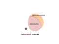
- Within the mesodermal layer either side of the notochord (axial mesoderm) lie the paraxial mesoderm strips.
- Below the unsegmented cranial paraxial mesoderm region the region that will later segment is described as presomitic mesoderm (PSM).
- This PSM is being "patterned" by two two molecular activities described as the "clock" and the "wavefront" (reviewed[2])
- Many different molecular factors are involved in this patterning effect.
- Hes7, FGF, Sprouty4, Notch, Shh
- notochord influences somite formation, notochord removal increases the period of molecular clock oscillations.[3]
Mesoderm to Somite

Mesoderm means the "middle layer" and it is from this layer that nearly all the bodies connective tissues are derived. In early mesoderm development a number of transient structures will form and then be lost as tissue structure is patterned and organised. Humans are vertebrates, with a "backbone", and the first mesoderm structure we will see form after the notochord will be somites.
- During segmentation the outer cell layer forms an epithelial layer over a still mesenchymal organization of cells at the core.
- The early forming somite has a cavity at its core called a "somitocoel" that later fills with proliferating mesoderm cells.

trilaminar embryo
mesoderm regions
somite coelom
neural tube and neural crest


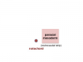
paraxial mesoderm
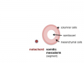
early somite


Somite to Sclerotome and Dermomyotome
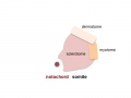
Somite initially forms 2 main regional components
- ventromedial region - sclerotome forms vertebral body and intervertebral disc
- dorsolateral region - dermomyotome forms dermis and skeletal muscle
sclerotome and dermomyotome
dermatome and myotome
epaxial and hypaxial muscles


![]()
Sclerotome
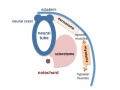
- The left and right sclerotomes from the same segmental level engulf the notochord.
- Each segmental level is then resegmented in a rostrocaudal direction.
Dermomyotome
- The dermomyotome is divided into a dorsal and ventral half.
- Dorsal - dermatome.
- Ventral - myotome, this will also divide into a dorsal and ventral half that contribute the epaxial and hypaxial skeletal muscle groups respectively.
Additional Images
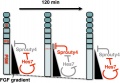
Model for Sprouty4 and FGF in mesoderm segmentation
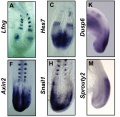
Mouse (E8.5-9.5) somitogenesis gene expression
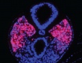
Mouse (E8.5) cleaved intracellular portion of Notch in unsegmented presomitic mesoderm (PSM)


References
Reviews
<pubmed>18482400</pubmed> <pubmed>21038776</pubmed> <pubmed>21038775</pubmed> <pubmed>17988868</pubmed> <pubmed>17643270</pubmed> <pubmed>17600784</pubmed> <pubmed>17024300</pubmed> <pubmed>15964269</pubmed>
Articles
<pubmed></pubmed>
Search PubMed
Search NLM Online Textbooks: "Somitogenesis" : Developmental Biology | The Cell- A molecular Approach | Molecular Biology of the Cell | Endocrinology
Search Pubmed: Somitogenesis | Formation | Sclerotome | Hes7
Embryo Week: Week 1 | Week 2 | Week 3 | Week 4 | Week 5 | Week 6 | Week 7 | Week 8 | Week 9
- Carnegie Stages: 1 | 2 | 3 | 4 | 5 | 6 | 7 | 8 | 9 | 10 | 11 | 12 | 13 | 14 | 15 | 16 | 17 | 18 | 19 | 20 | 21 | 22 | 23 | About Stages | Timeline
Glossary Links
- Glossary: A | B | C | D | E | F | G | H | I | J | K | L | M | N | O | P | Q | R | S | T | U | V | W | X | Y | Z | Numbers | Symbols | Term Link
Cite this page: Hill, M.A. (2024, May 2) Embryology Somitogenesis. Retrieved from https://embryology.med.unsw.edu.au/embryology/index.php/Somitogenesis
- © Dr Mark Hill 2024, UNSW Embryology ISBN: 978 0 7334 2609 4 - UNSW CRICOS Provider Code No. 00098G