Scanning Electron Microscopy
Introduction
The Scanning Electron Microscope (SEM) was a development of the electron microscope. Unlike a light microscope, using light, the electron microscope uses a focussed beam of electrons to image materials. The first version of this technology was the transmission electron microscope (TEM).
- On this current site the acronym "SEM" is used to denote a Scanning Electron Micrograph, the image produced by this form of microscopy.
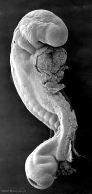
There are a series of beautiful SEM images made available by Prof Kathy Sulik of the early developing human embryo between week 3 to 5 (Carnegie stage 7 to 14) available: 7, 8, 9, 10, 11, 12, 13 and 14.
Microscopy Timeline
- 1665 - Robert Hooke publishes Micrographia, a collection of biological micrographs.
- 1674 - Anton van Leeuwenhoek improved simple microscope for biological specimens.
- 1833 - Brown published a microscopic observation of orchids, describing the cell nucleus.
- 1898 - Golgi first saw and described the Golgi apparatus by staining cells with silver nitrate.
- 1931 - Ernst Ruska first transmission electron microscope, (TEM).
- 1934 - Frits Zernike describes phase contrast microscopy.
- 1957 - Marvin Minsky patents principle of confocal imaging.
- 1953 - Frits Zernike receives the Nobel Prize in Physics for invention of the phase contrast microscope.
- 1955 - George Nomarski theoretical basis of Differential interference contrast microscopy.
- 1981 - Gerd Binnig and Heinrich Rohrer develop the canning Tunneling Microscope (STM).
- 1981 - Daniel Axelrod develop Total Internal Reflection Fluorescence Microscope (TIRFM).
- 1981 - Allen and Inoué perfected video-enhanced-contrast light microscopy.
- 1986 - Ernst Ruska, Gerd Binnig and Heinrich Rohrer receive the Nobel Prize in Physics for invention of the electron microscope (ER) and scanning tunneling microscope (GB and HR).
- 2000 - Hell and collaborators develop Stimulated Emission Depletion Microscopy (STED).
- 2008 - Freudiger and Wei Min develop Stimulated Raman Scattering Microscopy (SRS)
- Carnegie Stages: 1 | 2 | 3 | 4 | 5 | 6 | 7 | 8 | 9 | 10 | 11 | 12 | 13 | 14 | 15 | 16 | 17 | 18 | 19 | 20 | 21 | 22 | 23 | About Stages | Timeline
External Links
External Links Notice - The dynamic nature of the internet may mean that some of these listed links may no longer function. If the link no longer works search the web with the link text or name. Links to any external commercial sites are provided for information purposes only and should never be considered an endorsement. UNSW Embryology is provided as an educational resource with no clinical information or commercial affiliation.
Glossary Links
- Glossary: A | B | C | D | E | F | G | H | I | J | K | L | M | N | O | P | Q | R | S | T | U | V | W | X | Y | Z | Numbers | Symbols | Term Link
Cite this page: Hill, M.A. (2024, May 2) Embryology Scanning Electron Microscopy. Retrieved from https://embryology.med.unsw.edu.au/embryology/index.php/Scanning_Electron_Microscopy
- © Dr Mark Hill 2024, UNSW Embryology ISBN: 978 0 7334 2609 4 - UNSW CRICOS Provider Code No. 00098G
