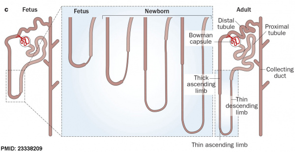
After nephron development has completed and concomitant with the development of the renal papilla in the newborn, the thin ascending limb of Henle’s loops is generated as an outgrowth from the S3 segment of the proximal tubule and from the distal tubule anlage of the nephron.
Endocrine Kidney
Covered also in Endocrine Development lecture
- Renin - Increase Angiotensin-aldosterone system
- Prostaglandins - decrease Na+ reabsorption
- Erythropoietin - Increase Erythrocyte (rbc) production
- 1,25 (OH)2 vitamin D - Calcium homeostasis
- Prekallikreins - (plasma protein inactive precursor of kallikrein) Increase kinin production (altered vascular permeability)
Cloaca
- hindgut region ending at the cloacal membrane
- divided (ventro-dorsally) by the urogenital septum
- ventral - common urogenital sinus
- dorsal - rectum
|
|
Common Urogenital Sinus
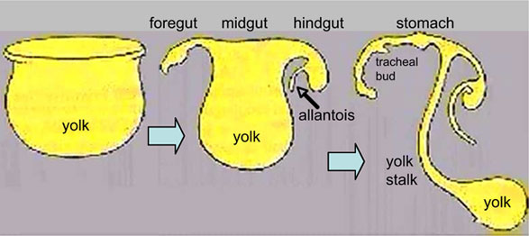
- superior end continuous with allantois
- common urogenital sinus and mesonephric duct fuse (connect)
- differentiates to form the bladder
- inferior end forms urethra
- this will be different in male and female development
Urinary Bladder
|
|
- early origins of the bladder at the superior end of the common urogenital sinus
- 8 open inferiorly to the cloaca and superiorly to the allantois
- Septation of the claoca - divides the anterior region to the primordial bladder component from the posterior rectal component.
- associated ureters and urethra
Dorsal view of developing bladder
- Ultrasound measurement of the bladder size can be used as a diagnostic tool for developmental abnormalities.
|
Bladder Structure
Can be described anatomically by its 4 layers from outside inward:
- Serous - the superior or abdominal surfaces and the lateral" surfaces of the bladder are covered by visceral peritoneum, the serous membrane (serosa) of the abdominal cavity, consisting of mesthelium and elastic fibrous connective tissue.
- Muscular - the detrusor muscle is the muscle of the urinary bladder wall.
- Submucosa - connects the muscular layer with the mucous layer.
- Mucosa - (mucus layer) a transitional epithelium layer formed into folds (rugae).
Detrusor Muscle
- The adult detrusor muscle consists of three layers of smooth (involuntary) muscle fibres.
- external layer - fibres arranged longitudinally
- middle layer - fibres arranged circularly
- internal layer - fibres arranged longitudinally
Ureter Development
- The adult ureter is a thick-walled muscular tube, 25 - 30 cm in length, running from the kidney to the urinary bladder.
- Anatomically can be described in two parts the abdominal part (pars abdominalis) and pelvic part (pars pelvina).
- The ureter is composed of three layers: outer fibrous layer (tunica adventitia), muscular layer (tunica muscularis) and mucous layer (tunica mucosa).
- The muscular layer can also be subdivided into 3 fibre layers: an external longitudinal, a middle circular, and an internal longitudinal.
Trigone Development
Kidney Ascent
|
|
- Pro-, Meso-, Meta- Early development descending
- Metanephros - initially pelvic, beside aorta
- Growth and straightening of body - Kidneys in abdomen and displace laterally
|
Renal Arteries
- Arise with ascent and inferior branches lost
- Sequential, 25% population have 2 or more renal arteries
- branch of abdominal aorta, divides into 4-5 branches
- each gives off small branches to suprarenal glands, ureter, surrounding cellular tissue and muscles
Note: Frequently a second renal artery (inferior renal) from abdominal aorta at a lower level, supplies lower portion of kidney
Abnormalities
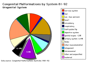 Australian renal abnormalities There are many different forms of renal development abnormalities associated with kidney, ureters, bladder and urethra. There are many genetic disorders associated with failure or abnormal renal development. Prenatal diagnosis of obstructive and renal agenesis/dysgenesis disorders are also important for early reproductive decisions by the parents. For example, with bilateral renal agenesis, failure of both kidneys to development, is not compatible with fetal/neonatal survival.
Because of their close developmental association, often described as the urogenital system, there can be an associated genital abnormalities.
More detailed information is available on the links below.
- Links: Renal Abnormalities | Genital Abnormalities
Horseshoe Kidney
- fusion of the lower poles of the kidney.
- During migration from the sacral region the two metanephric blastemas can come into contact, mainly at the lower pole.
- The ureters pass in front of the zone of fusion of the kidneys.
- The kidneys and ureters usually function adequately but there is an increased incidence of upper urinary tract obstruction or infection.
- Some horseshoe variations have been described as having associated ureter abnormalities including duplications.
Urorectal Septum Malformation
- thought to be a deficiency in caudal mesoderm which in turn leads to the malformation of the urorectal septum and other structures in the pelvic region.
- Recent research has also identified the potential presence of a persistent urachus prior to septation of the cloaca (common urogenital sinus).
Bladder
- absent or small bladder - associated with renal agenesis.
Bladder Exstrophy
- developmental abnormality associated with bladder development.
- origins appear to occur not just by abnormal bladder development, but by a congenital malformation of the ventral wall of abdomen (between umbilicus and pubic symphysis).
- There may also be other anomolies associated with failure of closure of abdominal wall and bladder (epispadias, pubic bone anomolies).
Ureter and Urethra
- Ureter - Duplex Ureter
- Urethra- Urethral Obstruction and Hypospadias
Polycystic Kidney Disease
- diffuse cystic malformation of both kidneys
- cystic malformations of liver and lung often associated, Often familial disposition
- Two types
- Infantile (inconsistent with prolonged survival)
- Adult (less severe and allows survival)
- Autosomal dominant PKD disease - recently identified at mutations in 2 different human genes encoding membrane proteins (possibly channels)
Wilms' Tumor
- (nephroblastoma) Named after Max Wilms, a German doctor who wrote first medical articles 1899
- most common type of kidney cancer children
- WT1 gene - encodes a zinc finger protein
- Both constitutional and somatic mutations disrupting the DNA-binding domain of WT1 result in a potentially dominant-negative phenotype
- some blastema cells (mass of undifferentiated cells) persist to form a ‘nephrogenic rest’
- Most rests become dormant or regress but others proliferate to form hyperplastic rests
- any type of rest can then undergo a genetic or epigenetic change to become a neoplastic rest
- can proliferate further to produce a benign lesion (adenomatous rest) or a malignant Wilms’ tumour
Prune Belly Syndrome
- lower urinary tract obstruction
- mainly male
- fetal urinary system ruptures leading to collapse and "prune belly" appearance.
Renal Cysts
The Bosniak classification system (Category I - IV) was designed to separate identified cystic renal masses by analysis of computed tomography (CT) features into surgical and nonsurgical categories.[11] Named after Morton Bosniak, Yale University School of Medicine, the developer of this classification system.
Molecular
| Abbreviation
|
Growth Factor
|
Renal Development
|
Expression Location
|
| BMP4
|
Bone Morphogenetic Protein 4
|
prevents ectopic ureteric bud outgrowth and extra ureteric bud divisions
|
mesenchymal cells surrounding mesonephric duct and stromal mesenchyme surrounding steric bud stalks
|
| BMP7
|
Bone Morphogenetic Protein 7
|
survival of metanephric mesenchyme
|
metanephric mesenchyme
|
| Fgf8
|
Fibroblast Growth Factor 8
|
transition of the induced cap mesenchyme into RVs
|
cap mesenchyme
|
| GDNF
|
Glial-cell derived neurotrophic factor
|
induces steric bud outgrowth from mesonephric duct, interacts with Ret
|
metanephric mesenchyme
|
| VEGF
|
Vascular endothelial growth factor
|
promotes endothelial cell proliferation, differentiation
|
s-shaped body
|
| Wnt4
|
Wingless-Type MMTV Integration Site Family, Member 4
|
mesenchymal-to-epithelial transition
|
cap metanephric mesenchyme, pre-tubular aggregate, nephron progenitors
|
| Wnt5a
|
Wingless-Type MMTV Integration Site Family, Member 5a
|
nephrogenesis induction, ectopic bud formation
|
steric bud, metanephric mesenchyme
|
| Wnt9b
|
Wingless-type MMTV integration site family, Member 9B
|
renewal and differentiation of nephron progenitors and normal ureteric bud branching, mesenchymal-to-epithelial transition
|
steric bud stalk epithelial cells
|
- Foxd1 - (Brain Factor-2) transcription factor that is a renal stroma specific gene.
- Links: Renal System - Molecular | OMIM Foxd1
References
- ↑ <pubmed>1546799</pubmed>
- ↑ <pubmed>17495859</pubmed>
- ↑ <pubmed>22253716</pubmed>
- ↑ <pubmed>25758227</pubmed>
- ↑ <pubmed>22253716</pubmed>
- ↑ <pubmed>21110022</pubmed>
- ↑ <pubmed>20810610</pubmed>
- ↑ 8.0 8.1 <pubmed>23338209</pubmed>
- ↑ <pubmed>25737276</pubmed>
- ↑ <pubmed>25685519</pubmed>| J Adv Res.
- ↑ <pubmed>16040900</pubmed>| Radiology
Textbooks
- The Developing Human: Clinically Oriented Embryology (8th Edition) by Keith L. Moore and T.V.N Persaud - Moore & Persaud Chapter 13 p303-346
- Larsen’s Human Embryology by GC. Schoenwolf, SB. Bleyl, PR. Brauer and PH. Francis-West - Chapter 10 p261-306
- Before We Are Born (5th ed.) Moore and Persaud Chapter14 p289-326
- Essentials of Human Embryology, Larson Chapter 10 p173-205
- Human Embryology, Fitzgerald and Fitzgerald Chapter 21-22 p134-152
Online Textbooks
Search Bookshelf intermediate mesoderm | kidney development | renal development | ureteric bud | nephron development | bladder development
Reviews
<pubmed>25737276</pubmed>
<pubmed>20691850</pubmed>
<pubmed>19906853</pubmed>
<pubmed>19828308</pubmed>
<pubmed>19615554</pubmed>
<pubmed>18184729</pubmed>
<pubmed>17442697</pubmed>
Forefronts Symposium on Nephrogenetics: from development to physiology March 8-11, 2007 Danvers, MA A meeting to synthesize an integrated view of the normal development and function of the kidney from the genetic standpoint.
<pubmed>16916378</pubmed>
Articles
<pubmed></pubmed>
<pubmed></pubmed>
<pubmed>24154527 </pubmed>
<pubmed>18846389</pubmed>
Search PubMed
Search Pubmed: Renal System Development | Renal Development | intermediate mesoderm | kidney development | renal development | ureteric bud | nephron development | bladder development
Additional Images
Stage 11 historic Atwell (1930)
Stage 11 historic Heuser (1930)
Nephrons - cortical and juxtamedullary
Kidney and adrenal gland (adult)
Fetal urogenital region most lateral right
Fetal urogenital region lateral right
Fetal urogenital region medial
Fetal urogenital region midline
Renal outflow obstruction
Mouse E12.5 kidney in vitro
Terms
Open table below to see list of renal terms.
| Renal Terms
|
- bladder exstrophy - A congenital malformation with bladder open to ventral wall of abdomen (between umbilicus and pubic symphysis) and may have other anomolies associated with failure of closure of abdominal wall and bladder (epispadias, pubic bone anomolies).
- blastema - Term used to describe a mass of undifferentiated cells. (More? Wilm's tumour)
- Bowman's capsule - (capsula glomeruli, glomerular capsule) Surrounds the glomerulus within the nephron with a vascular and urinary pole and is the beginning of the tubular component. Named in 1842 after Sir William Bowman (1816 – 1892) an English surgeon and anatomist.
- Brenner hypothesis - a clinical hypothesis that states, individuals with a congenital reduction in nephron number have a much greater likelihood of developing adult hypertension and subsequent renal failure. Developed in the 1980's by Barry Brenner at the Brigham and Women's Hospital, this also fits with the DOHAD hypothesis. (More? PubMed 3063284 | Barry Brenner)
- capillary loop - (C stage) The third stage in nephron development between 25-29 weeks. (stage sequence: V - S - C - M)
- diabetes insipidus - The disorder is related to the hormone antidiuretic hormone (ADH, also called vasopressin) its synthesis, secretion, receptors and signaling pathway. In diabetes insipidus there is an excretion of large amounts (up to 30 litres/day) of a watery urine and an unremitting thirst.
- fenestrated capillary - Specialised capillaries containing circular pores (fenestrae) that penetrate the endothelium, may be closed by a thin diaphragm.
- glomerulus - The capillary network (tuft) within Bowman's capsule of the nephron enters at the vascular pole (afferent and efferent arteriole).
- hydronephrosis - (congenital hydronephrosis, Greek, hydro = water) A kidney abnormality due to partial or complete obstruction at the pelvi-ureteric junction. This leads to a grossly dilated renal pelvis causing extensive renal damage before birth.
- hyperplastic rests - In kidney development, embryonic blastema cells can persist and proliferate to form a pool of cells, which under either genetic or epigenetic influence can then change to become a neoplastic rest. Normally the majority of nephrogenic rests either regress or become dormant.
- juxtaglomerular cells - Cells located at the vascular pole that secrete renin and form a part of the juxtaglomerular complex.
- loop of Henle - Nephron region spanning from the proximal convoluted tubule to the distal convoluted tubule. Named after Named after Friedrich Gustav Jakob Henle (1809–1885) a German anatomist.
- macula densa - Columnar cell cluster appearing as a dense row of cell nuclei where the straight portion of the distal tubule contacts the glomerulus. Region also in close contact with the efferent and afferent arterioles of the glomerulus and involved in sodium chloride regulation. (More? image)
- maturation stage - (M stage) The forth stage in nephron development in infants aged 1-6 months. (stage sequence: V - S - C - M)
- mesangial cells - Cells in the nephron glomerulus that form the connective tissue giving structural support to podocytes and vessels.
- mesonephros - The second temporary stage of kidney development (pro-, meso-, meta-). The intermediate mesonephros develops and disappears with the exception of its duct, the mesonephric duct, which will form the male reproductive duct system. In males, the mesonephric tubules go on to form the ducts of the testis. In females, these degenerate. A few mesonephric tubules remain as efferent ductules in the male and vestigial remnants in the female.
- mesonephric duct - (= Wollfian duct) An early developing urogenital duct running the length of the embryo that will differentiate and form the male reproductive duct system. In females this duct degenerates (some remnants may remain associated in broad ligament).
- metanephros - The adult kidney, third stage of mammalian kidney (pro-, meso-, meta-) development within the intermediate mesoderm.
- metanephric cap - (metanephric blastema) The intermediate mesoderm which surrounds the ureteric bud and will contribute most of the adult nephron.
- multicystic kidney - There is no functional kidney tissue present in the kidney and it is replaced by a multilocular cyst. This is non-familial and is produced by atresia of a ureter and is always unilateral.
- neoplastic rest - In kidney development, a neoplastic rest can develop under either genetic or epigenetic influence from a hyperplastic rest, originating from an embryonic blastema cell. Normally the majority of nephrogenic rests either regress or become dormant.
- nephrin - protein of the slit diaphragm of renal filtration barrier, located at the cell surface in the area between two podocytes. NPHS1 gene location 19q13.12, mutations in this gene are associated with Congenital Nephrotic Syndrome (Nephrotic syndrome). (More? renal abnormalities)
- nephrogenic rest - Used to describe the embryonic blastema cells which persist and under either genetic or epigenetic can change to become a neoplastic rest. These neoplastic rests can develop postnatally as a benign form (adenomatous rest) or a malignant Wilm's tumour form. The rests are further characterised by the time of generation leading to different anatomical kidney locations: early intralobar nephrogenic rests (within the renal lobe) and late pelilobar nephrogenic rests (periphery of the renal lobe)
- nephron - (Greek, nephros = kidney) The functional unit of the adult kidney.
- nephros - (Greek, nephros = kidney) Term used to describe features associated with the kidney. (pronephros, mesonephros, metanephros, nephric, nephron, nephroblastoma).
- Nephrotic syndrome - (CNS, Nephrotic syndrome) rare kidney disorder characterized by heavy proteinuria, hypoproteinemia, and edema starting soon after birth. Most cases are caused by genetic abnormalities in the components of the glomerular filtration barrier, especially nephrin and podocin. (More? renal abnormalities)
- parietal layer - Cells of the outer of Bowman's capsule that form a simple squamous epithelium. The inner layer is the visceral layer.
- podocin - protein of the slit diaphragm of renal filtration barrier, located at the cell surface in the area between two podocytes. NPHS2 gene location 1q25.2, mutations in this gene are associated with Congenital Nephrotic Syndrome (Nephrotic syndrome). (More? renal abnormalities)
- podocyte - (visceral epithelial cell) kidney glomerulus cell forming the main component of the glomerular filtration barrier. (glomerular podocyte) Kidney epithelial cell type in the nephron (kidney functional unit) located in the glomerulus. Podocytes form the visceral layer of Bowman's capsule and are at the filtration barrier between capillary blood and the nephron tubular system and function to ultrafiltrate blood, and support glomerular capillary pressures. The differentiation of podocytes involves the formation of cellular foot processes and then the slit membrane. (More? image)
- podocyte specific proteins - podocalyxin, glomerular epithelial protein-1, podocin, nephrin, synaptopodin, and alpha-actinin-4), podocyte synthesized proteins (vascular endothelial growth factor and novH), transcription factors (WT1 and PAX2).
- pronephros - (Greek, pro = before) The first temporary stage of kidney development (pro-, meso-, meta-). This forms the kidney of primitive fish and lower vertebrates. Kidney development occurs within the intermediate mesoderm interacting with endoderm. In humans, this very rudimentary kidney forms very early at the level of the neck. It is rapidly replaced by the mesonephros, intermediate stage kidney, differentiating in mesoderm beneath.
- proteinuria - The abnormal presence of protein in the urine and an indicator of diesease including diabetic kidney disease (DKD, diabetic nephropathy).
- proximal tubule - Portion of the nephron duct between Bowman's capsule to the loop of Henle, divided into the proximal convoluted tubule (PCT) and the proximal straight tubule (PST).
- renal - (Latin, renes = kidney) Term used in relation to the kidney and associated structures (renal pelvis, renal artery)
- S-shaped body - (S stage) The second stage in nephron development between 20-24 weeks. (stage sequence: V - S - C - M)
- transitional epithelium - (urothelium) Histological term to describe the epithelium lining the ureters and urinary bladder. (More? image)
- trigone - refers to the urinary bladder triangular region formed by the two ureters and the urethra.
- ureter - The two ureters are hollow tubes that link the kidney and the bladder and carry urine. They develop from the ureteric bud and are lined by a transitional epithelium with an outer muscular wall.
- urethra - The single muscular tube that links and carries urine from the bladder to the exterior. In humans, the urethral length differs between the sexes (male longer, female shorter).
- vascular pole - The side of nephron Bowman's capsule where the afferent arteriole and efferent arteriole enter the glomerulus. image
- visceral layer - Cells (podocytes) of the inner of Bowman's capsule that form extremely complex shapes. Cytoplasm form a fenestrated epithelium around the fenestrated capillaries of the glomerulus. The outer layer is the parietal layer.
- vesicle stage - (V stage) The first stage in nephron development between 13-19 weeks. (stage sequence: V - S - C - M)
- urinary - Term used to describe all components of the kidney system including the bladder, ureters and urethra.
- urinary pole - The side of nephron Bowman's capsule where the proximal convoluted tubule starts. image
- urine - Term used to describe the liquid waste produced by the kidney, stored in the bladder and excreted from teh body through the urethra.
- urorectal septum - (URS) The structure which develops to separate the cloaca (common urogenital sinus) into an anterior urinary part and a posterior rectal part.
- Wilms' tumour - A form of kidney/renal cancer (nephroblastoma) named after Dr Max Wilms who first described the tumor. This childhood kidney cancer is caused by the inactivation of a tumour suppressor gene (BRCA2) or Wilms tumor-1 gene (Wt1) and is one of the most common solid tumors of childhood, occurring in 1 in 10,000 children and accounting for 8% of childhood cancers. Wt1 also required at early stages of gonadal development. (More? OMIM - Wilm's tumour | Dr Max Wilms)
- Wilms' tumor 1-associating protein - (WTAP) protein expressed in extraembryonic tissues and required for the formation of embryonic mesoderm and endoderm.
- Wolffian duct - (= mesonephric duct, preferred terminology), runs from the mesonephros to cloaca, differentiates to form the male vas deferens and in the female regresses. Named after Caspar Friedrich Wolff (1733-1794), a German scientist and early embryology researcher and is said to have established the doctrine of germ layers. (More? Caspar Friedrich Wolff)
|
|
|
External Links
External Links Notice - The dynamic nature of the internet may mean that some of these listed links may no longer function. If the link no longer works search the web with the link text or name. Links to any external commercial sites are provided for information purposes only and should never be considered an endorsement. UNSW Embryology is provided as an educational resource with no clinical information or commercial affiliation.
Glossary Links
- Glossary: A | B | C | D | E | F | G | H | I | J | K | L | M | N | O | P | Q | R | S | T | U | V | W | X | Y | Z | Numbers | Symbols | Term Link
Cite this page: Hill, M.A. (2024, April 27) Embryology Renal System Development. Retrieved from https://embryology.med.unsw.edu.au/embryology/index.php/Renal_System_Development
- What Links Here?
- © Dr Mark Hill 2024, UNSW Embryology ISBN: 978 0 7334 2609 4 - UNSW CRICOS Provider Code No. 00098G
|


