File:Stage 11 historic-Davis1923-2a.jpg: Difference between revisions
No edit summary |
mNo edit summary |
||
| Line 16: | Line 16: | ||
'''Features:''' heart, neural tube, forebrain, midbrain, hindbrain, posterior neuropore, somites, sensory placodes | '''Features:''' heart, neural tube, forebrain, midbrain, hindbrain, posterior neuropore, somites, sensory placodes | ||
<br> | |||
{{Carnegie stage 11 links}} | |||
<br> | |||
{{Carnegie_stage_table_1}} | |||
<br> | |||
'''Image version links:''' [[:File:Stage 11 historic-Davis1923-2.jpg|Large 1000px]] | [[:File:Stage 11 historic-Davis1923-2a.jpg| 800px]] | | '''Image version links:''' [[:File:Stage 11 historic-Davis1923-2.jpg|Large 1000px]] | [[:File:Stage 11 historic-Davis1923-2a.jpg| 800px]] | | ||
[[:File:Stage 11 historic-Davis1923-2b.jpg|Medium 600px]]| [[:File:Stage 11 historic-Davis1923-2c.jpg|Small 400px]] | [[:File:Stage 11 historic-Davis1923-2b.jpg|Medium 600px]]| [[:File:Stage 11 historic-Davis1923-2c.jpg|Small 400px]] | ||
{{ | ===Reference=== | ||
{{Ref-Davis1923}} | |||
{{Footer}} | |||
[[Category:Historic Embryology]] [[Category:Cartoon]] | [[Category:Historic Embryology]] [[Category:Cartoon]] | ||
[[Category:Neural]] [[Category:Genital]] [[Category:Gastrointestinal Tract]] | [[Category:Neural]] [[Category:Genital]] [[Category:Gastrointestinal Tract]] | ||
Revision as of 03:22, 29 May 2017
Human Embryo
Historic drawing of the Carnegie stage 11, 20 somite pairs, approx 25 days.
Lateral sectional view, amnion and yolk sac removed
- Left hand image - showing neural tube, gastrointestinal tract, pericardial cavity, connecting stalk
- Right hand image - showing cardiovascular system, gastrointestinal tract, somites
About Carnegie stage 11
- day 23 to 26
- size 2.5 - 4.5mm CRL
- somite pairs number 13 - 20
Features: heart, neural tube, forebrain, midbrain, hindbrain, posterior neuropore, somites, sensory placodes
| Week: | 1 | 2 | 3 | 4 | 5 | 6 | 7 | 8 |
| Carnegie stage: | 1 2 3 4 | 5 6 | 7 8 9 | 10 11 12 13 | 14 15 | 16 17 | 18 19 | 20 21 22 23 |
Image version links: Large 1000px | 800px | Medium 600px| Small 400px
Reference
Davis CL. Description of a human embryo having twenty paired somites. (1923) Carnegie Instn. Wash. Publ. 332, Contrib. Embryol., 15: 1-51.
Cite this page: Hill, M.A. (2024, April 27) Embryology Stage 11 historic-Davis1923-2a.jpg. Retrieved from https://embryology.med.unsw.edu.au/embryology/index.php/File:Stage_11_historic-Davis1923-2a.jpg
- © Dr Mark Hill 2024, UNSW Embryology ISBN: 978 0 7334 2609 4 - UNSW CRICOS Provider Code No. 00098G
File history
Click on a date/time to view the file as it appeared at that time.
| Date/Time | Thumbnail | Dimensions | User | Comment | |
|---|---|---|---|---|---|
| current | 16:05, 3 September 2009 | 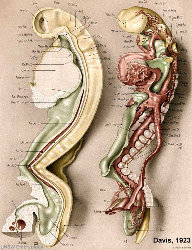 | 614 × 800 (83 KB) | S8600021 (talk | contribs) | Human Embryo Historic drawing of the Carnegie stage 11, 20 somite pairs, approx 25 days. Lateral sectional view, amnion and yolk sac removed :Left hand image - showing neural tube, gastrointestinal tract, pericardial cavity, connecting stalk :Right h |
You cannot overwrite this file.
File usage
There are no pages that use this file.