File:Stage7-bf1.jpg: Difference between revisions
No edit summary |
mNo edit summary |
||
| Line 7: | Line 7: | ||
Note the relative sizes of the extraembryonic coeloms (cavities) and the thin embryonic disc. | Note the relative sizes of the extraembryonic coeloms (cavities) and the thin embryonic disc. | ||
{{Carnegie stage 7 links}} | |||
<br> | |||
{{Carnegie_stage_table_1}} | |||
<br> | |||
{{Template:SEM}} | {{Template:SEM}} | ||
<br> | |||
[[Category:Human Embryo]] [[Category:Carnegie Stage]] [[Category:Carnegie Stage 7]] [[Category:Week 3]] [[Category:Gastrulation]] | [[Category:Human Embryo]] [[Category:Carnegie Stage]] [[Category:Carnegie Stage 7]] [[Category:Week 3]] [[Category:Gastrulation]] | ||
Latest revision as of 13:14, 24 February 2018
Human Embryo
Carnegie stage 7, 17 days, presomite
Embryonic disc (lateral view) within chorionic cavity (cut), yolk sac (below embryonic disc) and amniotic sac (above embryonic disc). Connecting stalk behind.
Note the relative sizes of the extraembryonic coeloms (cavities) and the thin embryonic disc.
| Stage 7 Links: Week 3 | Gastrulation | Lecture | Practical | Carnegie Embryos | Category:Carnegie Stage 7 | Next Stage 8 |
| Historic Papers: 1923 head-process | 1933 tubal | 1940 | 1949 |
| Week: | 1 | 2 | 3 | 4 | 5 | 6 | 7 | 8 |
| Carnegie stage: | 1 2 3 4 | 5 6 | 7 8 9 | 10 11 12 13 | 14 15 | 16 17 | 18 19 | 20 21 22 23 |
Image Source: Scanning electron micrographs of the Carnegie stages of the early human embryos are reproduced with the permission of Prof Kathy Sulik, from embryos collected by Dr. Vekemans and Tania Attié-Bitach. Images are for educational purposes only and cannot be reproduced electronically or in writing without permission.
File history
Click on a date/time to view the file as it appeared at that time.
| Date/Time | Thumbnail | Dimensions | User | Comment | |
|---|---|---|---|---|---|
| current | 12:47, 21 August 2009 | 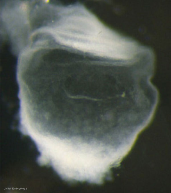 | 600 × 676 (34 KB) | MarkHill (talk | contribs) | Human Embryo Carnegie stage 7, 17 days, presomite Embryonic disc (lateral view) within chorionic cavity (cut), yolk sac (below embryonic disc) and amniotic sac (above embryonic disc). Connecting stalk behind. Note the relative sizes of the extraembryon |
You cannot overwrite this file.
File usage
The following 15 pages use this file:
- 2010 BGD Practical 3 - Week 3 Summary
- 2010 Lab 2
- BGDA Lecture - Development of the Embryo/Fetus 1
- BGDA Lecture - Development of the Embryo/Fetus 2
- BGDA Practical 3 - Week 3 Summary
- Carnegie stage 7
- Embryo Virtual Slides
- Human Embryo SEM
- Timeline human development
- Talk:Timeline human development
- Template:First Trimester Timeline
- Template:First Trimester Timeline collapsable table
- Template:SlideStage7bf1
- Template:VirtualSlidesStage7
- Template talk:First Trimester Timeline