File:Stage17 model 02.jpg: Difference between revisions
From Embryology
mNo edit summary |
|||
| (2 intermediate revisions by the same user not shown) | |||
| Line 3: | Line 3: | ||
This ventral view of the model shows a cross-section of the placental cord and the vessels and structures located within the cord. | This ventral view of the model shows a cross-section of the placental cord and the vessels and structures located within the cord. | ||
* <font color= | * <font color=green>'''Dark Green'''</font> - yolk sack | ||
* <font color= | * <font color=palegreen>'''Light Green'''</font> - Allantois | ||
* <font color= | * <font color=orangered>'''Red'''</font> - Placental Arteries | ||
* <font color= | * <font color=blue>'''Blue'''</font> - Placental Vein | ||
:'''Links:''' [[Placenta - Cord]] | [[Carnegie stage 17]] | [[Placenta Development]] | :'''Links:''' [[Placenta - Cord]] | [[Carnegie stage 17]] | [[Placenta Development]] | [[Embryology Models]] | ||
H261045 | H261045 | ||
<br> | |||
{{Carnegie stage 17 links}} | |||
<br> | |||
{{Carnegie_stage_table_1}} | |||
<br> | |||
{{Blechschmidt collection}} | |||
[[Category:Carnegie Stage 17]] | |||
Latest revision as of 15:59, 24 May 2017
Stage 17 Model - Placental Cord
This ventral view of the model shows a cross-section of the placental cord and the vessels and structures located within the cord.
- Dark Green - yolk sack
- Light Green - Allantois
- Red - Placental Arteries
- Blue - Placental Vein
H261045
| Week: | 1 | 2 | 3 | 4 | 5 | 6 | 7 | 8 |
| Carnegie stage: | 1 2 3 4 | 5 6 | 7 8 9 | 10 11 12 13 | 14 15 | 16 17 | 18 19 | 20 21 22 23 |
Image source: The Blechschmidt Collection images are reproduced with the permission of Prof. Christoph Viebahn, director of the Institute of Anatomy and Embryology, , University Medical Center Göttingen. Images are for educational purposes only and cannot be reproduced electronically or in writing without permission.
File history
Click on a date/time to view the file as it appeared at that time.
| Date/Time | Thumbnail | Dimensions | User | Comment | |
|---|---|---|---|---|---|
| current | 06:41, 16 December 2013 | 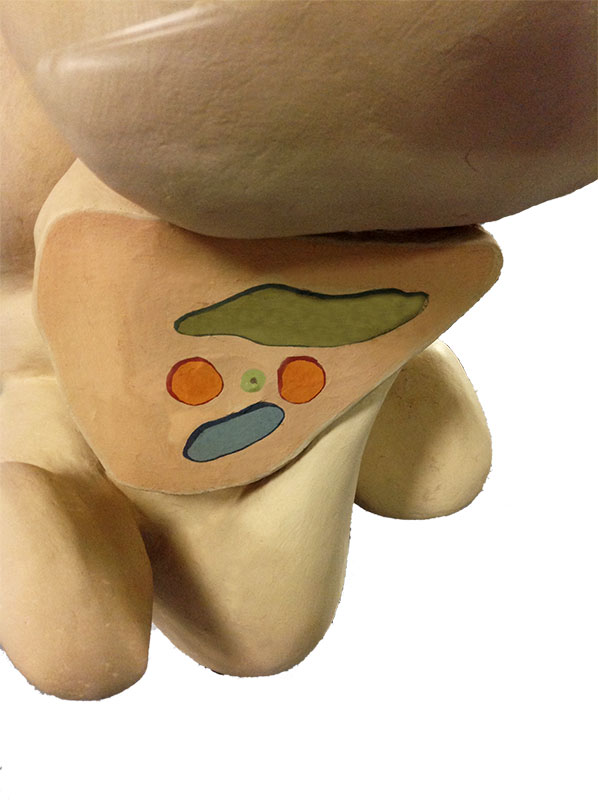 | 598 × 800 (0 bytes) | Z8600021 (talk | contribs) | H261045 {{Blechschmidt collection}} |
You cannot overwrite this file.
File usage
The following 6 pages use this file: