Category:Carnegie Stage 10: Difference between revisions
mNo edit summary |
|||
| Line 44: | Line 44: | ||
* '''9 somites''' - Embryo “Esch I,” Marburg. Elaborately described by Veit and Esch (1922), and cited, with illustrations, by Bartelmez and Evans (1926), who count 9 somites instead of 8 as stated by the original authors. Chorionic villi studied in detail by Ortmann (1938). Photographs and models are in the Carnegie Collection, No. 4251. | * '''9 somites''' - Embryo “Esch I,” Marburg. Elaborately described by Veit and Esch (1922), and cited, with illustrations, by Bartelmez and Evans (1926), who count 9 somites instead of 8 as stated by the original authors. Chorionic villi studied in detail by Ortmann (1938). Photographs and models are in the Carnegie Collection, No. 4251. | ||
* '''9 somites''' - Embryo “Du Ga,” Geneva. Described by Eternod (1896); models by Ziegler were distributed commercially. Cited by Bartelmez (1922) and Bartelmez and Evans (1926), with illustrations. Tracings made by H. M. Evans at Geneva and models are in the Carnegie Collection, No. 4439. | * '''9 somites''' - Embryo “Du Ga,” Geneva. Described by Eternod (1896); models by Ziegler were distributed commercially. Cited by Bartelmez (1922) and Bartelmez and Evans (1926), with illustrations. Tracings made by H. M. Evans at Geneva and models are in the Carnegie Collection, No. 4439. | ||
* '''About 9 somites | * '''About 9 somites''' - Embryo Unger, Keibel Collection, Freiburg i. Br., No. 4 of Keibel and Elze (1908). Listed by Bartelmez and Evans (1926). | ||
* '''9 somites''' - Embryo “Jacobsen,” formerly at Kiel (Graf Spee's collection was destroyed in World War II). Described by von Spee (1887). Listed by Bartelmez and Evans (1926) as having “at least” 9 somites. | * '''9 somites''' - Embryo “Jacobsen,” formerly at Kiel (Graf Spee's collection was destroyed in World War II). Described by von Spee (1887). Listed by Bartelmez and Evans (1926) as having “at least” 9 somites. | ||
* '''9 somites''' - Embryo Ca of Orts Llorca, Madrid. Various details described by Mari Martinez (1950) and Martinez Rovira (1953). | * '''9 somites''' - Embryo Ca of Orts Llorca, Madrid. Various details described by Mari Martinez (1950) and Martinez Rovira (1953). | ||
Revision as of 23:13, 16 September 2015
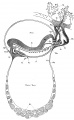
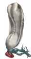
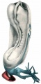
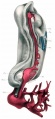
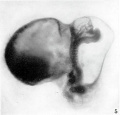
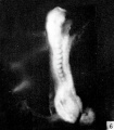
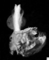
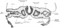
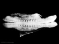
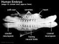
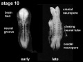
The Embryology pages and media listed below relate to human embryonic development during week 4, Carnegie Stage 10.
- Links: Carnegie stage 10 | Week 4
Facts: Week 4, 22 - 23 days, 2 - 3.5 mm, Somite number 4 - 12
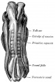
Features: Somite Number 4 - 12, rostral neuropore, neural folds in region of developing brain, neural tube, somites, caudal neuropore, neural fold fuses, remnant of amniotic sac
Ectoderm: Neural fold deeepens, edges approach midline, neural fold fuses, neural plate folds ventrally in brain region
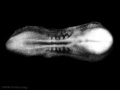
Mesoderm: Somitogenesis, continued segmentation of paraxial mesoderm (4 - 12 somite pairs)
| Week: | 1 | 2 | 3 | 4 | 5 | 6 | 7 | 8 |
| Carnegie stage: | 1 2 3 4 | 5 6 | 7 8 9 | 10 11 12 13 | 14 15 | 16 17 | 18 19 | 20 21 22 23 |
| Carnegie Collection - Stage 10 | ||||||||||
|---|---|---|---|---|---|---|---|---|---|---|
| Serial No. | Pairs of somites | Size (mm) | Grade | Fixative | Embedding Medium | Thinness (µm) | Stain | Year | Notes | |
| 391 | 8 | E, 2 Ch., 14 | Good | Formalin | P | 10 | Al. coch. | 1907 | Monograph by Dandy (1910)[1] | |
| 1201 | 7 | E,2 Ch.. 144 | Good | Formalin | P | 8 | H. & or. G. | 1915 | Univ. Chicago No. H 87 | |
| 2795 | 4-5 | E,2 | Poor | Alc. | P | 6 | Al coch,or.G. | 1919 | ||
| 3707 | 12 | E, 1 5 | Good | Formalin | P | 12.5 | I. H. | 1921 | Univ. Calif. No. H 197 | |
| 3709 | 4 | E. 1.4 Ch.. 14.8 | Poor | Formalin | P | 10 | Erythrosin | 1921 | Univ. Chicago No H 279 | |
| 3710 | 12 | E., 3.6 Ch., 19.0 | Good | Formalin | C-P | 10 | H. & or. G. | 1921 | Univ. Chicago No. H 392 | |
| 4216 | 8 | E, 2 Ch, 9.8 | Good | Formalin | P | 15 | ? | 1923 | Monograph by Payne (1925)[2] | |
| 5074 | 10 | E., 3.3 Ch., 10.8 | Exc. | Bouin | P | 10 | Al. coch. | 1925 | Univ. Rochester No. H 10. Monograph by Corner (1929)[3] | |
| 6330 | 7 | E, 2.83 | Good | P | 5 | Ehr. H. | 1931 | Univ. Chicago No. H 1404 | ||
| 6740 | 12 | E., 2.2 | Good | p | C-P | 8 | ? | 1933 | Litzenberg embryo. Studied by Boyden (1940) | |
| 7251 | 8 | E., 1.27 | Good | Formalin | C-P | 10 | (Stain - Haematoxylin Eosin) | 1941 | "Singapore embryo." Univ. Cambridge No. H 98. Studied by Wilson (1914)[4] | |
| 8244 | 6 | E., 1.55 Ch, 8,5 | Good | Alc. | C-P | 8 | (Stain - Haematoxylin Eosin) phlox. | 1944 | ||
| 9870 | 12 | Ch, ca. 8 | Good | Zenker | P | 5 | Various, chiefly carmine | 1952 | Univ. Chicago. No. H 637. Dicephaly | |
Abbreviations
| ||||||||||
Embryo Examples
- 4 somites - Carnegie No. 3709 (University of Chicago H 279). Characterized with outline sketches, by Bartelmez and Evans (1926).
- 4 somites - Histologisch-Embryologisches Institut, Embryo A, Vienna. Fully described by Sternberg (1927).
- 4 somites - Histologisch-Embryologisches Institut, Embryo Ca, Vienna. Fully described by Orts Llorca (1934).
- 4–5 somites - Florian's Embryo Bi II. The whereabouts of this, the following embryo, and the 10-somite Bi XI (see below) are not known; Florian's collection has not been found since his untimely death during World War II. The embryo Bi II was briefly characterized by Studnicka (1929), cited and partly illustrated by Florian (1928, 1930a).
- ‘’’4-5 somites - Florian's Embryo Bi III. (See note on previous embryo.) Briefly characterized by Studnicka (1929) and cited by Florian (1928).
- ‘’’4-5 somites - Carnegie No. 2795. Cited and briefly characterized by Bartelmez and Evans (1926). The specimen is distorted and somewhat macerated.
- 5 somites - Anatomisches Institut, Zürich, GM 1954. Described and illustrated by Schenck (1954).
- 5 somites - No. 103, Department of Anatomy, Tohoku University, Sendai. Distribution of alkaline phosphatase studied by Mori (1959a) in this and in another (No. 101), possibly 8-somite, embryo.
- 5–6 somites - Pfannenstiel “Klb” (originally at Giessen; was in Keibel's Institute at Freiburg i. Br. about 1911, may now be in Berlin). This well known embryo is No. 3 in the Keibel and Elze Normentafel (1908). Models by Kroemer (1903). A partial set of tracings made by H. M. Evans is in the Carnegie Collection, No. 5463.
- 6 somites - Carnegie No. 8244. Somewhat distorted; histologically fair.
- 6 somites - University of Michigan No. 71, Ann Arbor. Briefly described by Arey and Henderson (1943). A full description in an unpublished doctoral dissertation is in the files of L. B. Arey at Northwestern University, Chicago.
- 6 somites - Carnegie No 8818 (University of Chicago H 338) Pathological, not used in present study. Listed here because cited by Bartelmez and Evans (1926).
- 6–7 somites - His’s Embryo “SR.” Cited by His (1880) and by Bartelmez and Evans (1926). Has been studied only in the gross.
- 6–7 somites - Embryo LM (present location unknown). Cited here from manuscript notes at Carnegie laboratory, made from Russian text of Burow (1928). Condition said to be poor.
- 7 (?) somites - Embryo “Ludwig,” Berlin. Described by Streiter (1951). This specimen, which is somewhat macerated, is in certain characteristics considerably in advance of others of similar somitic number.
- 7 somites - Carnegie No. 6330 (University of Chicago H 1404). Extensive manuscript notes on this specimen, made under the supervision of G. W. Bartelmez, are in the files of the Carnegie laboratory.
- 8 somites - Carnegie No. 4216. Described by Payne (1925), and very frequently cited.
- 8 somites - Dublin. Described by West (1930); see also Arey (1938). Photographs and models are in the Carnegie Collection, No. 4923. Bartelmez (personal communication) thinks that this distorted embryo had only 5–6 somites.
- 8 somites - Carnegie No. 391. Described by Dandy (1910) and frequently cited (cf. Bartelmez and Evans, 1926, with additional illustrations). There were neither camera drawings nor photographs of the intact specimen, and therefore the reconstructions are not entirely satisfactory. The plaster models now at the Carnegie laboratory were made by O. Heard under the supervision of Bartelmez for the paper by Bartelmez and Evans (1926). The apparent lack of fusion of the neural folds described by Dandy is an artifact produced by a crack.
- 8 somites - Carnegie No. 1201 (University of Chicago H 87). Described briefly by Evans and Bartelmez (1917); cited, with illustrations, by Bartelmez and Evans (1926).
- 8 somites - Embryologisches Institut, Embryo Ct, Vienna. Fully described by Politzer (1930). Arey (1938) counts 8 paired somites in this embryo instead of 7 as stated by Politzer.
- 8 somites - University of Cambridge, Department of Anatomy H 98. Photographs and models in Carnegie Collection, No. 7251. Described by J. T. Wilson (1914). Cited by Bartelmez and Evans (1926), who consider it slightly abnormal in form although good histologically.
- 9 somites - Embryo “Esch I,” Marburg. Elaborately described by Veit and Esch (1922), and cited, with illustrations, by Bartelmez and Evans (1926), who count 9 somites instead of 8 as stated by the original authors. Chorionic villi studied in detail by Ortmann (1938). Photographs and models are in the Carnegie Collection, No. 4251.
- 9 somites - Embryo “Du Ga,” Geneva. Described by Eternod (1896); models by Ziegler were distributed commercially. Cited by Bartelmez (1922) and Bartelmez and Evans (1926), with illustrations. Tracings made by H. M. Evans at Geneva and models are in the Carnegie Collection, No. 4439.
- About 9 somites - Embryo Unger, Keibel Collection, Freiburg i. Br., No. 4 of Keibel and Elze (1908). Listed by Bartelmez and Evans (1926).
- 9 somites - Embryo “Jacobsen,” formerly at Kiel (Graf Spee's collection was destroyed in World War II). Described by von Spee (1887). Listed by Bartelmez and Evans (1926) as having “at least” 9 somites.
- 9 somites - Embryo Ca of Orts Llorca, Madrid. Various details described by Mari Martinez (1950) and Martinez Rovira (1953).
- ‘’’9-10 somites - Embryo R. Meyer 335. (Robert Meyer's collection was purchased by the late Hedwig Frey and bequeathed by him to the Anatomisches Institut, University of Zurich.) Listed by Bartelmez and Evans (1926), and cited by Felix (1912).
- 10 somites - Da2, Anatomical Institute, Basel. Described by Ludwig (1929). Plastic reconstructions. Neural groove closure extends rostral to otic discs.
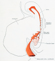
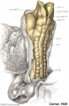
- 10 somites - Carnegie No. 5074 (University of Rochester H 10). Fully described by Corner (1929), and subjected to volumetric analysis by Boyden (1940). Excellent specimen.
- 10 somites - Grosser's Embryo Schwz (present location unknown). Briefly described, without illustrations, by Treutler (1931). Preservation said to be not altogether satisfactory.
- 10 somites - Florian’s Embryo Bi XI. (See note on Bi II above.) Briefly described, with illustrations, by Politzer and Sternberg (1930); cited and partly illustrated by Florian (1930a).
- 10 somites - Anatomy Department, University of South Wales, Cardiff. Partly described and illustrated by Baxter and Boyd (1939).[5]
- 11 somites - Embryo T 152, University of Toronto, Department of Anatomy. Cited by Arey (1938).
- 11 somites - Embryo G-dt, Uppsala. Described by Holmdahl (1943) as having 11 well-differentiated pairs of somites - with beginning delimitation of 4 more.
- 11–12 somites - Carnegie No. 8970 (University of Chicago H 637). Somewhat damaged. Cited, with illustrations, by Bartelmez (1922) and Bartelmez and Evans (1926).
- 12 somites - Carnegie No. 3710 (University of Chicago H 392). Cited by Bartelmez (1922) and Bartelmez and Evans (1926).
- 12 somites - Carnegie No. 3707 (University of California H 197). “Legge embryo.” Cited, with illustrations, by Bartelmez and Evans (1926). The coital history accompanying this specimen, which was declared to be reliable, would give it a postovulatory age of either 18 or 39 days; the former seems rather brief but the latter is much too long.
- 12 somites - Litzenberg embryo, University of Minnesota, Minneapolis. Briefly described by J. C. Litzenberg (1933); characterized and subjected to volumetric analysis by Boyden (1940), who counts 12 somites instead of 13–14 as in the original description. Photographs and model in Carnegie Collection, No. 6740.
- 12 somites - M. 24, University of Michigan, Ann Arbor. Cited by Arey (1938).
- ↑ Dandy WE. A human embryo with seven pairs of somites measuring about 2 mm in length. (1910) Amer. J Anat. 10: 85-109.
- ↑ Payne, F. 1925. General description of a 7-somite human embryo. Carnegie Instn. Wash. Publ. 361, Contrib. Embryol., 16,115-124.
- ↑ Corner GW. A well-preserved human embryo of 10 somites. (1929) Contrib. Embryol., Carnegie Inst. Wash. Publ. 394, 20:81-102.
- ↑ Wilson JT. Observations upon young human embryos. (1914) J Anat Physiol., 48(3): 315-51 PMID 17233002 PMC1288949
- ↑ Baxter JS. and Boyd JD. Observations on The Neural Crest of a Ten-Somite Human Embryo (1939) J Anat. 73:318–326. PMID 17104759
Subcategories
This category has the following 18 subcategories, out of 18 total.
C
Pages in category 'Carnegie Stage 10'
The following 62 pages are in this category, out of 62 total.
C
- Template:Carnegie Collection stage 10 table
- Carnegie Embryo 4439
- Template:Carnegie Embryo Stage10
- Carnegie stage 10
- Carnegie stage 10 gallery
- Template:Carnegie stage 10 links
- Template:CE1201
- Template:CE2795
- Template:CE3707
- Template:CE3709
- Template:CE3710
- Template:CE391
- Template:CE4216
- Template:CE5074
- Template:CE6330
- Template:CE7251
- Template:CE8244
- Template:CE8818
- Template:CE8970
- Template:CE9870
- Template:CS10
P
- Paper - A human embryo of seven to eight somites (1917)
- Paper - A Human Embryo with Seven Pairs of Somites Measuring about 2 mm in Length
- Paper - A volumetric analysis of young human embryos of the 10- and 12-somite stage
- Paper - A well-preserved human embryo of 10 somites (1929)
- Paper - A young human ovum of the early somite period
- Paper - Development of the human embryo during the period of somite formation with 2 to 16 pairs of somites
- Paper - Development of the human embryo during the period of somite formation, including embryos with 2 to 16 pairs of somites
- Paper - Developmental horizons in human embryos group X
- Paper - Normal development of early human embryos: Observation of 90 specimens at Carnegie stages 7 to 13
- Paper - Observations on the neural crest of a ten-somite human embryo (1939)
- Paper - The first appearance of the neural tube and optic primordium in the human embryo at stage 10
- Paper - The Huber six-somite human embryo (M 71)
- Paper - The human brain at stages 18-20 including the choroid plexuses and the amygdaloid and septal nuclei (1990)
- Template:Pfannenstiel III
R
- Template:Ref-AnsonCauldwellPick1943
- Template:Ref-AreyHenderson1943
- Template:Ref-Bartelmez1926
- Template:Ref-Baxter1939
- Template:Ref-Corner1925
- Template:Ref-Corner1929
- Template:Ref-Dandy1910
- Template:Ref-Eternod1896
- Template:Ref-EvansBartelmez1917
- Template:Ref-Heuser1957
- Template:Ref-Litzenberg1933
- Template:Ref-MullerO'Rahilly1990
- Template:Ref-PMID4051192
- Template:Ref-Sternberg1927
- Template:Ref-Sternberg1929
- Template:Ref-Veit1918
- Template:Ref-West1930
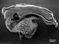
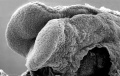
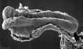
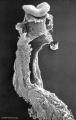
Media in category 'Carnegie Stage 10'
The following 84 files are in this category, out of 84 total.
 Bailey083.jpg 924 × 774; 129 KB
 Bailey375.jpg 929 × 785; 130 KB
 Bartelmez1923 fig02.jpg 1,295 × 2,189; 218 KB
 Bartelmez1923 fig03.jpg 1,105 × 1,350; 202 KB
 Bartelmez1923 fig04.jpg 1,416 × 1,155; 170 KB
 Bartelmez1923 fig05.jpg 1,102 × 815; 195 KB
 BaxterBoyd1939-fig01.jpg 361 × 736; 43 KB
 BaxterBoyd1939-fig02.jpg 459 × 851; 65 KB
 BaxterBoyd1939-fig03.jpg 732 × 1,000; 170 KB
 BaxterBoyd1939-fig04.jpg 642 × 615; 109 KB
 BaxterBoyd1939-fig05.jpg 1,000 × 864; 263 KB
 BaxterBoyd1939-fig06.jpg 489 × 917; 141 KB
 BaxterBoyd1939-fig07.jpg 795 × 917; 242 KB
 BaxterBoyd1939-plate01.jpg 1,680 × 2,400; 786 KB
 BaxterBoyd1939-plate02.jpg 1,681 × 2,400; 860 KB
 BaxterBoyd1939-text-fig01.jpg 1,283 × 1,000; 138 KB
 BaxterBoyd1939-text-fig02.jpg 1,200 × 861; 133 KB
 Corner1929 fig10-11.jpg 1,200 × 1,438; 730 KB
 Dandy1910-plate01.jpg 1,738 × 2,359; 541 KB
 Dandy1910-plate02.jpg 1,754 × 2,400; 951 KB
 Dandy1910-plate03.jpg 1,000 × 1,617; 224 KB
 Dandy1910-plate04.jpg 1,000 × 1,875; 174 KB
 Dandy1910-plate05.jpg 1,000 × 2,034; 238 KB
 Dandy1910-plate06.jpg 1,000 × 2,166; 265 KB
 Gray0020.jpg 1,038 × 1,536; 446 KB
 Heart Tube Fusion.jpg 1,551 × 1,139; 125 KB
 Heuser1957 fig03.jpg 898 × 989; 47 KB
 Heuser1957 fig04.jpg 754 × 989; 89 KB
 Heuser1957 fig05.jpg 898 × 860; 64 KB
 Heuser1957 fig06.jpg 753 × 862; 76 KB
 Heuser1957 fig07.jpg 1,056 × 1,296; 103 KB
 Keibel Mall 2 444.jpg 1,280 × 608; 114 KB
 Keibel Mall 2 539.jpg 1,280 × 1,414; 185 KB
 Stage 10 historic-Corner1929-1.jpg 654 × 1,000; 145 KB
 Stage10 bf1.jpg 1,000 × 748; 39 KB
 Stage10 bf10.jpg 2,048 × 2,048; 531 KB
 Stage10 bf1a.jpg 800 × 598; 29 KB
 Stage10 bf1b.jpg 600 × 449; 19 KB
 Stage10 bf1c.jpg 400 × 299; 10 KB
 Stage10 bf2.jpg 1,000 × 809; 29 KB
 Stage10 bf2c.jpg 400 × 323; 8 KB
 Stage10 bf3.jpg 836 × 1,000; 35 KB
 Stage10 bf4.jpg 1,000 × 750; 46 KB
 Stage10 bf4a.jpg 800 × 600; 33 KB
 Stage10 bf4b.jpg 600 × 450; 21 KB
 Stage10 bf4c.jpg 400 × 300; 11 KB
 Stage10 bf5.jpg 1,000 × 750; 54 KB
 Stage10 bf5a.jpg 800 × 600; 37 KB
 Stage10 bf5b.jpg 600 × 450; 23 KB
 Stage10 bf5c.jpg 400 × 300; 7 KB
 Stage10 bf6.jpg 1,000 × 750; 91 KB
 Stage10 bf6a.jpg 800 × 600; 63 KB
 Stage10 bf6b.jpg 600 × 450; 41 KB
 Stage10 bf6c.jpg 400 × 300; 22 KB
 Stage10 bf7.jpg 459 × 500; 24 KB
 Stage10 bf8.jpg 459 × 500; 33 KB
 Stage10 bf9.jpg 459 × 500; 26 KB
 Stage10 dorsal1.jpg 387 × 279; 6 KB
 Stage10 K12202-01.jpg 1,307 × 1,307; 240 KB
 Stage10 K12202-02.jpg 1,307 × 1,307; 242 KB
 Stage10 K12202-03.jpg 1,434 × 1,105; 191 KB
 Stage10 K12202-04.jpg 1,434 × 1,105; 227 KB
 Stage10 K12202-05.jpg 1,307 × 1,307; 278 KB
 Stage10 K12202-06.jpg 1,307 × 1,307; 299 KB
 Stage10 neural sm.jpg 665 × 499; 22 KB
 Stage10 SEM1.jpg 277 × 450; 28 KB
 Stage10 sem1.jpg 1,000 × 484; 46 KB
 Stage10 sem10.jpg 1,000 × 740; 68 KB
 Stage10 sem10a.jpg 800 × 592; 48 KB
 Stage10 sem10b.jpg 600 × 444; 32 KB
 Stage10 sem10c.jpg 400 × 296; 16 KB
 Stage10 sem11.jpg 1,000 × 636; 76 KB
 Stage10 sem2.jpg 1,000 × 590; 60 KB
 Stage10 sem3.jpg 634 × 1,000; 55 KB
 Stage10 sem4.jpg 1,000 × 814; 83 KB
 Stage10 sem5.jpg 845 × 1,000; 83 KB
 Stage10 sem6 annotated.jpg 720 × 960; 122 KB
 Stage10 sem6.jpg 614 × 1,000; 57 KB
 Stage10 sem7.jpg 1,243 × 1,000; 116 KB
 Stage10 sem8.jpg 648 × 1,000; 84 KB
 Stage10 sem9.jpg 740 × 1,000; 72 KB
 Stage10 sem9a.jpg 592 × 800; 51 KB
 Stage10 sem9b.jpg 444 × 600; 32 KB
 Stage10 sem9c.jpg 296 × 400; 16 KB


