Carnegie stage 9
| Embryology - 26 Apr 2024 |
|---|
| Google Translate - select your language from the list shown below (this will open a new external page) |
|
العربية | català | 中文 | 中國傳統的 | français | Deutsche | עִברִית | हिंदी | bahasa Indonesia | italiano | 日本語 | 한국어 | မြန်မာ | Pilipino | Polskie | português | ਪੰਜਾਬੀ ਦੇ | Română | русский | Español | Swahili | Svensk | ไทย | Türkçe | اردو | ייִדיש | Tiếng Việt These external translations are automated and may not be accurate. (More? About Translations) |
Introduction

Facts
Human embryonic stage 9 occurs during week 3 between 19 to 21 days.
Gestational Age GA - week 5
The embryo is now 1.5 to 2.5 mm in size and somites have begun to form and number between 1 to 3 somite pairs during this stage.
The initial images are displayed unlabeled to allow you to explore the embryo for yourself, linked labeled versions are also available for some images.
See also Carnegie stage 9 image gallery.
| Week: | 1 | 2 | 3 | 4 | 5 | 6 | 7 | 8 |
| Carnegie stage: | 1 2 3 4 | 5 6 | 7 8 9 | 10 11 12 13 | 14 15 | 16 17 | 18 19 | 20 21 22 23 |
- Carnegie Stages: 1 | 2 | 3 | 4 | 5 | 6 | 7 | 8 | 9 | 10 | 11 | 12 | 13 | 14 | 15 | 16 | 17 | 18 | 19 | 20 | 21 | 22 | 23 | About Stages | Timeline
Summary
- Ectoderm - Neural plate brain region continues to expand, neural plate begins folding over the notochord. Gastrulation continues through the primitive streak region.
- Mesoderm - Paraxial mesoderm segmentation into somites begins (1 - 3 somite pairs). Lateral plate mesoderm begins to vacuolate, dividing it into somatic and splanchnic mesoderm and to later form the intra-embryonic coelom. Prechordal splanchnic mesoderm begins to form the cardiogenic region, from which the primordial heart will develop.
- Endoderm - Notochordal plate still visible which will form the notochord. Endoderm is still widely open to the yolk sac and germ cells form part of this layer. Extra-embryonic mesoderm on the yolk sac surface begins to form "blood islands".
See also Events
Identify
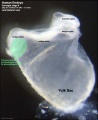
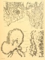
- Neural groove and neural folds, the mesoderm, which segments beside the neural groove to form somites but extends laterally to margin of embryonic disc lateral plate mesoderm, where it merges with the covering extraembryonic mesoderm.
- The intra-embryonic coelom develops in the middle of the lateral plate mesoderm. Note amniotic ectoderm covered by extra-emebryonic mesoderm (empty spaces above and below the mesoderm are artefacts, as are the lateral folds in the ectoderm).
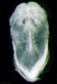
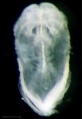
The first two images using bright field microscopy approximate the orientation of the scanning electron micrographs below. There are additional scanning electron micrographs showing selected features in detail. Carnegie_stage_9 image gallery
- Links: Week 3 | Gastrulation | Lecture | Practical | 1920 Carnegie No.1878 | Stage 10
Bright Field Lateral
| Lateral View | Ventrolateral View |
|---|---|

|

|
| Lateral View 1 - Large | 800px | Medium | Small | Ventrolateral View 2 - Large | 800px | Medium | Small |
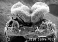
Scanning EM Lateral
| Lateral View | Ventrolateral View |
|---|---|

|

|
| Lateral View 1 - Small | Medium | Large | Ventrolateral View 2 - Small | Medium | Large |
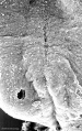
Notochordal plate

Notochordal plate - Small | Medium | Large
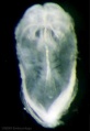
Bright Field Dorsal
| Dorsal | Ventral |
|---|---|

|

|
| Lateral View 1 - Small | Medium | Large | Ventrolateral View 2 - Small | Medium | Large |
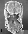
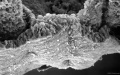
Scanning EM Dorsal


Kyoto Collection
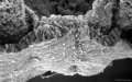
| Dorsal View | Ventral View |
|---|---|

|

|
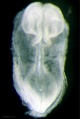
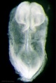
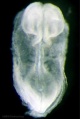
| Embryonic disc, showing the epiblast viewed from the amniotic (dorsal) side. | Embryonic disc, showing the epiblast, connecting stalk and brain fold. |
Image source: UNSW Embryology page Created: 19.03.1999
Image source: The Kyoto Collection images are reproduced with the permission of Prof. Kohei Shiota and Prof. Shigehito Yamada, Anatomy and Developmental Biology, Kyoto University Graduate School of Medicine, Kyoto, Japan for educational purposes only and cannot be reproduced electronically or in writing without permission.
Events
References
- ↑ Bartelmez GW. The origin of the otic and optic primordia in man. (1922) J. Comp. Neural., 34: 201-232.
- ↑ Ingalls NW. A human embryo at the beginning of segmentation, with special reference to the vascular system. (1920) Contrib. Embryol., Carnegie Inst. Wash. Publ. 274, 11: 61-90.
Additional Images
Primordial germ cell region
Primordial germ cell region

Light Images
Large 1000px

800px
Medium 600px
Small 400px


Large 1000px

800px
Medium 600px
Small 400px


Scanning EM Images
SEM Dorsal

Large 1000px
800px

Medium 600px

Small 400px


SEM Cranial Neural fold

Large 1000px
800px

Medium 600px

Small 400px


SEM Caudal Region

Large 1000px

800px
Medium 600px

Small 400px


SEM Caudal Region cross section

Large 1000px
800px

Medium 600px
Small 400px


Image Source: Scanning electron micrographs of the Carnegie stages of the early human embryos are reproduced with the permission of Prof Kathy Sulik, from embryos collected by Dr. Vekemans and Tania Attié-Bitach. Images are for educational purposes only and cannot be reproduced electronically or in writing without permission.
Historic Images
| Historic Disclaimer - information about historic embryology pages |
|---|
 |
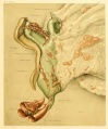
1920 Carnegie No.1878 A Human Embryo at the Beginning of Segmentation, with Special Reference to the Vascular System. By N. William Ingalls
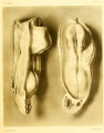
Left lateral and dorsal view
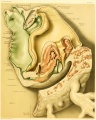
Lateral view of the vasculature
Views of the heart and vasculature

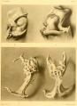
Gray Fig. 31


- Carnegie Stages: 1 | 2 | 3 | 4 | 5 | 6 | 7 | 8 | 9 | 10 | 11 | 12 | 13 | 14 | 15 | 16 | 17 | 18 | 19 | 20 | 21 | 22 | 23 | About Stages | Timeline
Cite this page: Hill, M.A. (2024, April 26) Embryology Carnegie stage 9. Retrieved from https://embryology.med.unsw.edu.au/embryology/index.php/Carnegie_stage_9
- © Dr Mark Hill 2024, UNSW Embryology ISBN: 978 0 7334 2609 4 - UNSW CRICOS Provider Code No. 00098G