Carnegie stage 8
Introduction
 |
Gastrulation is continuing as cells migrate from the epiblast, continuing to form mesoderm. Mesoderm lies between the ectoderm and endoderm as a continuous sheet except at the buccopharyngeal and cloacal membranes. These membranes have ectoderm and endoderm only and will lie at the rostral (head) and caudal (tail) of the gastrointestinal tract. From the primitive node a tube extends under the ectoderm in the opposite direction to the primitive streak. This tube forms first the axial process then notochordal process, then finally the notochord. The notochord is a key to embryonic folding and regulation of ectoderm and mesoderm differentiation. It lies in the rostrocordal axis and the embryonic disc will fold either side ventrally, pinching off a portion of the yolk sac to form the lining of the gastrointestinal tract. FactsHuman embryonic stage 8 occurs during week 3 between 17 to 19 days. The embryo is now 1.0 - 1.5 mm in size. Identify
|
- Carnegie Stages: 1 | 2 | 3 | 4 | 5 | 6 | 7 | 8 | 9 | 10 | 11 | 12 | 13 | 14 | 15 | 16 | 17 | 18 | 19 | 20 | 21 | 22 | 23 | About Stages | Timeline
Bright Field


Scanning EM


Image Source: Scanning electron micrographs of the Carnegie stages of the early human embryos are reproduced with the permission of Prof Kathy Sulik, from embryos collected by Dr. Vekemans and Tania Attié-Bitach. Images are for educational purposes only and cannot be reproduced electronically or in writing without permission.
Kyoto Collection

View: embryonic disc, showing the epiblast viewed from the amniotic (dorsal) side. Amniotic membrane removed, connecting stalk to the left.
Image source: Embryology page Created: 19.03.1999
Image source: The Kyoto Collection images are reproduced with the permission of Prof. Kohei Shiota and Prof. Shigehito Yamada, Anatomy and Developmental Biology, Kyoto University Graduate School of Medicine, Kyoto, Japan for educational purposes only and cannot be reproduced electronically or in writing without permission.
Carnegie Collection

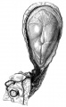
Dorsal view. Primitive node appears as a conspicuous opaque region with the notochordal process extending rostrally. |

Ventral view. Notochordal plate visible. |
Serial Sections

|

|
Other Images
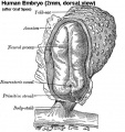
Human Embryo neural plate historic drawing 2 mm embryo dorsal view
Dorsal view of Carnegie embryo No. 5960. (drawn by James F. Didusch from a reconstruction).


- Carnegie Stages: 1 | 2 | 3 | 4 | 5 | 6 | 7 | 8 | 9 | 10 | 11 | 12 | 13 | 14 | 15 | 16 | 17 | 18 | 19 | 20 | 21 | 22 | 23 | About Stages | Timeline
Cite this page: Hill, M.A. (2024, May 3) Embryology Carnegie stage 8. Retrieved from https://embryology.med.unsw.edu.au/embryology/index.php/Carnegie_stage_8
- © Dr Mark Hill 2024, UNSW Embryology ISBN: 978 0 7334 2609 4 - UNSW CRICOS Provider Code No. 00098G