Carnegie stage 3
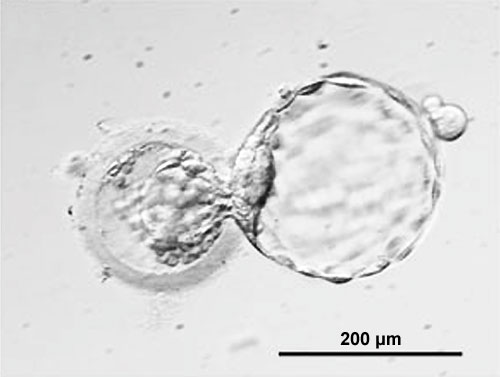
Human Blastocyst "hatching" from zona pellucida, in early Embryonic Development designated as Carnegie stage 3. Blastocyst is too the right of image and Zona pellucida is shown to the left of the image. Note: the small opening in the zona pellucida through which the blastocyst is hatching the flattened trophoblast cells forming the outer cell layer of the blastocyst the inner cell mass shown in the centre of the image and on the left-hand wall of the blastocyst the blastocoel forming a large fluid-filled space within the blastocyst
Facts: Week 1, 4 - 5 days, size 0.1-0.2 mm
Features: zona pellucida, trophoblast shell, inner cell mass, blastoceol
Image source: Klimanskaya I, Chung Y, Becker S, Lu SJ, Lanza R. Human embryonic stem cell lines derived from single blastomeres. Nature. 2006 Aug 23 PMID:16929302 Embryology page Created: 19.03.1999
- Carnegie Stages: 1 | 2 | 3 | 4 | 5 | 6 | 7 | 8 | 9 | 10 | 11 | 12 | 13 | 14 | 15 | 16 | 17 | 18 | 19 | 20 | 21 | 22 | 23 | About Stages | Timeline
Cite this page: Hill, M.A. (2024, April 30) Embryology Carnegie stage 3. Retrieved from https://embryology.med.unsw.edu.au/embryology/index.php/Carnegie_stage_3
- © Dr Mark Hill 2024, UNSW Embryology ISBN: 978 0 7334 2609 4 - UNSW CRICOS Provider Code No. 00098G
