Carnegie stage 19: Difference between revisions
mNo edit summary |
mNo edit summary |
||
| Line 25: | Line 25: | ||
:'''Links:''' [[Week 7]] | [[Human_System_Development|System Development]] | [[Lecture_-_Limb_Development|Lecture - Limb]] | [[Lecture_-_Head_Development|Lecture - Head Development]] | [[Lecture_-_Sensory_Development|Lecture - Sensory]] | [[ | :'''Links:''' [[Week 7]] | [[Human_System_Development|System Development]] | [[Lecture_-_Limb_Development|Lecture - Limb]] | [[Lecture_-_Head_Development|Lecture - Head Development]] | [[Lecture_-_Sensory_Development|Lecture - Sensory]] | [[ANAT2341_Lab_6|Science Practical - Head]] | [[ANAT2341_Lab_10|Science Practical - Sensory]] | [[ANAT2341_Lab_8|Science Practical - Urogenital]] | [[:Category:Carnegie_Stage_19|Category:Carnegie Stage 19]] | [[Carnegie_stage_20|Stage 20]] | ||
{{ | |||
{{Carnegie_stages}} | |||
==Bright Field== | ==Bright Field== | ||
Revision as of 12:08, 7 February 2014
| Embryology - 19 May 2024 |
|---|
| Google Translate - select your language from the list shown below (this will open a new external page) |
|
العربية | català | 中文 | 中國傳統的 | français | Deutsche | עִברִית | हिंदी | bahasa Indonesia | italiano | 日本語 | 한국어 | မြန်မာ | Pilipino | Polskie | português | ਪੰਜਾਬੀ ਦੇ | Română | русский | Español | Swahili | Svensk | ไทย | Türkçe | اردو | ייִדיש | Tiếng Việt These external translations are automated and may not be accurate. (More? About Translations) |
Introduction

Facts
Week 7, 48 - 51 days, 16 - 18 mm
Events
Ectoderm: sensory placodes, lens pit, otocyst, nasal pits moved ventrally, fourth ventricle of brain
Mesoderm: heart prominence, ossification continues
Head: forebrain, eye, external acoustic meatus
Body: straightening of trunk, heart, liver, umbilical cord
Features
eyelid, eye, external acoustic meatus, auricle of external ear, digital ray, wrist, liver prominence
Identify: straightening of trunk, pigmented eye, eyelid, external acoustic meatus, digital rays, liver prominance, thigh, ankle, foot plate, umbilical cord
- Links: Week 7 | System Development | Lecture - Limb | Lecture - Head Development | Lecture - Sensory | Science Practical - Head | Science Practical - Sensory | Science Practical - Urogenital | Category:Carnegie Stage 19 | Stage 20
- Carnegie Stages: 1 | 2 | 3 | 4 | 5 | 6 | 7 | 8 | 9 | 10 | 11 | 12 | 13 | 14 | 15 | 16 | 17 | 18 | 19 | 20 | 21 | 22 | 23 | About Stages | Timeline
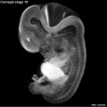
Bright Field

- Facts: Week 7, 48 - 51 days, 16 - 18 mm
- View: Lateral view. Amniotic membrane removed.
- Features: eyelid, eye, external acoustic meatus, auricle of external ear, digital ray, wrist, liver prominence
- Identify: straightening of trunk, pigmented eye, eyelid, external acoustic meatus, digital rays, liver prominance, thigh, ankle, foot plate, umbilical cord
Image: Dr Steven O'Connor (Houston, Texas) - Other embryo images.
- Carnegie Stages: 1 | 2 | 3 | 4 | 5 | 6 | 7 | 8 | 9 | 10 | 11 | 12 | 13 | 14 | 15 | 16 | 17 | 18 | 19 | 20 | 21 | 22 | 23 | About Stages | Timeline
Cite this page: Hill, M.A. (2024, May 19) Embryology Carnegie stage 19. Retrieved from https://embryology.med.unsw.edu.au/embryology/index.php/Carnegie_stage_19
- © Dr Mark Hill 2024, UNSW Embryology ISBN: 978 0 7334 2609 4 - UNSW CRICOS Provider Code No. 00098G
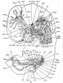
Scanning EM

Ventral view of head showing upper lip, maxilla and nasal region.
Image Source: Prof Virginia Diewert
Kyoto Collection

View: This is a dorsolateral view of embryo. Amniotic membrane removed.
Image source: Embryology page Created: 19.03.1999
Image source: The Kyoto Collection images are reproduced with the permission of Prof. Kohei Shiota and Prof. Shigehito Yamada, Anatomy and Developmental Biology, Kyoto University Graduate School of Medicine, Kyoto, Japan for educational purposes only and cannot be reproduced electronically or in writing without permission.
Carnegie Collection


- Carnegie stage 19: 4501 Right | 4501 Anterior | 4501 Left | 6824 Right | 6824 Anterior | 6824 Left | 8092 Right | 8092 Anterior | 8092 Left
| iBook - Carnegie Embryos | |
|---|---|

|
|
Photographs

|

|
| Image - Ed Uthman (pathologist in Houston, Texas)
Image version links: ExtraLarge 2054x3081px | Large 683 x 1024px | Medium 333 x 500px | Small 160 x 240px |
Image - Dr Steven O'Connor (Houston, Texas)
Image version links: ExtraLarge 387×2592px | Large 1024x685px | Medium 500x334px | Small 240x161px |
Additional Images
Stage 19 Optical Projection Tomography

External ear Stages 14-23 and adult
- Human Embryo 17.8mm.jpg
historic drawing of human embryo
Human Embryo 17.8 mm


- Carnegie Stages: 1 | 2 | 3 | 4 | 5 | 6 | 7 | 8 | 9 | 10 | 11 | 12 | 13 | 14 | 15 | 16 | 17 | 18 | 19 | 20 | 21 | 22 | 23 | About Stages | Timeline
Cite this page: Hill, M.A. (2024, May 19) Embryology Carnegie stage 19. Retrieved from https://embryology.med.unsw.edu.au/embryology/index.php/Carnegie_stage_19
- © Dr Mark Hill 2024, UNSW Embryology ISBN: 978 0 7334 2609 4 - UNSW CRICOS Provider Code No. 00098G