BGDA Practical Placenta - Villi Development: Difference between revisions
mNo edit summary |
|||
| (51 intermediate revisions by the same user not shown) | |||
| Line 32: | Line 32: | ||
* ''' | * '''anchoring villi''' - or stem villi cytotrophoblast cells attached to maternal tissue. | ||
* '''branched villi''' - grow from sides of | * '''branched villi''' - grow from sides of anchoring villi, region of main exchange, surrounded by maternal blood in intervillous spaces. | ||
* '''terminal villi''' - not active | * '''terminal villi''' - not active outgrow is caused by proliferation of the trophoblast. Passive protrusions induced by capillary coiling due to growth of the fetal capillaries within the mature intermediate villi (third trimester). | ||
* '''chorionic plate''' - region of membrane at the base of the villi through which placental arteries and vein passes. | * '''chorionic plate''' - region of membrane at the base of the villi through which larger placental arteries and vein passes. | ||
==Placental Circulation== | |||
{| | {| | ||
| Placental | | Schematic representation of blood flow through the placenta and surrounding tissue.{{#pmid:29230859|PMID29230859}} | ||
<br><br> | |||
* Blue and red arrows show the flow directions of oxygenated (red) and deoxygenated (blue) fetal blood through the placental vasculature. | |||
* Only the largest villi are included (normal placentas terminal villi make up 40% of villous tree volume) | |||
* Dashed white arrows show idealized flow lines through intervillous space for maternal blood. | |||
* Relative oxygenation states shown by the red to blue colour gradient. | |||
* Maternal septa divide vascular spaces into the placental cotyledons. | |||
| width=5px| | |||
| [[File:Placental blood flow 01.jpg|700px]] | |||
|} | |||
==Anchoring Villi Histology== | |||
Please note that there are additional slides listed in the current set, only the first placenta slide and the cord cross-section will be covered in detail in the practical class. | |||
{| | |||
| [[File:Moodle icon2.jpg|link=http://moodle.telt.unsw.edu.au/mod/book/view.php?id=853422&chapterid=106721]] | |||
| [http://moodle.telt.unsw.edu.au/mod/book/view.php?id=853422&chapterid=106721 Moodle Placenta Virtual Slides] | |||
(you must be logged in to Moodle) | |||
|} | |||
{| | |||
| [[File: | | width=250px| | ||
{| | |||
| [[File:Moodle icon2.jpg|40px|link=https://www.best.edu.au/s/share?url=kaw1n2zn%2Fqhwg56f5&data=8%401!9%4033600!10%40-14702.5&version=1]] | |||
| [https://www.best.edu.au/s/share?url=kaw1n2zn%2Fqhwg56f5&data=8%401!9%4033600!10%40-14702.5&version=1 Slide - Human Placenta] | |||
|} | |||
| width=250px| | |||
{| | |||
| [[File:Moodle icon2.jpg|40px|link=https://www.best.edu.au/s/share?url=r7xatnc7%2Fcrxpmx49&data=8%405!9%409298!10%40-5546&version=1]] | |||
| [https://www.best.edu.au/s/share?url=r7xatnc7%2Fcrxpmx49&data=8%405!9%409298!10%40-5546&version=1 Placenta - mid-term (fetal side)] | |||
|} | |||
| width=250px| | |||
{| | |||
| [[File:Moodle icon2.jpg|40px|link=https://www.best.edu.au/s/share?url=r7xatnc7%2Fcrxpmx49&data=8%401!9%4012030.5!10%40-7689.5&version=1]] | |||
| [https://www.best.edu.au/s/share?url=r7xatnc7%2Fcrxpmx49&data=8%401!9%4012030.5!10%40-7689.5&version=1 Placenta - (late))] | |||
|} | |||
|- | |||
| | |||
{| | |||
| [[File:Moodle icon2.jpg|40px|link=https://www.best.edu.au/s/share?url=q5fwwpm7%2Fcukuegma&data=8%401!9%4012464!10%40-3712&version=1]] | |||
| [https://www.best.edu.au/s/share?url=q5fwwpm7%2Fcukuegma&data=8%401!9%4012464!10%40-3712&version=1 Slide - Human Placental Cord] | |||
|} | |||
| | |||
{| | |||
| [[File:Moodle icon2.jpg|40px|link=https://www.best.edu.au/s/share?url=2rnd8dka%2Fx8t443kf&data=8%400!9%409057!10%40-6658&version=1]] | |||
| [https://www.best.edu.au/s/share?url=2rnd8dka%2Fx8t443kf&data=8%400!9%409057!10%40-6658&version=1 Placenta - mid-term (maternal side)] | |||
|} | |||
|} | |} | ||
<br> | |||
[[File:Placenta_anchoring_villi.jpg]] | |||
==Villi Development== | ==Villi Development== | ||
| Line 73: | Line 117: | ||
We will now look at an example of first trimester placentation in a Virtual Slide. | We will now look at an example of first trimester placentation in a Virtual Slide. | ||
{| | |||
[ | | [[File:Moodle icon2.jpg|40px|link=https://www.best.edu.au/s/share?url=kaw1n2zn%2Fqhwg56f5&data=8%401!9%4033600!10%40-14702.5&version=1]] | ||
| [https://www.best.edu.au/s/share?url=kaw1n2zn%2Fqhwg56f5&data=8%401!9%4033600!10%40-14702.5&version=1 Slide - Human Placenta] | |||
|} | |||
Please note that there are additional slides listed in the current set, only the first placenta slide and the cord cross-section will be covered in detail in the practical class. | Please note that there are additional slides listed in the current set, only the first placenta slide and the cord cross-section will be covered in detail in the practical class. | ||
| Line 86: | Line 131: | ||
{| | {| | ||
| [[File:Embryo-membranes_stage_11.jpg|300px]] | | [[File:Embryo-membranes_stage_11.jpg|300px]] | ||
| [[File:Stage13_bf4.jpg| | | [[File:Stage13_bf4.jpg|300px]] | ||
|- | |- | ||
| <center>Week 4 (Stage 11)</center> | | <center>Week 4 (Stage 11)</center> | ||
| <center>Week 4-5 (Stage 13)</center> | | <center>Week 4-5 (Stage 13)</center> | ||
|- | |||
| [[File:Stage18 bf10.jpg|300px]] | |||
| [[File:Placental membranes.jpg|300px]] | |||
|- | |||
| <center>Week 7 (Stage 18)</center> | |||
| <center>Week 7 (Stage 18)</center> | | <center>Week 7 (Stage 18)</center> | ||
|} | |} | ||
| Line 96: | Line 145: | ||
{| | {| | ||
! colspan=4|Virtual slides | |||
|- | |||
| valign=bottom|{{SlideStage13bf04}} | | valign=bottom|{{SlideStage13bf04}} | ||
| valign=bottom|{{SlideStage14bf18}} | | valign=bottom|{{SlideStage14bf18}} | ||
| valign=bottom|{{SlideStage14bf21}} | | valign=bottom|{{SlideStage14bf21}} | ||
| valign=bottom|{{SlideStage18bf12}} | |||
|} | |} | ||
==Terms== | ==Terms== | ||
{{Placenta terms}} | |||
<br> | |||
{{BGDA Practical 14 - Villi Interactive}} | |||
<br> | |||
{{BGDALabPlacenta}} | {{BGDALabPlacenta}} | ||
==Additional Information== | ==Additional Information== | ||
{{Med Prac additional Information}} | {{Med Prac additional Information}} | ||
| Line 168: | Line 190: | ||
Mesenchymal villi continuously form out of the trophoblastic sprouts throughout pregnancy and have been considered the basis for growth and differentiation of the villous trees. | Mesenchymal villi continuously form out of the trophoblastic sprouts throughout pregnancy and have been considered the basis for growth and differentiation of the villous trees. | ||
=== | ===Third Trimester Placental Blood Flow=== | ||
{| | |||
|+ Diffusion Tensor Magnetic Resonance Imaging (MRI) | |||
|- | |||
| width=605px|[[File:Placenta MRI01.jpg|600px]] | |||
| valign=top| | |||
<br><br>This is a third trimester placenta ({{GA}} 35 weeks + 6 days) showing the mean diffusivity maps for three slices of a single placental diffusion-weighted MRI scan.{{#pmid:29230859|PMID29230859}} | |||
<br><br><br> | |||
* Red and black outline the uterine wall and placenta ROIs respectively. | |||
* Colour represents mean diffusivity in mm<sup>2</sup> s<sup>-1</sup> (note that the scale is a factor of 10 higher for the b = 4 40 s mm<sup>-2</sup> maps). | |||
< | <br><br> | ||
{{MRI}} Diffusion tensor imaging (DTI) techniques provide information on the microstructural processes and has also been used to study brain development. Note that clinically, Doppler ultrasound has also been used to measure placental blood flow. | |||
|} | |||
===Human Villi Timeline=== | |||
Detailed overview of villi development. | |||
{{Placenta Villi Timeline table}} | |||
===Historic Images=== | |||
- | {| | ||
! Decidua and villi location | |||
! Chorionic villi | |||
|- | |||
| [[File:Gray0034.jpg|400px]] | |||
| [[File:Bailey495.jpg|400px]] | |||
|} | |||
==References== | |||
<references/> | |||
Latest revision as of 21:27, 2 June 2019
Chorionic Villi
Primary villiWeek 2 - first stage of chorionic villi development. Trophoblastic shell cells (syncitiotrophoblasts and cytotrophoblasts) form finger-like extensions into maternal decidua. |

|
Secondary villiWeek 3 - second stage of chorionic villi development Extra-embryonic mesoderm grows into villi, covers the entire surface of chorionic sac. Basal region will form chorionic plate. |

|
Tertiary villiWeek 4 - third stage of chorionic villi development. Mesenchyme differentiates into blood vessels and cells, forms arteriocapillary network, fuse with placental vessels, developing in connecting stalk. |

|
- anchoring villi - or stem villi cytotrophoblast cells attached to maternal tissue.
- branched villi - grow from sides of anchoring villi, region of main exchange, surrounded by maternal blood in intervillous spaces.
- terminal villi - not active outgrow is caused by proliferation of the trophoblast. Passive protrusions induced by capillary coiling due to growth of the fetal capillaries within the mature intermediate villi (third trimester).
- chorionic plate - region of membrane at the base of the villi through which larger placental arteries and vein passes.
Placental Circulation
| Schematic representation of blood flow through the placenta and surrounding tissue.[1]
|

|
Anchoring Villi Histology
Please note that there are additional slides listed in the current set, only the first placenta slide and the cord cross-section will be covered in detail in the practical class.

|
Moodle Placenta Virtual Slides
(you must be logged in to Moodle) |
|
|
| ||||||
|
|

Villi Development
| First Trimester | Third Trimester |
|---|---|

|

|


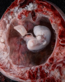
Placenta anchoring villi
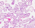
Villi first trimester
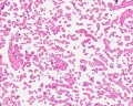
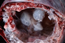
Villi at term
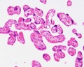
Villi at term
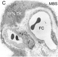
EM haemomonochorial placenta


Virtual Slide
We will now look at an example of first trimester placentation in a Virtual Slide.
| Slide - Human Placenta |
Please note that there are additional slides listed in the current set, only the first placenta slide and the cord cross-section will be covered in detail in the practical class.
Chorionoic Villi Location
Originally villi cover entire chorionic surface and then become restricted to decidua basalis region forming 2 regions:
- Frondosum - "leafy" where villi are mainly located.
- Capsularis - smooth chorion, where villi are absent or not abundant.

|

|

|

|
| Virtual slides | |||||||||||||||
|---|---|---|---|---|---|---|---|---|---|---|---|---|---|---|---|
|
|
|
| ||||||||||||


Terms
| Placenta Terms (expand to view) |
|---|
with an incidence of about 2.8 per 1,000 pregnancies, there is also a rarer form of extra-abdominal varices.PMID 24883288
with an incidence of about 2.8 per 1,000 pregnancies, there is also a rarer form of extra-abdominal varices. PMID 24883288
|
| Other Terms Lists |
|---|
| Terms Lists: ART | Birth | Bone | Cardiovascular | Cell Division | Endocrine | Gastrointestinal | Genital | Genetic | Head | Hearing | Heart | Immune | Integumentary | Neonatal | Neural | Oocyte | Palate | Placenta | Radiation | Renal | Respiratory | Spermatozoa | Statistics | Tooth | Ultrasound | Vision | Historic | Drugs | Glossary |
Placental Villi Interactive Component
| Attempt the Quiz - Placental Villi | ||
|---|---|---|

Here are a few simple Quiz questions that relate to Placental Villi from the practical.
|
Additional Information
| Additional Information - Content shown under this heading is not part of the material covered in this class. It is provided for those students who would like to know about some concepts or current research in topics related to the current class page. |

2013 Meeting Presentation - Placenta Embryology and Circulation
Cytotrophoblast Layer
There is a new interpretation of the changes that are occuring in the cytotrophoblast (CTB) layer during early to full-term human placenta development. Traditionally the interpretation was that the cytotrophoblast layer thinned and became discontinuous towards term. The thinning is thought due to the epithelium surface expanding at a faster rate than its volume. Two recent studies suggest that while the cytotrophoblast layer does indeed thin, it does not become discontinuous.
Syncytiotrophoblast Layer
The syncytiotrophoblast (STB) layer forms the epithelial covering of the entire villous tree. These cells are multinucleated, terminally-differentiated syncytium formed by the fusion of the underlying progenitor cytotrophoblast (CTB) cells. The process is described as "syncytialization" and is mediated by syncytin-1, an envelope protein of a human endogenous retrovirus W (HERV-W). The differentiation is regulated by chorionic gonadotropin (hCG) and the fusion of cytotrophoblast cells is ongoing during placental development.
Cellular parts derived from the syncytiotrophoblasts (apoptotic nuclei and microparticulate debris) can be shed into the maternal blood in which they are bathed. The apototic process appears to be part of the fusion mechanism between cytotrophoblast and the overlying multinucleate syncytiotrophoblast layer.
Studies have suggested that these cells are transcriptionally inactive. A recent study using a number of different detection techniques now suggests that at least some of the cells nuclei may still be transcriptionally inactive.
Mesenchymal Villi
Mesenchymal villi generate all other villous types:
- immature intermediate villi
- stem villi
- mature intermediate villi
- terminal villi
Mesenchymal villi continuously form out of the trophoblastic sprouts throughout pregnancy and have been considered the basis for growth and differentiation of the villous trees.
Third Trimester Placental Blood Flow

|
|
Human Villi Timeline
Detailed overview of villi development.
| Fertilization Age
(weeks) |
Gestational Age
(weeks) |
Vessel Lumen Diameter
(range in microns) |
|
| 3 to 4 | 5 and 6 | 10 - 15 |
|
| 5 to 6 | 7 and 8 | 10 - 26 |
|
| 7 to 8 | 9 and 10 | 60 - 75 two central vessels
26 - 34 capillary network |
|
| 9 to 10 | 11 and 12 | 70 - 90 two central vessels
26 - 34 capillary network |
|
| Term | Terminal villi
| ||
| Table data[2] Paper uses clinical gestational age (GA) table corrected also for post-conception (fertilization) age. | |||
Historic Images
| Decidua and villi location | Chorionic villi |
|---|---|

|

|
References
- ↑ 1.0 1.1 Slator PJ, Hutter J, McCabe L, Gomes ADS, Price AN, Panagiotaki E, Rutherford MA, Hajnal JV & Alexander DC. (2018). Placenta microstructure and microcirculation imaging with diffusion MRI. Magn Reson Med , 80, 756-766. PMID: 29230859 DOI.
- ↑ Lisman BA, van den Hoff MJ, Boer K, Bleker OP, van Groningen K & Exalto N. (2007). The architecture of first trimester chorionic villous vascularization: a confocal laser scanning microscopical study. Hum. Reprod. , 22, 2254-60. PMID: 17545656 DOI.