BGDA Practical 3 - Early Cell Division: Difference between revisions
mNo edit summary |
|||
| (37 intermediate revisions by 2 users not shown) | |||
| Line 1: | Line 1: | ||
{{ | {{BGDALab3}} | ||
==Introduction== | ==Introduction== | ||
[[File:Blackberry.jpg|thumb| | [[File:Blackberry.jpg|thumb|150px|Latin, ''morus'' = mulberry]] | ||
Following fertilization, the zygote undergoes a series of mitotic cell divisions during the first week of development | Following fertilization, the zygote undergoes a series of mitotic cell divisions forming initially a solid ball of cells a '''morula''', then during the first week of development the formation of a hollow ball of cells, the 1-chambered conceptus or '''blastocyst''' (defined by the presence of this cavity the '''blastocoel'''). | ||
== From Oocyte to Blastocyst == | == From Oocyte to Blastocyst == | ||
| Line 12: | Line 12: | ||
This early mitosis is a unique embryonic cell cycle (M, S, M phases) compared to adult (M, G<sub>1</sub>, S, G<sub>2</sub>, M phase). With virtually no G<sub>1</sub> or G<sub>2</sub> phases this results in a reduction in cytoplasmic volume of each daughter cell with each cell division. See also [[:File:Human-oocyte_to_blastocyst.jpg|Human oocyte to blastocyst]] | This early mitosis is a unique embryonic cell cycle (M, S, M phases) compared to adult (M, G<sub>1</sub>, S, G<sub>2</sub>, M phase). With virtually no G<sub>1</sub> or G<sub>2</sub> phases this results in a reduction in cytoplasmic volume of each daughter cell with each cell division. See also [[:File:Human-oocyte_to_blastocyst.jpg|Human oocyte to blastocyst]] | ||
|} | |} | ||
'''Facts:''' Week 1, 2-3 days, size 0.1-0.2 mm '''Features:''' | '''Facts:''' Week 1, 2-3 days, size 0.1-0.2 mm '''Features:''' {{zona pellucida}}, [[B#blastomeres|blastomeres]] | ||
{| | |||
|- | |||
| [[File:Human_fertilization_01.gif]] | |||
| <html5media height="500" width="640">File:Human fertilization 01.mp4</html5media> | |||
[[Media:Human fertilization 01.mp4|'''Click Here''' to play on mobile device]] | |||
|- | |||
| colspan=2|{{Human Fertilization Movie 1 frame table}} | |||
|} | |||
| Line 32: | Line 45: | ||
[[File:Human_blastocyst_day_1-6.jpg|link= | [[File:Human_blastocyst_day_1-6.jpg|link=Blastocyst Day 3-6 Movie]] | ||
Movie shows ''in vitro'' development of a human morula to blastocyst stage occurring between day 3 and day 6 post-fertilization. | |||
{| border='0px' | |||
|- | |||
| width=505px|<html5media height="470" width="500">File:Human_blastocyst_day_3-6.mp4</html5media> | |||
[[Media:Human_blastocyst_day_3-6.mp4|'''Click Here''' to play on mobile device]] | |||
| valign="top" |Timing shown below are "movie times" not the "developmental days". | |||
'''00:00''' day 3 - Initially a solid mass of cells {{morula}} stage enclosed by the {{zona pellucida}}. | |||
'''00:18''' day 4 - blastocoel formation | |||
'''00:22''' - inner cell mass (embryoblast) | |||
'''00:29''' - maximal expansion of blastocyst in ZP | |||
'''00:31''' - contraction | |||
'''00:32''' - re-expansion | |||
'''00:37''' day 5 - contraction | |||
'''00:38''' - re-expansion | |||
'''00:39''' - contraction (collapse) | |||
'''00:40''' - re-expansion (blastocoel enlargement) | |||
'''00:41''' - commence blastocyst hatching (1 o'clock position) | |||
'''00:44''' day 6 - complete hatching, blastocyst expansion (inner cell mass 2 o'clock position) | |||
'''00:47''' - trophoblast layer thickens | |||
'''00:53''' movie finishes | |||
|- | |||
|} | |||
== Blastocyst Hatching == | == Blastocyst Hatching == | ||
| Line 46: | Line 99: | ||
'''Assisted Hatching''' - in vitro fertilization techniques use zona thinning, zona drilling, zona slitting or laser-assisted hatching to help blastocyst hatching and increase the probability of implantation occurring. | '''Assisted Hatching''' - in vitro fertilization techniques use zona thinning, zona drilling, zona slitting or laser-assisted hatching to help blastocyst hatching and increase the probability of implantation occurring. | ||
[[File:CSt3.jpg]] Zona pellucida (left) with Blastocyst (right) hatching through a small opening in the wall. (More? [[Carnegie stage 3]]) | [[File:CSt3.jpg]] | ||
Zona pellucida (left) with Blastocyst (right) hatching through a small opening in the wall. (More? [[Carnegie stage 3]]) | |||
| Line 53: | Line 108: | ||
{{BGDA Practical 3 - Early Cell Division Interactive}} | |||
{{ | {{BGDALab3}} | ||
<br><br> | |||
==Additional Information== | |||
{{Med Prac additional Information}} | |||
{| | |||
| <html5media height="480" width="380">File:Mouse_zygote_division_02.mp4</html5media> | |||
[[Media:Mouse_zygote_division_02.mp4|'''Click Here''' to play on mobile device]] | |||
| width=405px|<html5media height="480" width="400">File:Mouse_zygote_division.mp4</html5media> | |||
[[Media:Mouse_zygote_division.mp4|'''Click Here''' to play on mobile device]] | |||
|- | |||
| Movie shows mitotic division of the early mouse embryo starting at the zygote stage. | |||
| {{#pmid:19995936}} | |||
|} | |||
== | ===Errors in Mitosis=== | ||
{{#pmid:28457629}} | |||
:"Along with errors in meiosis, mitotic errors during post-zygotic cell division contribute to pervasive aneuploidy in human embryos. Relatively little is known, however, about the genesis of these errors or their fitness consequences. Rapid technological advances are helping to close this gap, revealing diverse molecular mechanisms contributing to mitotic error. These include altered cell cycle checkpoints, aberrations of the centrosome, and failed chromatid cohesion, mirroring findings from cancer biology. Recent studies are challenging the idea that mitotic error is abnormal, emphasizing that the fitness impacts of mosaicism depend on its scope and severity. In light of these findings, technical and philosophical limitations of various screening approaches are discussed, along with avenues for future research." | |||
===Dizygotic Twinning=== | ===Dizygotic Twinning=== | ||
Dizygotic twins (fraternal, non-identical) arise from separate [[F#fertilization|fertilization]] events involving two separate [[O#oocyte|oocyte]] (egg, ova) and [[S#spermatozoa|spermatozoa]] (sperm). | Dizygotic twins (fraternal, non-identical) arise from separate [[F#fertilization|fertilization]] events involving two separate [[O#oocyte|oocyte]] (egg, ova) and [[S#spermatozoa|spermatozoa]] (sperm). | ||
=== | ===Blastomere Isolation=== | ||
{| | |||
| [[File:Blastomere isolation.jpg|300px]] | |||
| This is early morula stage still enclosed by the zona pellucida. | |||
* A single blastomere cell is being drawn into the pipette (right) from the solid cellular mass. | |||
* A second larger pipette (left) holds the morula in place. | |||
One of the new methods for generating embryonic stem cells (ESCs) involves using these single blastomere cells. | |||
| | |} | ||
===Blastocyst Mechanics=== | |||
A recent review{{#pmid28681376|PMID28681376}} of the initial {{morula}} to {{blastocyst}} formation, based upon animal models, identifies important mechanical steps: | |||
# '''Compaction''' - {{morula}} blastomeres packing tightly (microfilament cytoskeleton) | |||
# '''Cleavage planes''' - spindle direction of dividing cells ({{mitosis}}). | |||
# '''Polarisation''' - blastomeres apico-basal ({{Hippo}} pathway, Yes-associated protein (Yap) | |||
# '''Cavitation''' - blastocoel formation with cycles of cavity expansion and collapse. | |||
## epiblast cells - contact the polar trophectoderm | |||
## primitive endoderm - facing the cavity | |||
== Terms == | |||
* '''bilaminar'''- having 2 layers, usually referring to the epiblast and hypoblast layers that appear in week 2 of development. | |||
* '''{{blastocyst}}''' - the developmental stage following morula, as this stage matures, the zona pellucia is lost allowing the conceptus to adplant and then implant into the uterine wall. | |||
* '''blastomeres''' - the cells resulting from the initial rounds of mitotic division of the zygote. These cells become smaller (in cytoplasmic volume) with each division. | |||
* '''corona radiata''' - layer of follicle cells of cumulus oophorus remaining attached to zona pellucida of oocyte after ovulation. | |||
* '''{{inner cell mass}}''' - the clump of cells found inside the blastocyst. These cells will go in to form the embryo, these are the "stem cells" (we here about in the media) that are totipotential, they can form any tissue in the embryo. Mature oocyte-the female germ cell released at ovulation from the ovary. | |||
* '''{{morula}}''' - (L. morus = mulberry) early stage of development (12-15 cells) Followed by formation of a cavity in the mass (blastocyst stage). (More? [[Week 1]]) | |||
''' | * '''parental genomes''' - the male (sperm) and female (oocyte) DNA which contributes to the embryo's cells. | ||
* '''{{polar body}}''' - 3 exclusion bodies formed that contain the DNA not used by the embryo. Contributed to initially by the meiotic division of the oocyte. | |||
* '''pronuclei''' - the male (sperm) and female (oocyte) nuclei within the fertilized oocyte, prior to their combination to form the new embryo's nuclei. | |||
* '''trilaminar embryonic disc'''- the 3 layered embryo stage (ectoderm, mesoderm, endoderm) that appear in week 3 of development. | |||
* '''{{trophoblast}}''' - (Gr. trophe = nutrition) outer layer of cells on blastocyst that will generate the embryonic part of the placenta. | |||
* '''uterine wall''' - the site of normal blastocyst implantation. | |||
* '''{{zona pellucida}}''' - glycoprotein shell that surrounds the oocyte through to blastula stage of development. | |||
* '''{{zygote}}''' - The first cell stage following fertilization of the oocyte by the sperm. This is the first cell of the conceptus which will divide into blastomeres. | |||
== | ===References=== | ||
<references/> | |||
== Online Resources == | == Online Resources == | ||
{{ | {{BGDAFooter}} | ||
Latest revision as of 10:04, 8 May 2019
Introduction

Following fertilization, the zygote undergoes a series of mitotic cell divisions forming initially a solid ball of cells a morula, then during the first week of development the formation of a hollow ball of cells, the 1-chambered conceptus or blastocyst (defined by the presence of this cavity the blastocoel).
From Oocyte to Blastocyst

|
First cell divisions of the zygote forming initially 2 blastomeres and continuing to divide to form the morula. | This early mitosis is a unique embryonic cell cycle (M, S, M phases) compared to adult (M, G1, S, G2, M phase). With virtually no G1 or G2 phases this results in a reduction in cytoplasmic volume of each daughter cell with each cell division. See also Human oocyte to blastocyst |
Facts: Week 1, 2-3 days, size 0.1-0.2 mm Features: zona pellucida, blastomeres

|
<html5media height="500" width="640">File:Human fertilization 01.mp4</html5media> | |||||||||||||||||||||||||||||||||||
| ||||||||||||||||||||||||||||||||||||


|

|

|
| Human Embryo (day 2) | Human Embryo (day 3) | Human Embryo (day 5) |

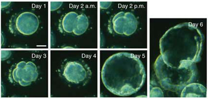
Movie shows in vitro development of a human morula to blastocyst stage occurring between day 3 and day 6 post-fertilization.
| <html5media height="470" width="500">File:Human_blastocyst_day_3-6.mp4</html5media> | Timing shown below are "movie times" not the "developmental days".
00:00 day 3 - Initially a solid mass of cells morula stage enclosed by the zona pellucida. 00:18 day 4 - blastocoel formation 00:22 - inner cell mass (embryoblast) 00:29 - maximal expansion of blastocyst in ZP 00:31 - contraction 00:32 - re-expansion 00:37 day 5 - contraction 00:38 - re-expansion 00:39 - contraction (collapse) 00:40 - re-expansion (blastocoel enlargement) 00:41 - commence blastocyst hatching (1 o'clock position) 00:44 day 6 - complete hatching, blastocyst expansion (inner cell mass 2 o'clock position) 00:47 - trophoblast layer thickens 00:53 movie finishes |
Blastocyst Hatching
By the end of the first week the blastocyst now consists of a ball of cells containing a large hollow fluid-filled (blastoceol) space.
Trophoblast Layer - Continued expansion of the blastoceol and cell division has led to the single layer of cells located at the periphery now being pressed against the inflexible zona pellucida wall and becoming flattened (squamous). This peripheral layer of cells is now called the trophoblast layer. Later in development this later will be involved in implantation and form part of the placenta.
Inner Cell Mass - On one wall of the blastoceol there is present a second layer of non-flattened cells. These inner cells are called the inner cell mass. Later in development from this layer the embryo will be formed.
Hatching - a combination of lysins (from the blastocyst or the uterus) and physical expansion, reduces the thickness and weakens the zona pelludica wall in preparation for hatching. Typically the blastocyst will hatch through a small opening (potentially at the site of fertilization) in the zona pellucida. The blastocyst is now ready to begin implantation.
Assisted Hatching - in vitro fertilization techniques use zona thinning, zona drilling, zona slitting or laser-assisted hatching to help blastocyst hatching and increase the probability of implantation occurring.

Zona pellucida (left) with Blastocyst (right) hatching through a small opening in the wall. (More? Carnegie stage 3)
Links: MBoC - Mouse Blastocyst Development
Early Cell Division Interactive Component
| Attempt the Quiz - Early Cell Division |
|---|

Here are a few simple Quiz questions that relate to Early Cell Division from the lecture and practical. See your Quiz Result - Answer all the questions, then click "submit" to complete. The page will reload and you can then reopen this table to see your result and feedback.
|
Additional Information
| Additional Information - Content shown under this heading is not part of the material covered in this class. It is provided for those students who would like to know about some concepts or current research in topics related to the current class page. |
| <html5media height="480" width="380">File:Mouse_zygote_division_02.mp4</html5media> | <html5media height="480" width="400">File:Mouse_zygote_division.mp4</html5media> |
| Movie shows mitotic division of the early mouse embryo starting at the zygote stage. | Hayashi-Takanaka Y, Yamagata K, Nozaki N & Kimura H. (2009). Visualizing histone modifications in living cells: spatiotemporal dynamics of H3 phosphorylation during interphase. J. Cell Biol. , 187, 781-90. PMID: 19995936 DOI. |
Errors in Mitosis
McCoy RC. (2017). Mosaicism in Preimplantation Human Embryos: When Chromosomal Abnormalities Are the Norm. Trends Genet. , 33, 448-463. PMID: 28457629 DOI.
- "Along with errors in meiosis, mitotic errors during post-zygotic cell division contribute to pervasive aneuploidy in human embryos. Relatively little is known, however, about the genesis of these errors or their fitness consequences. Rapid technological advances are helping to close this gap, revealing diverse molecular mechanisms contributing to mitotic error. These include altered cell cycle checkpoints, aberrations of the centrosome, and failed chromatid cohesion, mirroring findings from cancer biology. Recent studies are challenging the idea that mitotic error is abnormal, emphasizing that the fitness impacts of mosaicism depend on its scope and severity. In light of these findings, technical and philosophical limitations of various screening approaches are discussed, along with avenues for future research."
Dizygotic Twinning
Dizygotic twins (fraternal, non-identical) arise from separate fertilization events involving two separate oocyte (egg, ova) and spermatozoa (sperm).
Blastomere Isolation

|
This is early morula stage still enclosed by the zona pellucida.
One of the new methods for generating embryonic stem cells (ESCs) involves using these single blastomere cells. |
Blastocyst Mechanics
A recent review{{#pmid28681376|PMID28681376}} of the initial morula to blastocyst formation, based upon animal models, identifies important mechanical steps:
- Compaction - morula blastomeres packing tightly (microfilament cytoskeleton)
- Cleavage planes - spindle direction of dividing cells (mitosis).
- Polarisation - blastomeres apico-basal (Hippo pathway, Yes-associated protein (Yap)
- Cavitation - blastocoel formation with cycles of cavity expansion and collapse.
- epiblast cells - contact the polar trophectoderm
- primitive endoderm - facing the cavity
Terms
- bilaminar- having 2 layers, usually referring to the epiblast and hypoblast layers that appear in week 2 of development.
- blastocyst - the developmental stage following morula, as this stage matures, the zona pellucia is lost allowing the conceptus to adplant and then implant into the uterine wall.
- blastomeres - the cells resulting from the initial rounds of mitotic division of the zygote. These cells become smaller (in cytoplasmic volume) with each division.
- corona radiata - layer of follicle cells of cumulus oophorus remaining attached to zona pellucida of oocyte after ovulation.
- inner cell mass - the clump of cells found inside the blastocyst. These cells will go in to form the embryo, these are the "stem cells" (we here about in the media) that are totipotential, they can form any tissue in the embryo. Mature oocyte-the female germ cell released at ovulation from the ovary.
- morula - (L. morus = mulberry) early stage of development (12-15 cells) Followed by formation of a cavity in the mass (blastocyst stage). (More? Week 1)
- parental genomes - the male (sperm) and female (oocyte) DNA which contributes to the embryo's cells.
- polar body - 3 exclusion bodies formed that contain the DNA not used by the embryo. Contributed to initially by the meiotic division of the oocyte.
- pronuclei - the male (sperm) and female (oocyte) nuclei within the fertilized oocyte, prior to their combination to form the new embryo's nuclei.
- trilaminar embryonic disc- the 3 layered embryo stage (ectoderm, mesoderm, endoderm) that appear in week 3 of development.
- trophoblast - (Gr. trophe = nutrition) outer layer of cells on blastocyst that will generate the embryonic part of the placenta.
- uterine wall - the site of normal blastocyst implantation.
- zona pellucida - glycoprotein shell that surrounds the oocyte through to blastula stage of development.
- zygote - The first cell stage following fertilization of the oocyte by the sperm. This is the first cell of the conceptus which will divide into blastomeres.
References
Online Resources
BGDA: Lecture 1 | Lecture 2 | Practical 3 | Practical 6 | Practical 12 | Lecture Neural | Practical 14 | Histology Support - Female | Male | Tutorial
Glossary Links
- Glossary: A | B | C | D | E | F | G | H | I | J | K | L | M | N | O | P | Q | R | S | T | U | V | W | X | Y | Z | Numbers | Symbols | Term Link
Cite this page: Hill, M.A. (2024, May 21) Embryology BGDA Practical 3 - Early Cell Division. Retrieved from https://embryology.med.unsw.edu.au/embryology/index.php/BGDA_Practical_3_-_Early_Cell_Division
- © Dr Mark Hill 2024, UNSW Embryology ISBN: 978 0 7334 2609 4 - UNSW CRICOS Provider Code No. 00098G