Category:Fetal
This Embryology category shows pages and media related to fetal development.
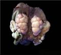
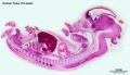
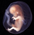
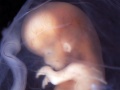
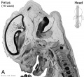
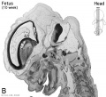
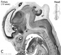
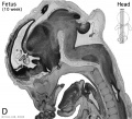
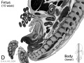
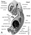
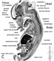
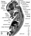
In human development the fetal period occurs after the end of the embryonic period at post-fertilisation week 8 or GA week 10. Late first and the entire second and third trimester are the fetal development period.
| Fetal Links: fetal | Week 10 | Week 12 | second trimester | third trimester | fetal neural | Fetal Blood Sampling | fetal growth restriction | birth | birth weight | preterm birth | Developmental Origins of Health and Disease | macrosomia | BGD Practical | Medicine Lecture | Science Lecture | Lecture Movie | Category:Human Fetus | Category:Fetal | |||
|
| Links: human timeline | first trimester timeline | second trimester timeline | third trimester timeline | ||
| Event | ||
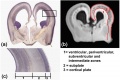
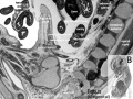
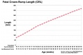
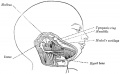
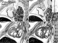
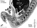
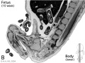
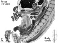
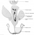
| Clinical second trimester |  Week 12 - CRL 85 mm, femur length 15 mm, biparietal diameter 25 mm Week 12 - CRL 85 mm, femur length 15 mm, biparietal diameter 25 mm
Hearing Week 12-16 - Capsule adjacent to membranous labrynth undegoes vacuolization to form a cavity (perilymphatic space) around membranous labrynth and fills with perilymph
Respiratory Month 3-6 - lungs appear glandular, end month 6 alveolar cells type 2 appear and begin to secrete surfactant Tongue Week 12 - first differentiated epithelial cells (Type II and III) Genital female genital canal (80 days) formed with absorption of the median septum | |
| tongue Week 12 to 13 - maximum synapses between cells and afferent nerve fibers
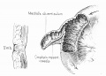
hearing outer ear Week 13 - Meatal plug disc-like, innermost surface in contact with the primordial malleus, contributes to the formation of the tympanic membrane. | ||
| tongue Week 14 to 15 - taste pores develop, mucous
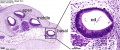
ovary 100 days - primary follicles present nail toenails appear Head Development facial skeleton remodelling begins Hearing - Inner Ear Development Week 14 GA 16 - neural-crest-derived melanocytes, now intermediate cells of the stria vascularis, tightly integrate with Na+ /K+ -ATPase-positive marginal cells, which started to express KCNQ1 in their apical membrane.[1] | ||
| Pancreas glucagon detectable in fetal plasma.
spleen Week 15 -alpha-smooth muscle actin (alpha-SMA)-positive reticulum cells scattered around the arterioles.[2] | ||
| 14 cm | 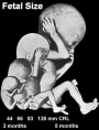 Hearing Week 16-24 - Centres of ossification appear in remaining cartilage of otic capsule form petrous portion of temporal bone. Continues to ossify to form mastoid process of temporal bone. Hearing Week 16-24 - Centres of ossification appear in remaining cartilage of otic capsule form petrous portion of temporal bone. Continues to ossify to form mastoid process of temporal bone.
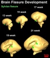
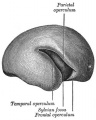
pituitary adenohypophysis fully differentiated respiratory Week 16 to 25 lung histology - canalicular Hearing - Outer Ear Development Week 16.5 - External auditory meatus is fully patent throughout its length, lumen is still narrow and curved. Hearing - Inner Ear Development Week 16 GA 18 - cells in the outer sulcus express KCNJ10 and gap junction proteins GJB2/CX26 and GJB6/CX30, but these are not expressed in the spiral ligament.[1] gap junction cartoon neural - Cerebrum development of the periinsular sulci (week 16-17, GA 18-19 weeks)[3]
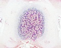
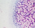
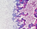
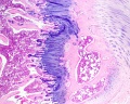
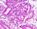
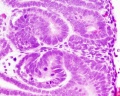
primary follicles begin to form in the ovary and are characterized by an oocyte glandular urethra forms and skin folds present | |
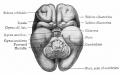
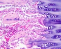
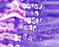
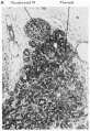
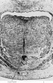
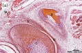
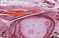
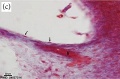
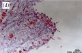
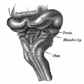
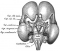
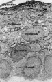
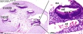
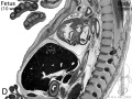
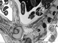
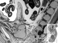
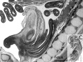
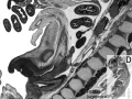
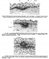
 Neural - Brain development histology week 17 Neural - Brain development histology week 17
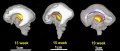
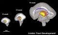
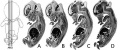
Cerebellum Magnetic Resonance Imaging (MRI) can study the developing cerebellum from 17 to 18 weeks (GA 19 to 20 weeks). tooth Week 17 - First papilla of the permanent dentition appear (first molar) immediately behind the second milk molar, milk teeth are well advanced (Fetus 180 mm). | ||
 tongue Week 18 - substance P detected in dermal papillae, not in taste bud primordia tongue Week 18 - substance P detected in dermal papillae, not in taste bud primordia
integumentary vernix caseosa covers skin spleen Week 18 - alpha-SMA-positive reticulum cells increase in number and began to form a reticular framework. An accumulation of T and B lymphocytes occurred within the framework, and a primitive white pulp was observed around the arterioles.[2] Hearing - Outer Ear Development week 18 - External auditory meatus is already fully expanded to its complete form. neural - Cerebrum central sulci and opercularization of the insula (week 18-20, GA 20-22 weeks)[3] | ||
| neural week 19 neuronal migration ends and the radial glial cells that aided the migration now become transformed into astrocytes and astrocytic precursors.[4] | ||
| pituitary week 20 to 24 growth hormone levels peak, then decline
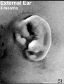
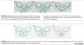
integumentary lanugo, skin hair integumentary 5 months - Hair growth initiated at base of cord, lateral outgrowths form associated sebaceous glands; Other cords elongate and coil to form sweat glands; Cords in mammary region branch as they elongate to form mammary glands. touch pacinian corpuscle begin to develop[5] | ||
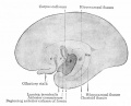
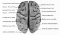
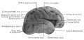
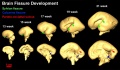
 Neural brain cortical sulcation - sylvian fissure, interhemispheric fissure, callosal sulcus, parietooccipital fissure, and hippocampic fissures present[6] Neural brain cortical sulcation - sylvian fissure, interhemispheric fissure, callosal sulcus, parietooccipital fissure, and hippocampic fissures present[6]
spleen - Week 22 - antigenic diversity of the reticular framework was observed, and T and B lymphocytes were segregated in the framework. T lymphocytes were sorted into the alpha-smooth muscle actin-positive reticular framework, and the periarteriolar lymphoid sheath (PALS) was formed around the arteriole. B lymphocytes aggregated in eccentric portions to the PALS and formed the lymph follicle (LF). The reticular framework of the LF was alpha-SMA-negative. [2] neural - Cerebrum covering of the posterior insula (week 22-24, GA 24-26 weeks)[3] | ||
| respiratory Week 24 to 40 lung histology - terminal sac
spleen Week 24 - marginal zone appeared in the alpha-smooth muscle actin-positive reticular framework around the white pulp.[2] tooth Week 24 - Permanent incisors and canines appear. Earliest potential survival expected if born ovary follicles can consist of growing oocytes surrounded by several layers of granulosa cells | ||
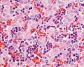
| respiratory end month 6 alveolar cells type 2 appear and begin to secrete surfactant
neural - Cerebrum closure of the laeteral sulcus (Sylvian fissure or lateral fissure) (week 25-26, GA 27-28 weeks)[3] | ||
| touch pacinian corpuscle well developed[5] | ||
| Links: human timeline | first trimester timeline | second trimester timeline | third trimester timeline | ||
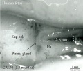
| Event | ||
| Clinical third trimester | hearing 3rd Trimester - vibration acoustically of maternal abdominal wall induces startle respone in fetus.
| |
| respiratory Month 7 - respiratory bronchioles proliferate and end in alveolar ducts and sacs | ||
|
tooth Week 29 - Permanent premolars (correspond to the milk molars) appear. | ||
|
Genital male gonad (testes) descending | ||
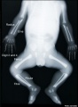
| nail fingernails reach digit tip | ||
| neural brain cortical sulcation - primary sulci present[6] | ||
| neural brain cortical sulcation - insular, cingular, and occipital secondary sulci present[6] | ||
 Nail Development toenails reach digit tip Nail Development toenails reach digit tip
Lens Development - lens growth and interocular distance plateaus after 36 weeks of gestation[7] | ||
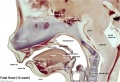
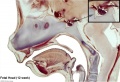
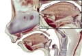
| Birth |  Clinical Week 40 Clinical Week 40
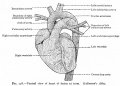
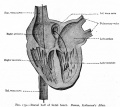
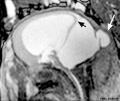
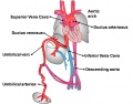
Heart pressure difference closes foramen ovale leaving a fossa ovalis thyroid TSH levels increase, thyroxine (T3) and T4 levels increase to 24 h, then 5-7 days postnatal decline to normal levels adrenal - zona glomerulosa, zona fasiculata present | |
| Carnegie Collection - Fetal | |||||||||||||||||||||||||||||||||||||||||||||||||||||||||||||||||||||||||||||||||||||||||||||||||||||||||||||||||||||||||||||||||||||||||||||||||||||||||||||||||||||||||||||||||||||||||||||||||||||||||||||||||||||||||
|---|---|---|---|---|---|---|---|---|---|---|---|---|---|---|---|---|---|---|---|---|---|---|---|---|---|---|---|---|---|---|---|---|---|---|---|---|---|---|---|---|---|---|---|---|---|---|---|---|---|---|---|---|---|---|---|---|---|---|---|---|---|---|---|---|---|---|---|---|---|---|---|---|---|---|---|---|---|---|---|---|---|---|---|---|---|---|---|---|---|---|---|---|---|---|---|---|---|---|---|---|---|---|---|---|---|---|---|---|---|---|---|---|---|---|---|---|---|---|---|---|---|---|---|---|---|---|---|---|---|---|---|---|---|---|---|---|---|---|---|---|---|---|---|---|---|---|---|---|---|---|---|---|---|---|---|---|---|---|---|---|---|---|---|---|---|---|---|---|---|---|---|---|---|---|---|---|---|---|---|---|---|---|---|---|---|---|---|---|---|---|---|---|---|---|---|---|---|---|---|---|---|---|---|---|---|---|---|---|---|---|---|---|---|---|---|---|---|
| Serial No. | Size CRL (mm) | Grade | Fixative | Embedding Medium | Plane | Thinness (µm) | Stain | Point Score | Sex | Year | Notes | ||||||||||||||||||||||||||||||||||||||||||||||||||||||||||||||||||||||||||||||||||||||||||||||||||||||||||||||||||||||||||||||||||||||||||||||||||||||||||||||||||||||||||||||||||||||||||||||||||||||||||||||
| 95 | 40 | catalogued as CRL 40 but development suggests 50 stage. Spinal cord - Kunitomo (1920)[8] Colon - Lineback (1920)[9] | |||||||||||||||||||||||||||||||||||||||||||||||||||||||||||||||||||||||||||||||||||||||||||||||||||||||||||||||||||||||||||||||||||||||||||||||||||||||||||||||||||||||||||||||||||||||||||||||||||||||||||||||||||||||
| 96 | 50 | Brain venous sinuses - Streeter (1915)[10] Spinal cord - Kunitomo (1920)[8] Brain vascular - Streeter (1921)[11] Brain weight - Jenkins (1921)[12] | |||||||||||||||||||||||||||||||||||||||||||||||||||||||||||||||||||||||||||||||||||||||||||||||||||||||||||||||||||||||||||||||||||||||||||||||||||||||||||||||||||||||||||||||||||||||||||||||||||||||||||||||||||||||
| 142 | 125 | Spinal cord - Kunitomo (1920)[8] | |||||||||||||||||||||||||||||||||||||||||||||||||||||||||||||||||||||||||||||||||||||||||||||||||||||||||||||||||||||||||||||||||||||||||||||||||||||||||||||||||||||||||||||||||||||||||||||||||||||||||||||||||||||||
| 145 | 33 | Spinal cord - Kunitomo (1920)[8] | |||||||||||||||||||||||||||||||||||||||||||||||||||||||||||||||||||||||||||||||||||||||||||||||||||||||||||||||||||||||||||||||||||||||||||||||||||||||||||||||||||||||||||||||||||||||||||||||||||||||||||||||||||||||
| 184 | 50 | 34 vertebrae, 31 spinal ganglia, Spinal cord - Kunitomo (1920)[8] | |||||||||||||||||||||||||||||||||||||||||||||||||||||||||||||||||||||||||||||||||||||||||||||||||||||||||||||||||||||||||||||||||||||||||||||||||||||||||||||||||||||||||||||||||||||||||||||||||||||||||||||||||||||||
| 211 | 33 | 34 vertebra, 31 spinal ganglia, Spinal cord - Kunitomo (1920)[8] | |||||||||||||||||||||||||||||||||||||||||||||||||||||||||||||||||||||||||||||||||||||||||||||||||||||||||||||||||||||||||||||||||||||||||||||||||||||||||||||||||||||||||||||||||||||||||||||||||||||||||||||||||||||||
| 217 | 45 | Male | Genital - Spaulding (1921)[13] | ||||||||||||||||||||||||||||||||||||||||||||||||||||||||||||||||||||||||||||||||||||||||||||||||||||||||||||||||||||||||||||||||||||||||||||||||||||||||||||||||||||||||||||||||||||||||||||||||||||||||||||||||||||||
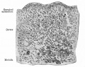
| 300 | 73 | 85 days, Bone ossification - Mall (1906)[14] | |||||||||||||||||||||||||||||||||||||||||||||||||||||||||||||||||||||||||||||||||||||||||||||||||||||||||||||||||||||||||||||||||||||||||||||||||||||||||||||||||||||||||||||||||||||||||||||||||||||||||||||||||||||||
| 362 | 30 | Spinal cord - Kunitomo (1920)[8] | |||||||||||||||||||||||||||||||||||||||||||||||||||||||||||||||||||||||||||||||||||||||||||||||||||||||||||||||||||||||||||||||||||||||||||||||||||||||||||||||||||||||||||||||||||||||||||||||||||||||||||||||||||||||
| 448 | 52 | Colon - Lineback (1920)[9] | |||||||||||||||||||||||||||||||||||||||||||||||||||||||||||||||||||||||||||||||||||||||||||||||||||||||||||||||||||||||||||||||||||||||||||||||||||||||||||||||||||||||||||||||||||||||||||||||||||||||||||||||||||||||
| 449 | 36 | Spinal cord - Kunitomo (1920)[8] | |||||||||||||||||||||||||||||||||||||||||||||||||||||||||||||||||||||||||||||||||||||||||||||||||||||||||||||||||||||||||||||||||||||||||||||||||||||||||||||||||||||||||||||||||||||||||||||||||||||||||||||||||||||||
| 538 | |||||||||||||||||||||||||||||||||||||||||||||||||||||||||||||||||||||||||||||||||||||||||||||||||||||||||||||||||||||||||||||||||||||||||||||||||||||||||||||||||||||||||||||||||||||||||||||||||||||||||||||||||||||||||
| 590 | 21 to 23 | Male | Genital - Spaulding (1921)[13] | ||||||||||||||||||||||||||||||||||||||||||||||||||||||||||||||||||||||||||||||||||||||||||||||||||||||||||||||||||||||||||||||||||||||||||||||||||||||||||||||||||||||||||||||||||||||||||||||||||||||||||||||||||||||
| 607 | 37 | Male | Genital - Spaulding (1921)[13] | ||||||||||||||||||||||||||||||||||||||||||||||||||||||||||||||||||||||||||||||||||||||||||||||||||||||||||||||||||||||||||||||||||||||||||||||||||||||||||||||||||||||||||||||||||||||||||||||||||||||||||||||||||||||
| 625 | 220 | Temporomandibular joint - Moffatt (1957)[15] | |||||||||||||||||||||||||||||||||||||||||||||||||||||||||||||||||||||||||||||||||||||||||||||||||||||||||||||||||||||||||||||||||||||||||||||||||||||||||||||||||||||||||||||||||||||||||||||||||||||||||||||||||||||||
| 662 | 80 | Spinal cord - Kunitomo (1920)[8] | |||||||||||||||||||||||||||||||||||||||||||||||||||||||||||||||||||||||||||||||||||||||||||||||||||||||||||||||||||||||||||||||||||||||||||||||||||||||||||||||||||||||||||||||||||||||||||||||||||||||||||||||||||||||
| 693 | 45 | Male | Genital - Spaulding (1921)[13] | ||||||||||||||||||||||||||||||||||||||||||||||||||||||||||||||||||||||||||||||||||||||||||||||||||||||||||||||||||||||||||||||||||||||||||||||||||||||||||||||||||||||||||||||||||||||||||||||||||||||||||||||||||||||
| 847 | 58.8 | Male | Genital - Spaulding (1921)[13] | ||||||||||||||||||||||||||||||||||||||||||||||||||||||||||||||||||||||||||||||||||||||||||||||||||||||||||||||||||||||||||||||||||||||||||||||||||||||||||||||||||||||||||||||||||||||||||||||||||||||||||||||||||||||
| 858 | 57.25 | Temporomandibular joint - Moffatt (1957)[15] | |||||||||||||||||||||||||||||||||||||||||||||||||||||||||||||||||||||||||||||||||||||||||||||||||||||||||||||||||||||||||||||||||||||||||||||||||||||||||||||||||||||||||||||||||||||||||||||||||||||||||||||||||||||||
| 922 | 37 | ||||||||||||||||||||||||||||||||||||||||||||||||||||||||||||||||||||||||||||||||||||||||||||||||||||||||||||||||||||||||||||||||||||||||||||||||||||||||||||||||||||||||||||||||||||||||||||||||||||||||||||||||||||||||
| 928 | 120 | Spinal cord - Kunitomo (1920)[8] | |||||||||||||||||||||||||||||||||||||||||||||||||||||||||||||||||||||||||||||||||||||||||||||||||||||||||||||||||||||||||||||||||||||||||||||||||||||||||||||||||||||||||||||||||||||||||||||||||||||||||||||||||||||||
| 948 | 45 | Male | Genital - Spaulding (1921)[13] | ||||||||||||||||||||||||||||||||||||||||||||||||||||||||||||||||||||||||||||||||||||||||||||||||||||||||||||||||||||||||||||||||||||||||||||||||||||||||||||||||||||||||||||||||||||||||||||||||||||||||||||||||||||||
| 972 | 37 | 34 vertebrae, 30 spinal ganglia, Spinal cord - Kunitomo (1920)[8] | |||||||||||||||||||||||||||||||||||||||||||||||||||||||||||||||||||||||||||||||||||||||||||||||||||||||||||||||||||||||||||||||||||||||||||||||||||||||||||||||||||||||||||||||||||||||||||||||||||||||||||||||||||||||
| 1318 | 37 | Temporomandibular joint - Moffatt (1957)[15] | |||||||||||||||||||||||||||||||||||||||||||||||||||||||||||||||||||||||||||||||||||||||||||||||||||||||||||||||||||||||||||||||||||||||||||||||||||||||||||||||||||||||||||||||||||||||||||||||||||||||||||||||||||||||
| 1388 | 51 | Female | Genital - Spaulding (1921)[13] | ||||||||||||||||||||||||||||||||||||||||||||||||||||||||||||||||||||||||||||||||||||||||||||||||||||||||||||||||||||||||||||||||||||||||||||||||||||||||||||||||||||||||||||||||||||||||||||||||||||||||||||||||||||||
| 1455 | 78.5 | Temporomandibular joint - Moffatt (1957)[15] | |||||||||||||||||||||||||||||||||||||||||||||||||||||||||||||||||||||||||||||||||||||||||||||||||||||||||||||||||||||||||||||||||||||||||||||||||||||||||||||||||||||||||||||||||||||||||||||||||||||||||||||||||||||||
| 1591 | 36 | subcutaneous vascular plexus - Finley (1923)[16] | |||||||||||||||||||||||||||||||||||||||||||||||||||||||||||||||||||||||||||||||||||||||||||||||||||||||||||||||||||||||||||||||||||||||||||||||||||||||||||||||||||||||||||||||||||||||||||||||||||||||||||||||||||||||
| 1656 | 67 | 34 vertebrae, Spinal cord - Kunitomo (1920)[8] | |||||||||||||||||||||||||||||||||||||||||||||||||||||||||||||||||||||||||||||||||||||||||||||||||||||||||||||||||||||||||||||||||||||||||||||||||||||||||||||||||||||||||||||||||||||||||||||||||||||||||||||||||||||||
| 1686 | 40 | Male | Genital - Spaulding (1921)[13] | ||||||||||||||||||||||||||||||||||||||||||||||||||||||||||||||||||||||||||||||||||||||||||||||||||||||||||||||||||||||||||||||||||||||||||||||||||||||||||||||||||||||||||||||||||||||||||||||||||||||||||||||||||||||
| 3990 | 49 | Temporomandibular joint - Moffatt (1957)[15] | |||||||||||||||||||||||||||||||||||||||||||||||||||||||||||||||||||||||||||||||||||||||||||||||||||||||||||||||||||||||||||||||||||||||||||||||||||||||||||||||||||||||||||||||||||||||||||||||||||||||||||||||||||||||
| 4473 | 43 | 20 | Spinal cord meninges - Sensenig (1951)[17] | ||||||||||||||||||||||||||||||||||||||||||||||||||||||||||||||||||||||||||||||||||||||||||||||||||||||||||||||||||||||||||||||||||||||||||||||||||||||||||||||||||||||||||||||||||||||||||||||||||||||||||||||||||||||
| 4475 | 48 | 20 | Spinal cord meninges - Sensenig (1951)[17] | ||||||||||||||||||||||||||||||||||||||||||||||||||||||||||||||||||||||||||||||||||||||||||||||||||||||||||||||||||||||||||||||||||||||||||||||||||||||||||||||||||||||||||||||||||||||||||||||||||||||||||||||||||||||
| 5652 | 49 | Temporomandibular joint - Moffatt (1957)[15] | |||||||||||||||||||||||||||||||||||||||||||||||||||||||||||||||||||||||||||||||||||||||||||||||||||||||||||||||||||||||||||||||||||||||||||||||||||||||||||||||||||||||||||||||||||||||||||||||||||||||||||||||||||||||
| 6581 | 75 | Temporomandibular joint - Moffatt (1957)[15] | |||||||||||||||||||||||||||||||||||||||||||||||||||||||||||||||||||||||||||||||||||||||||||||||||||||||||||||||||||||||||||||||||||||||||||||||||||||||||||||||||||||||||||||||||||||||||||||||||||||||||||||||||||||||
| 7218 | 80 | 20 um | Spinal cord meninges - Sensenig (1951)[17] | ||||||||||||||||||||||||||||||||||||||||||||||||||||||||||||||||||||||||||||||||||||||||||||||||||||||||||||||||||||||||||||||||||||||||||||||||||||||||||||||||||||||||||||||||||||||||||||||||||||||||||||||||||||||
| 1597b | 47 | Female | Genital - Spaulding (1921)[13] | ||||||||||||||||||||||||||||||||||||||||||||||||||||||||||||||||||||||||||||||||||||||||||||||||||||||||||||||||||||||||||||||||||||||||||||||||||||||||||||||||||||||||||||||||||||||||||||||||||||||||||||||||||||||
| 2250a | 40 | Female | Genital - Spaulding (1921)[13] | ||||||||||||||||||||||||||||||||||||||||||||||||||||||||||||||||||||||||||||||||||||||||||||||||||||||||||||||||||||||||||||||||||||||||||||||||||||||||||||||||||||||||||||||||||||||||||||||||||||||||||||||||||||||
| 2250b | 36 | Female | Genital - Spaulding (1921)[13] | ||||||||||||||||||||||||||||||||||||||||||||||||||||||||||||||||||||||||||||||||||||||||||||||||||||||||||||||||||||||||||||||||||||||||||||||||||||||||||||||||||||||||||||||||||||||||||||||||||||||||||||||||||||||
| This table currently contains only has embryo number information.
Abbreviations
| |||||||||||||||||||||||||||||||||||||||||||||||||||||||||||||||||||||||||||||||||||||||||||||||||||||||||||||||||||||||||||||||||||||||||||||||||||||||||||||||||||||||||||||||||||||||||||||||||||||||||||||||||||||||||
References
| |||||||||||||||||||||||||||||||||||||||||||||||||||||||||||||||||||||||||||||||||||||||||||||||||||||||||||||||||||||||||||||||||||||||||||||||||||||||||||||||||||||||||||||||||||||||||||||||||||||||||||||||||||||||||
| |||||||||||||||||||||||||||||||||||||||||||||||||||||||||||||||||||||||||||||||||||||||||||||||||||||||||||||||||||||||||||||||||||||||||||||||||||||||||||||||||||||||||||||||||||||||||||||||||||||||||||||||||||||||||
Subcategories
This category has the following 71 subcategories, out of 71 total.
C
- Carnegie Embryo 1161
- Carnegie Embryo 1163
- Carnegie Embryo 1183
- Carnegie Embryo 131
- Carnegie Embryo 1318
- Carnegie Embryo 1388
- Carnegie Embryo 142
- Carnegie Embryo 145
- Carnegie Embryo 1455
- Carnegie Embryo 1474b
- Carnegie Embryo 1591
- Carnegie Embryo 1597b
- Carnegie Embryo 1656
- Carnegie Embryo 1686
- Carnegie Embryo 1705a
- Carnegie Embryo 172
- Carnegie Embryo 184
- Carnegie Embryo 1852
- Carnegie Embryo 2026
- Carnegie Embryo 211
- Carnegie Embryo 217
- Carnegie Embryo 2250a
- Carnegie Embryo 2250b
- Carnegie Embryo 23
- Carnegie Embryo 234a
- Carnegie Embryo 300
- Carnegie Embryo 34
- Carnegie Embryo 362
- Carnegie Embryo 3990
- Carnegie Embryo 4473
- Carnegie Embryo 4475
- Carnegie Embryo 448
- Carnegie Embryo 449
- Carnegie Embryo 458
- Carnegie Embryo 48
- Carnegie Embryo 538
- Carnegie Embryo 5652
- Carnegie Embryo 590
- Carnegie Embryo 607
- Carnegie Embryo 625
- Carnegie Embryo 6581
- Carnegie Embryo 662
- Carnegie Embryo 693
- Carnegie Embryo 7218
- Carnegie Embryo 834
- Carnegie Embryo 847
- Carnegie Embryo 858
- Carnegie Embryo 922
- Carnegie Embryo 928
- Carnegie Embryo 948
- Carnegie Embryo 95
- Carnegie Embryo 96
- Carnegie Embryo 972
Pages in category 'Fetal'
The following 200 pages are in this category, out of 270 total.
(previous page) (next page)B
- BGDA Practical - Fetal Development
- Talk:BGDA Practical - Fetal Development
- BGDA Practical - Fetal Development - Quiz
- BGDA Practical - Fetal Development Interactive
- BGDA Practical 12 - Abnormalities
- Template:BGDA Practical 12 - Abnormalities Interactive
- BGDA Practical 12 - Birth
- Template:BGDA Practical 12 - Birth Interactive
- BGDA Practical 12 - Embryo to Fetus
- Template:BGDA Practical 12 - Embryo to Fetus Interactive
- BGDA Practical 12 - Neonatal
- Template:BGDA Practical 12 - Neonatal Interactive
- BGDA Practical 12 - Second Trimester
- Template:BGDA Practical 12 - Second Trimester Interactive
- BGDA Practical 12 - Third Trimester
- Template:BGDA Practical 12 - Third Trimester Interactive
- Template:BGDALab12
- BGDB Gastrointestinal - Fetal
- Template:BGDB Practical 6 - Abnormalities Interactive
- Template talk:BGDB Practical 6 - Abnormalities Interactive
- Template:BGDB Practical 6 - Fetal Interactive
- Template:BGDB Practical 6 - Postnatal Interactive
- Template:BGDB Sexual Differentiation - Fetal Interactive
- Template:Birth weight
- Template:Birthweight
- Book - Human embryos of different ages examined in median sections - a contribution to the mechanics of development
- Book - Physiology of the Fetus
- Book - Physiology of the Fetus 1
- Book - Physiology of the Fetus 10
- Book - Physiology of the Fetus 11
- Book - Physiology of the Fetus 12
- Book - Physiology of the Fetus 13
- Book - Physiology of the Fetus 14
- Book - Physiology of the Fetus 15
- Book - Physiology of the Fetus 16
- Book - Physiology of the Fetus 2
- Book - Physiology of the Fetus 3
- Book - Physiology of the Fetus 4
- Book - Physiology of the Fetus 5
- Book - Physiology of the Fetus 6
- Book - Physiology of the Fetus 7
- Book - Physiology of the Fetus 8
- Book - Physiology of the Fetus 9
C
- Template:Canalicular stage
- Template:Carnegie Collection fetal table
- Template talk:Carnegie Collection fetal table
- Template:CE1161
- Template:CE1206
- Template:CE1282b
- Template:CE131
- Template:CE1318
- Template:CE1358
- Template:CE1388
- Template:CE142
- Template:CE1449
- Template:CE145
- Template:CE1455
- Template:CE1474b
- Template:CE1476
- Template:CE1561
- Template:CE1591
- Template:CE1597b
- Template:CE1656
- Template:CE1686
- Template:CE1702
- Template:CE1705a
- Template:CE1708
- Template:CE1716
- Template:CE1724
- Template:CE1742
- Template:CE1782
- Template:CE1811
- Template:CE1831
- Template:CE184
- Template:CE1840a
- Template:CE1845
- Template:CE1852
- Template:CE1858
- Template:CE190
- Template:CE1915
- Template:CE1980
- Template:CE2003
- Template:CE2066
- Template:CE2075
- Template:CE2079
- Template:CE2095
- Template:CE210
- Template:CE2118
- Template:CE2144
- Template:CE2163
- Template:CE217
- Template:CE2170
- Template:CE218
- Template:CE2185
- Template:CE219
- Template:CE2250a
- Template:CE2250b
- Template:CE2274
- Template:CE2328
- Template:CE234a
- Template:CE237
- Template:CE300
- Template:CE301
- Template:CE362
- Template:CE3669
- Template:CE389a
- Template:CE3990
- Template:CE4021
- Template:CE4229
- Template:CE4291
- Template:CE4473
- Template:CE4475
- Template:CE448
- Template:CE449
- Template:CE458
- Template:CE481
- Template:CE484
- Template:CE490
- Template:CE491
- Template:CE4985
- Template:CE508
- Template:CE509
- Template:CE538
- Template:CE5652
- Template:CE5712
- Template:CE5762
- Template:CE607
- Template:CE625
- Template:CE642
- Template:CE6581
- Template:CE662
- Template:CE6658
- Template:CE693
- Template:CE7218
- Template:CE834
- Template:CE84
- Template:CE847
- Template:CE858
- Template:CE886
- Template:CE922
- Template:CE928
- Template:CE948
- Template:CE95
- Template:CE9526
- Template:CE96
- Template:CE972
- Template:CE981
F
H
P
- Paper - A quantitative study of the fetal growth changes in the parts of the human stomach wall
- Paper - Blood supply of the otic capsule of a 150 mm (C.R.) human fetus
- Paper - Cell columns in the spinal cord of a human foetus of fourteen weeks (1941)
- Paper - Early fetal activity in mammals
- Paper - Fetal growth in man
- Paper - Ossification of the otic capsule in human fetuses
- Paper - Perichondrial ossification and the fate of the perichondrium with special reference to that of the otic capsule
- Paper - Significant superficial anastomoses in the arterial blood supply to the human brain (1959)
- Paper - Simple methods of correlating crown-rump and crown-heel lengths of the human fetus
- Paper - Some Gross Structural and Quantitative Aspects of the Developmental Anatomy of the Human Embryonic, Fetal and Circumnatal Skeleton
- Paper - The arrangement of the bursae in the superior extremities of the full-time foetus
- Paper - The connexions of the posterior commissure in the human foetus and young infant
- Paper - The development of muscle in the human foetus
- Paper - The development of nerve endings in the human foetus
- Paper - The development of the human prostate gland with reference to the development of other structures at the neck of the urinary bladder (1912)
- Paper - The development of the islands of Langerhans in the human embryo (1903)
- Paper - The development of the seminal vesicles in man
- Paper - The Internal Genital Organs of a Female Foetus of 15 cm Length
- Paper - The physiological descent of the ovaries in the human foetus
- Paper - The right lung of a human foetus of 152 millimeters CRL
- Paper - The sexual differences of the fetal pelvis
- Paper - The sexual differences of the fetal pelvis (1899)
Media in category 'Fetal'
The following 200 files are in this category, out of 313 total.
(previous page) (next page)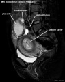 Abdominal ectopic pregnancy MRI.jpg 553 × 700; 45 KB
 Adrenal histology 005.jpg 1,280 × 1,024; 298 KB
 Adrenal histology 006.jpg 1,280 × 1,024; 313 KB
 Anencephaly ultrasound.jpg 900 × 658; 108 KB
 Anson1948 fig03.jpg 1,280 × 1,815; 358 KB
 Anson1948 fig07.jpg 1,280 × 1,801; 219 KB
 AnsonKarabinMartin1939 fig13-15.jpg 1,280 × 1,519; 171 KB
 Bailey094.jpg 376 × 689; 27 KB
 Bailey095.jpg 409 × 635; 52 KB
 Bailey113.jpg 813 × 553; 128 KB
 Bailey114.jpg 690 × 433; 63 KB
 Bailey141.jpg 761 × 323; 66 KB
 Bailey178.jpg 913 × 653; 125 KB
 Bailey179.jpg 892 × 794; 114 KB
 Bailey330.jpg 680 × 539; 109 KB
 Bailey332.jpg 637 × 356; 53 KB
 Bailey334.jpg 857 × 558; 77 KB
 Bailey356.jpg 767 × 533; 103 KB
 Bailey357.jpg 949 × 395; 88 KB
 Bailey443.jpg 666 × 539; 49 KB
 Bailey444.jpg 777 × 590; 127 KB
 Bailey448.jpg 722 × 416; 55 KB
 Bailey449.jpg 777 × 374; 45 KB
 Bailey450.jpg 680 × 419; 45 KB
 Bailey467.jpg 946 × 515; 159 KB
 Bast1931 plate01.jpg 1,280 × 871; 113 KB
 BeatonAnson1940 fig01-2.jpg 1,652 × 2,147; 652 KB
 BGDA2011-Embryo Lab 010611-01.mp3 ; 4.61 MB
- BGDA2011-Embryo Lab 010611-02.mp3 ; 3.23 MB
- BGDA2011-Embryo Lab 010611-03.mp3 ; 4.25 MB
- BGDA2011-Embryo Lab 010611-04.mp3 ; 1.79 MB
- BGDA2011-Embryo Lab 010611-05.mp3 ; 2.23 MB
 Bone histology 014.jpg 1,280 × 1,024; 541 KB
 Bone histology 015.jpg 1,280 × 1,024; 519 KB
 Bone histology 016.jpg 1,280 × 1,024; 379 KB
 Bone histology 017.jpg 1,280 × 1,024; 442 KB
 Bone histology 018.jpg 1,280 × 1,024; 336 KB
 Bone histology 019.jpg 1,280 × 1,024; 275 KB
 Bone histology 020.jpg 1,280 × 1,024; 272 KB
 Bone histology 021.jpg 1,280 × 1,024; 254 KB
 Boyd1950 fig03a.jpg 600 × 872; 135 KB
 Boyd1950 fig03b.jpg 600 × 996; 152 KB
 Boyd1950 fig04.jpg 800 × 703; 102 KB
 Boyd1950 fig06.jpg 1,000 × 1,002; 272 KB
 Boyd1950 fig08.jpg 1,000 × 1,553; 341 KB
 Boyd1950 fig09.jpg 801 × 900; 163 KB
 Boyd1950 fig10.jpg 1,000 × 453; 114 KB
 Brain fissure development 01.jpg 1,157 × 502; 47 KB
 Brain fissure development 02.jpg 1,000 × 583; 52 KB
 Brain fissure development 03.jpg 600 × 691; 33 KB
 Brain tract development 01.jpg 720 × 1,023; 76 KB
 Brain tract development 02.jpg 1,000 × 424; 29 KB
 Brain tract development 06.jpg 1,000 × 605; 35 KB
 Brain ventricles and ganglia development 01.jpg 994 × 564; 45 KB
 Brain ventricles and ganglia development 02.jpg 1,000 × 760; 49 KB
 Brain ventricles and ganglia development 03.jpg 989 × 583; 36 KB
 Brain week 17 histology.jpg 1,050 × 697; 61 KB
 Brain-tract-development-03.jpg 1,000 × 424; 31 KB
 Brain-tract-development-04.jpg 1,000 × 424; 30 KB
 Brain-tract-development-05.jpg 1,000 × 424; 29 KB
 Crowder1957 fig07.jpg 848 × 642; 300 KB
 Cullen1916 fig14.jpg 1,280 × 1,293; 434 KB
 Cullen1916 fig15.jpg 1,000 × 1,227; 314 KB
 Cullen1916 fig16.jpg 1,280 × 1,686; 621 KB
 Cullen1916 fig17.jpg 1,280 × 1,134; 365 KB
 Cullen1916 fig19.jpg 1,280 × 1,098; 330 KB
 Cullen1916 fig22.jpg 1,224 × 1,501; 324 KB
 Cullen1916 fig23.jpg 1,000 × 1,266; 346 KB
 Cullen1916 fig33.jpg 1,154 × 2,052; 422 KB
 Cullen1916 fig92.jpg 885 × 637; 109 KB
 Dandy Walker malformation MRI 01.jpg 600 × 506; 37 KB
 External Ear 5 months-icon.jpg 352 × 464; 63 KB
 Fawcett1911 fig06.jpg 1,000 × 680; 161 KB
 Fetal 10wk urogenital 1.jpg 800 × 600; 109 KB
 Fetal 10wk urogenital 2.jpg 800 × 600; 110 KB
 Fetal 10wk urogenital 3.jpg 800 × 600; 107 KB
 Fetal 10wk urogenital 4.jpg 800 × 600; 105 KB
 Fetal 5 month x-ray.jpg 1,500 × 2,298; 308 KB
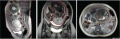 Fetal 9 month MRI 01.jpg 1,501 × 483; 145 KB
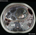 Fetal 9 month MRI 02.jpg 619 × 600; 67 KB
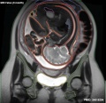 Fetal 9 month MRI 03.jpg 619 × 600; 64 KB
 Fetal 9 month MRI 04.jpg 619 × 600; 54 KB
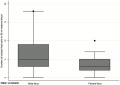 Fetal cells maternal blood graph.jpg 600 × 426; 18 KB
 Fetal Circulation Pathway.jpg 1,371 × 1,069; 112 KB
 Fetal corpus cavernosum and corpus spongiosum 01.jpg 1,795 × 2,082; 919 KB
 Fetal ductus venosus pressure wave 01.jpg 706 × 755; 52 KB
 Fetal ductus venosus ultrasound 01.jpg 783 × 1,000; 68 KB
 Fetal gonad retinoid receptor expression 01.jpg 1,004 × 1,000; 226 KB
 Fetal head growth circumference graph01.jpg 905 × 613; 58 KB
 Fetal head growth circumference graph02.jpg 800 × 650; 44 KB
 Fetal head lateral.jpg 632 × 447; 34 KB
 Fetal head medial.jpg 632 × 447; 34 KB
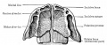
 Fetal head section 01.jpg 1,200 × 821; 186 KB
 Fetal head section 02.jpg 1,200 × 821; 171 KB
 Fetal head section 03.jpg 1,200 × 821; 174 KB
 Fetal head section 04.jpg 1,200 × 821; 188 KB
 Fetal head section.jpg 1,200 × 821; 167 KB
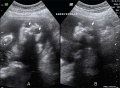
 Fetal kidney MRI 01.jpg 797 × 880; 68 KB
 Fetal kidney MRI 02.jpg 797 × 880; 64 KB
 Fetal kidney.jpg 689 × 623; 43 KB
 Fetal length change.jpg 972 × 648; 72 KB
 Fetal limb X-ray-01.jpg 685 × 931; 57 KB
 Fetal liver erythroblasts 01.jpg 905 × 534; 69 KB
 Fetal mandible and lower lip 01.jpg 1,028 × 557; 57 KB
 Fetal pineal gland 01.jpg 700 × 603; 67 KB
 Fetal polyhydramnios MRI-01.jpg 768 × 932; 60 KB
 Fetal rabbit neuroepithelial body 01.jpg 793 × 1,200; 98 KB
 Fetal temporomandibular joint 01.jpg 600 × 391; 107 KB
 Fetal temporomandibular joint 02.jpg 600 × 390; 109 KB
 Fetal temporomandibular joint 03.jpg 600 × 393; 48 KB
 Fetal temporomandibular joint 04.jpg 600 × 390; 67 KB
 Fetal temporomandibular joint 05.jpg 600 × 392; 71 KB
 Fetal temporomandibular joint 06.jpg 600 × 389; 50 KB
 Fetal thymus.jpg 450 × 600; 122 KB
 Gillilan1959-fig03.jpg 1,042 × 1,366; 317 KB
 Gilmour1941 fig02.jpg 1,000 × 526; 36 KB
 Gilmour1941 fig10.jpg 511 × 907; 105 KB
 Gray0034.jpg 800 × 613; 98 KB
 Gray0038.jpg 462 × 600; 45 KB
 Gray0043.jpg 800 × 496; 50 KB
 Gray0070.jpg 800 × 796; 182 KB
 Gray0071.jpg 700 × 440; 104 KB
 Gray0649.jpg 698 × 700; 72 KB
 Gray0654.jpg 402 × 500; 39 KB
 Gray0655.jpg 500 × 419; 39 KB
 Gray0658.jpg 361 × 450; 29 KB
 Greater-omentum.jpg 537 × 419; 48 KB
 Histology-fetal liver HEx100.jpg 1,280 × 1,024; 214 KB
 Histology-fetal liver HEx40.jpg 1,000 × 800; 281 KB
 Human 15 weeks - terminal nerve and vomeronasal organ nerves.jpg 940 × 403; 306 KB
 Human cochlea fetal development cartoon.jpg 592 × 1,200; 96 KB
 Human embryo skin 24 week EGA.jpg 596 × 939; 165 KB
 Human fetal cochlea 01.jpg 1,270 × 532; 266 KB
 Human fetal cochlea 02.jpg 1,270 × 532; 271 KB
 Human fetal gonad retinoid receptor expression.jpg 1,004 × 1,000; 447 KB
 Human fetal kidney histology 01.jpg 1,280 × 1,024; 481 KB
 Human fetal kidney histology 02.jpg 1,280 × 1,024; 322 KB
 Human fetal kidney histology 03.jpg 1,280 × 1,024; 333 KB
 Human fetal kidney histology 04.jpg 1,280 × 1,024; 307 KB
 Human fetal neural aneuploidy.jpg 1,000 × 1,400; 134 KB
 Human fetal uterus myometrium.jpg 500 × 554; 86 KB
 Human week 10 fetus 01.jpg 2,300 × 1,327; 448 KB
 Human week 10 fetus 02.jpg 800 × 462; 83 KB
 Human week 10 fetus 03.jpg 1,600 × 1,200; 370 KB
 Human week 10 fetus 04.jpg 1,600 × 1,200; 534 KB
 Human week 10 fetus 05.jpg 1,600 × 1,200; 612 KB
 Human week 10 fetus 06.jpg 1,200 × 900; 251 KB
 Human week 10 fetus 07.jpg 1,200 × 900; 283 KB
 Human week 10 fetus 08.jpg 1,200 × 900; 323 KB
 Human week 10 fetus 09.jpg 1,200 × 900; 345 KB
 Human week 10 fetus 10.jpg 1,200 × 900; 291 KB
 Human week 10 fetus 11.jpg 1,200 × 900; 304 KB
 Human week 10 fetus 12.jpg 1,200 × 900; 349 KB
 Human week 10 fetus 23.jpg 1,600 × 1,200; 393 KB
 Human week 10 fetus 26.jpg 1,200 × 900; 262 KB
 Human- fetal week 10 bf01.jpg 1,200 × 1,214; 119 KB
 Human- fetal week 10 bf02.jpg 1,200 × 900; 91 KB
 Human- fetal week 10 cerebellum A.jpg 347 × 284; 24 KB
 Human- fetal week 10 cerebellum B.jpg 347 × 284; 21 KB
 Human- fetal week 10 cerebellum C.jpg 347 × 284; 25 KB
 Human- fetal week 10 cerebellum D.jpg 347 × 284; 23 KB
 Human- fetal week 10 head A.jpg 600 × 544; 113 KB
 Human- fetal week 10 head A1.jpg 1,200 × 1,088; 159 KB
 Human- fetal week 10 head B.jpg 600 × 544; 66 KB
 Human- fetal week 10 head C.jpg 600 × 544; 118 KB
 Human- fetal week 10 head D.jpg 600 × 544; 111 KB
 Human- fetal week 10 heart ABCD.jpg 600 × 450; 133 KB
 Human- fetal week 10 lower body A.jpg 600 × 450; 96 KB
 Human- fetal week 10 lower body B.jpg 600 × 450; 93 KB
 Human- fetal week 10 lower body C.jpg 600 × 450; 94 KB
 Human- fetal week 10 lower body D.jpg 600 × 450; 91 KB
 Human- fetal week 10 sagittal plane A.jpg 500 × 573; 96 KB
 Human- fetal week 10 sagittal plane B.jpg 500 × 573; 99 KB
 Human- fetal week 10 sagittal plane C.jpg 500 × 573; 98 KB
 Human- fetal week 10 sagittal plane D.jpg 500 × 573; 105 KB
 Human- fetal week 10 sagittal planes.jpg 600 × 250; 29 KB
 Human- fetal week 10 upper body A.jpg 600 × 450; 104 KB
 Human- fetal week 10 upper body B.jpg 600 × 450; 105 KB
 Human- fetal week 10 upper body C.jpg 600 × 450; 109 KB
 Human- fetal week 10 upper body D.jpg 600 × 450; 106 KB
 Human- fetal week 10 urogenital A.jpg 600 × 450; 109 KB
 Human- fetal week 10 urogenital B.jpg 600 × 450; 109 KB
 Human- fetal week 10 urogenital C.jpg 600 × 450; 105 KB
 Human- fetal week 10 urogenital D.jpg 600 × 450; 101 KB
 Hydronephrosis.jpg 600 × 345; 64 KB
 Keibel Mall 144.jpg 678 × 607; 40 KB
 Keibel Mall 148.jpg 604 × 909; 93 KB
 Keibel Mall 2 302.jpg 1,278 × 803; 149 KB
 Keibel Mall 2 429.jpg 1,280 × 652; 204 KB
 Keibel Mall 2 583.jpg 1,280 × 1,044; 259 KB
 Keibel Mall 2 584.jpg 464 × 800; 68 KB
 Keibel Mall 2 585.jpg 1,280 × 1,144; 277 KB
 Keibel Mall 2 621.jpg 1,280 × 1,842; 589 KB
 Keibel Mall 2 626.jpg 850 × 679; 143 KB
 Keibel Mall 2 627.jpg 1,000 × 1,252; 169 KB
 Keibel Mall 2 632.jpg 1,000 × 684; 100 KB
 Keibel Mall 2 643.jpg 975 × 1,000; 95 KB
 Keibel Mall 203-205.jpg 838 × 1,000; 129 KB
 Keibel Mall 209.jpg 458 × 990; 89 KB
 Keibel Mall 210.jpg 340 × 542; 41 KB


