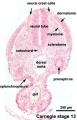
Carnegie stage 13
Introduction

Facts
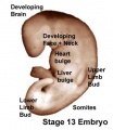
Week 4-5, 26 - 30 days, 3 - 5 mm, Somite Number 21 - 29
Events
Ectoderm: Neural tube continues to close, Caudal neuropore closes, forebrain
Mesoderm: continued segmentation of paraxial mesoderm (21 - 29 somite pairs), heart prominence
Head: 1st, 2nd and 3rd pharyngeal arch, forebrain, site of lens placode, site of otic placode, stomodeum
Body:
heart, liver, umbilical, early upper limb bulge
Features
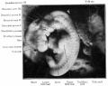
day 26, 27 somites, forebrain, site of lens placode, site of otic placode , stomodeum, 1st pharyngeal arch, 2nd pharyngeal arch, 3rd pharyngeal arch, heart prominence, somites
Identify: forebrain, site of lens placode, site of otic placode, stomodeum, 1st pharyngeal arch, 2nd pharyngeal arch, 3rd pharyngeal arch, heart prominence, somite
- Links: Week 4 | Somitogenesis | Placodes | Head | Lecture - Early Vascular | Lecture - Gastrointestinal | Lecture - Head Development | Science Practical - Gastrointestinal | Science Practical - Head | Category:Carnegie Stage 13 | Stage 14
- Carnegie Stage 13 Movies: Gastrointestinal Tract | Cardiovascular | Central Nervous System | Carnegie Stage 13 | Stage 22 Movies | Movies
- The original animations were part of the UNSW Independent Learning Project (ILP) prepared by Aashish Kumar (2006).
- Carnegie Stages: 1 | 2 | 3 | 4 | 5 | 6 | 7 | 8 | 9 | 10 | 11 | 12 | 13 | 14 | 15 | 16 | 17 | 18 | 19 | 20 | 21 | 22 | 23 | About Stages | Timeline
Bright Field


- Embryo Links: Embryo and Placenta | Embryo | Embryo (label) | Embryo (animated) | Embryo (animated large) | Head region | Head region (label) | Body region | Body region (label) | Carnegie stage 13
Scanning EM


Kyoto Collection

Image source: Embryology page Created: 19.03.1999
Image source: The Kyoto Collection images are reproduced with the permission of Prof. Kohei Shiota and Prof. Shigehito Yamada, Anatomy and Developmental Biology, Kyoto University Graduate School of Medicine, Kyoto, Japan for educational purposes only and cannot be reproduced electronically or in writing without permission.
Carnegie Collection


- Carnegie stage 13: 6473 left | 6473 dorsal | 6473 right | 6469 right | 8066 dorsal | 8066 left | 8119 left | 7433 right | 7433 ventral
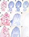
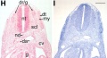
Stage 13 Serial Section Images
These are the original serial images prepared from embryo sections for Embryology practical classes and transferred online in 1996.
- Links: Carnegie stage 13 - serial sections | Carnegie stage 22 - serial sections | Carnegie stage 22
Labeled Sections

|

|

|

|

| ||
| A1L | A2L | A3L | A4L | A5L | A6L | A7L |

|

|

|

|

|

|

|
| B1L | B2L | B3L | B4L | B5L | B6L | B7L |

|

|

|

|

|

|

|
| C1L | C2L | C3L | C4L | C5L | C6L | C7L |

|

|

|

|

|

|

|
| D1L | D2L | D3L | D4L | D5L | D6L | D7L |

|

|

|

|

|

| |
| E1L | E2L | E3L | E4L | E5L | E6L | E7L |

|

|

|

|

|

|

|
| F1L | F2L | F3L | F4L | F5L | F6L | F7L |

|

|

|

|

|

|

|
| G1L | G2L | G3L | G4L | G5L | G6L | G7L |
Unlabeled Sections

|

|

|

| |||
| A1 | A2 | A3 | A4 | A5 | A6 | A7 |

|

|

|

|

|

|

|
| B1 | B2 | B3 | B4 | B5 | B6 | B7 |

|

|

|

|

|

|

|
| C1 | C2 | C3 | C4 | C5 | C6 | C7 |

|

|

|

|

|

|

|
| D1 | D2 | D3 | D4 | D5 | D6 | D7 |

|

|

|

|

|

| |
| E1 | E2 | E3 | E4 | E5 | E6 | E7 |

|

|

|

|

|

|

|
| F1 | F2 | F3 | F4 | F5 | F6 | F7 |

|

|

|

|

|

|

|
| G1 | G2 | G3 | G4 | G5 | G6 | G7 |
Stage 13 Movies
|
|
|
Additional Images
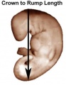
Stage 13 crown rump length
Stage 13 surface bulges
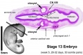
Stage 13 Optical Projection Tomography
Stage 13 otocyst
Stage 13 caudal trunk
Cross-section showing neural tube, notochord, limb bud
Selected image of above cross-section showing neural tube, notochord, limb bud
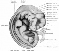
Historic - Human embryo with twenty-seven primitive segments (7 mm., 26 days)
Historic - Human embryo with 28 primitive segments (7.5 mm)


- Carnegie Stages: 1 | 2 | 3 | 4 | 5 | 6 | 7 | 8 | 9 | 10 | 11 | 12 | 13 | 14 | 15 | 16 | 17 | 18 | 19 | 20 | 21 | 22 | 23 | About Stages | Timeline
Image Source: Scanning electron micrographs of the Carnegie stages of the early human embryos are reproduced with the permission of Prof Kathy Sulik, from embryos collected by Dr. Vekemans and Tania Attié-Bitach. Images are for educational purposes only and cannot be reproduced electronically or in writing without permission.
Cite this page: Hill, M.A. (2024, June 21) Embryology Carnegie stage 13. Retrieved from https://embryology.med.unsw.edu.au/embryology/index.php/Carnegie_stage_13
- © Dr Mark Hill 2024, UNSW Embryology ISBN: 978 0 7334 2609 4 - UNSW CRICOS Provider Code No. 00098G