Category:Mouse E8.5
This Embryology category shows pages and media related to mouse embryonic day 8.5 E8.5 of development. This staging by "days" relate to in the female presence of a vaginal plug indicating that the mating occurred, see timed pregnancy.
- Mouse Stages: E1 | E2.5 | E3.0 | E3.5 | E4.5 | E5.0 | E5.5 | E6.0 | E7.0 | E7.5 | E8.0 | E8.5 | E9.0 | E9.5 | E10 | E10.5 | E11 | E11.5 | E12 | E12.5 | E13 | E13.5 | E14 | E14.5 | E15 | E15.5 | E16 | E16.5 | E17 | E17.5 | E18 | E18.5 | E19 | E20 | Timeline | About timed pregnancy
| Carnegie | Stage | |||||||||||||||||||||||
| Human | Days | 1 | 2-3 | 4-5 | 5-6 | 7-12 | 13-15 | 15-17 | 17-19 | 20 | 22 | 24 | 28 | 30 | 33 | 36 | 40 | 42 | 44 | 48 | 52 | 54 | 55 | 58 |
| Mouse | Days | 1 | 2 | 3 | E4.5 | E5.0 | E6.0 | E7.0 | E8.0 | E9.0 | E9.5 | E10 | E10.5 | E11 | E11.5 | E12 | E12.5 | E13 | E13.5 | E14 | E14.5 | E15 | E15.5 | E16 |
| Rat | Days | 1 | 3.5 | 4-5 | 5 | 6 | 7.5 | 8.5 | 9 | 10.5 | 11 | 11.5 | 12 | 12.5 | 13 | 13.5 | 14 | 14.5 | 15 | 15.5 | 16 | 16.5 | 17 | 17.5 |
| Note these Carnegie stages are only approximate day timings for average of embryos. Links: Carnegie Stage Comparison | ||||||||||||||||||||||||
| ||||||||||||||||||||||||
| Timeline Links: human timeline | mouse timeline | mouse detailed timeline | chicken timeline | rat timeline | Medaka | Category:Timeline |
Events
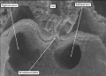
- gall bladder - majority of gallbladder progenitors in 9-11-somite-stage embryos are located in the lateral-most domain of the foregut endoderm at the first intersomite junction level along the anteroposterior axis.[1]
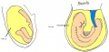
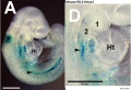
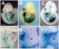
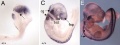
- neural - Formation and closure of anterior neuropore. The rostral extremity of the neural tube closes in embryos with usually about 15-18 somite pairs and defines this stage. The otic pit becomes progressively more indented but not closed, the mandibular process of the 1st branchial arch is clearly visible. The 3rd branchial arch becomes visible late in the stage.
- limb - An increasingly prominent ridge on the lateral body wall, approximately at the level of the 8th-12th somite, indicates the site of the future forelimb bud. Absent: forelimb bud. Embryonic age = 9 dpc (range 8.5-9.75 dpc) 13-20 somite pairs
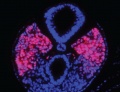
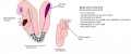
- heart - Heart tube undergoes looping at E8.5. The second heart field (pharyngeal mesoderm cells) contributes to parts of the right ventricle, the interventricular septum, the venous pole, and the base of the outflow tract.[2] Three heart regions can be identified by the bulging morphology: bulbus cordis (future right ventricle), primitive left ventricle, and common atrial chamber (behind the primitive left ventricle).[3]
References
- ↑ Uemura M, Igarashi H, Ozawa A, Tsunekawa N, Kurohmaru M, Kanai-Azuma M & Kanai Y. (2015). Fate mapping of gallbladder progenitors in posteroventral foregut endoderm of mouse early somite-stage embryos. J. Vet. Med. Sci. , 77, 587-91. PMID: 25648459 DOI.
- ↑ Buckingham M, Meilhac S & Zaffran S. (2005). Building the mammalian heart from two sources of myocardial cells. Nat. Rev. Genet. , 6, 826-35. PMID: 16304598 DOI.
- ↑ Savolainen SM, Foley JF & Elmore SA. (2009). Histology atlas of the developing mouse heart with emphasis on E11.5 to E18.5. Toxicol Pathol , 37, 395-414. PMID: 19359541 DOI.
Media in category 'Mouse E8.5'
The following 17 files are in this category, out of 17 total.
 Anderson2016-fig05.jpg 796 × 573; 66 KB
 Cervical intersomitic vessels.png 600 × 462; 308 KB
 Day 8.5 Turning of embryo.JPG 1,173 × 557; 65 KB
 Embryo left-right asymmetry pathway.jpg 800 × 458; 46 KB
 Mouse cleaved intracellular portion of Notch E8.5.jpg 532 × 408; 38 KB
 Mouse E8.5 Hoxa3.jpg 1,000 × 690; 210 KB
 Mouse E8.5-E10.5 Hoxa3.jpg 1,210 × 1,000; 356 KB
 Mouse head-neural crest 01.jpg 900 × 339; 48 KB
 Mouse pax7 neural fold 01.jpg 513 × 915; 100 KB
 Mouse somitogenesis gene expression E8.5-9.5.jpg 1,000 × 971; 130 KB
 Mouse yolk sac 01.jpg 1,002 × 669; 115 KB
 Mouse- placenta Hox13 expression.jpg 1,000 × 1,070; 150 KB
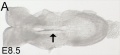 Mouse-E8.5 dorsal view.jpg 788 × 361; 28 KB
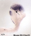 Mouse-E8.5-Sox10.jpg 400 × 463; 20 KB
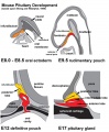 Mouse-pituitary development.jpg 660 × 800; 85 KB
 Otic placode embryo.jpg 500 × 461; 23 KB
 Theiler 13...JPG 1,030 × 427; 50 KB


