File:Keibel Mall 2 171.jpg
Original file (1,000 × 696 pixels, file size: 84 KB, MIME type: image/jpeg)
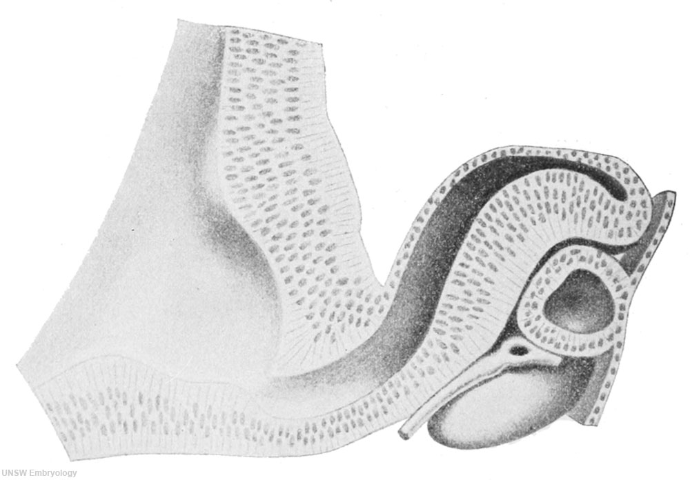
Fig. 171. Apical half of one of the optic anlagen of an embryo of 7 mm
Seen from behind. After one of Hochstetter's models prepared by F. Dedekind of Innsbruck. X 100. For explanation see text.
Development of the Eye: Fig 158 | Fig 159 | Fig 160 | Fig 161 | Fig 162 | Fig 163 | Fig 164 | Fig 165 | Fig 166 | Fig 167 | Fig 168 | Fig 169 | Fig 170 | Fig 171 | Chapter XVI. The Development of the Sense Organs | Vision Development
| Embryology - 23 May 2024 |
|---|
| Google Translate - select your language from the list shown below (this will open a new external page) |
|
العربية | català | 中文 | 中國傳統的 | français | Deutsche | עִברִית | हिंदी | bahasa Indonesia | italiano | 日本語 | 한국어 | မြန်မာ | Pilipino | Polskie | português | ਪੰਜਾਬੀ ਦੇ | Română | русский | Español | Swahili | Svensk | ไทย | Türkçe | اردو | ייִדיש | Tiếng Việt These external translations are automated and may not be accurate. (More? About Translations) |
Keibel F. The Development of the Sense Organs. (1912) chapter 16, vol. 2, in Keibel F. and Mall FP. Manual of Human Embryology II. (1912) J. B. Lippincott Company, Philadelphia.

File history
Click on a date/time to view the file as it appeared at that time.
| Date/Time | Thumbnail | Dimensions | User | Comment | |
|---|---|---|---|---|---|
| current | 10:27, 21 February 2014 | | 1,000 × 696 (84 KB) | Z8600021 (talk | contribs) | ==Fig. 171. Apical half of one of the optic anlagen of an embryo of 7 mm== Seen from behind. After one of Hochstetter's models prepared by F. Dedekind of Innsbruck. X 100. For explanation see text. {{Human Embryology Manual 2 19}} |
You cannot overwrite this file.
File usage
The following page uses this file: