Category:Carnegie Stage 12
From Embryology
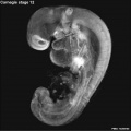
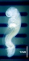
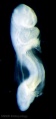
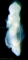
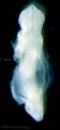
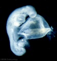
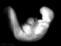
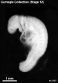
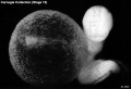
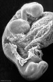
Carnegie Stage 12
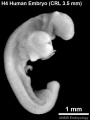
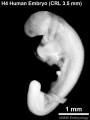
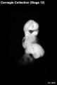
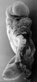
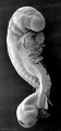
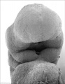
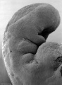
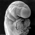
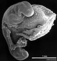
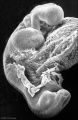
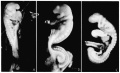
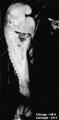
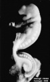
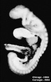
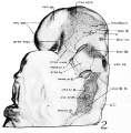
This Embryology category shows pages and media related to embryonic development in week 4, 26 - 30 days, GA week 6. The embryos have a crown rump length (CRL) of 3 - 5 mm and somite number 21 - 29 pairs.
There is also a specific Carnegie stage 12 resource page.
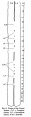
| Week: | 1 | 2 | 3 | 4 | 5 | 6 | 7 | 8 |
| Carnegie stage: | 1 2 3 4 | 5 6 | 7 8 9 | 10 11 12 13 | 14 15 | 16 17 | 18 19 | 20 21 22 23 |
- Carnegie Stages: 1 | 2 | 3 | 4 | 5 | 6 | 7 | 8 | 9 | 10 | 11 | 12 | 13 | 14 | 15 | 16 | 17 | 18 | 19 | 20 | 21 | 22 | 23 | About Stages | Timeline
Embryo Examples
Sorted by Somite Number
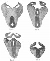
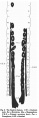
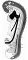
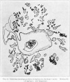
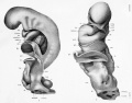
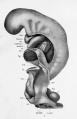
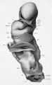
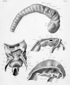
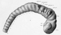
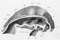
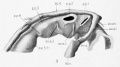
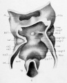
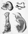
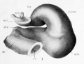
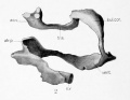
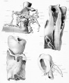
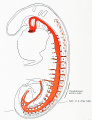
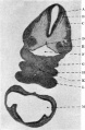
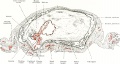
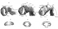
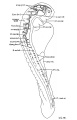
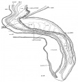
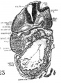
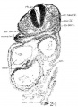
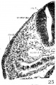
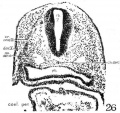
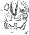
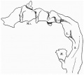
- 22 somites, Girgis embryo, Royal School of Medicine, Cairo. Apparently a normal embryo, studied and reconstructed by the wax-plate method at the Institute of Anatomy, University College, London by Girgis (1926). [1]Otic vesicle and caudal neuropore both still open. Three pharyngeal arches are present. Umbilical vesicle opens rather widely into gut. Morphology of central nervous system, digestive system, vascular system, and excretory system described in detail. The specimen had been kept in alcohol for a prolonged period, and apparently is not suitable for cytological minutiae.
- 22 somites, Carnegie No. 8963 (University of Chicago No. H 1093). Described by Wen (1928) with particular reference to the nervous system.
- 23 somites, Van den Broek embryo “A,” Zentral-Institut für Hirnforschung, Amsterdam.Two similar embryos are described together by Van den Broek (1911). The description refers almost * entirely to “A,” which is the better preserved of the two. It is evidently normal, and judging from the form of the brain, open otocyst, and liver it corresponds to about a 23-somite embryo, although it is said to have 21–22 somites.
- 23 somites, R Meyer, No. 300. The Meyer collection was transferred from Berlin to the University of Zürich in 1922, and placed in charge of H. Frey. This valuable specimen was described in monographic form by Thompson (1907, 1908). Also described and figured in the Keibel and Elze Normentafein (1908). It constitutes a type specimen.
- 23 somites, Hertwig embryo “Wolff II,” Anatomisches-biologisches Institut, Berlin. Normal, well-preserved specimen, having three pharyngeal arches and no trace of rostral neuropore. Described by Keibel and Elze (1908). Specimen has been reconstructed.
- 23 somites, Carnegie No. 8964 (University of Chicago No. H 984). Described by Wen (1928) with particular reference to the nervous system.
- 24 somites, Johnson, Harvard Collection, Boston. Complete description, based on many models and histological study, published in monographic form by Johnson (1917). Rostral and caudal neuropores closed. Otic vesicle retains a narrow opening to surface. Three pharyngeal arches are present. Hepatic trabeculae invading framework of liver. Umbilical vesicle is compressed rostrocaudally, i.e., early umbilical stalk. Probably originally 25 somites (Arey, 1938).
- 24 somites, Homo Nürnberger, Anatomisches Institut, Universität Köln. Described in detail and excellently illustrated by Rosenbauer (1955), with particular reference to the cardiovascular system.
- 25 somites, West embryo, University College, Cardiff. A 3-mm embryo much like the Johnson specimen and a good representative of the middle period of this stage (West, 1937)[2]. By means of profile and wax reconstructions the main organ systems are outlined, including an excellent study of the nephric system. The success attained by West in the orientation of his sections and the consequent accuracy in profile outlines is explained by the stained margins of the squarely trimmed paraffin block, which served as guides.
- 25 somites, Carnegie No. 6097. A graphic reconstruction was published by Müller and O'Rahilly (1980a).
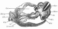
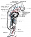
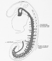
- 26 somites, His embryo M, Basel, H. h. 1. One of the group of embryos carefully studied by His for surface anatomy, and then cut in serial sections for microscopical examination, setting a new standard in human embryology (His, 1880–1885). From the development of the liver, lungs, heart, brain, and otocysts, and the absence of upper limb buds, it is estimated that it belongs in the 26-somite group.
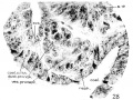
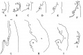
- 27 somites described by Waterston (1914)[3]
- 28 somites, His embryo Lr., Leipzig No. 67. Although used to good purpose by His, this specimen is probably not entirely normal (His, 1880–1885). The estimate of 28 somites is based on the narrowed umbilical stalk and the beginning upper limb buds.
- 28 somites, Hammar embryo (Nystroem), Anatomisches Institut, Uppsala. Described by Hammar in Keibel and Elze (1908). Digestive system described by Forssner (1907). The number of somites given above is estimated on the basis of three pharyngeal arches, full convex back, small opening in otic vesicles to surface, hepatic trabeculae, and appearance of section through cardiac region. Central nervous system shows folding of wall which characterizes imperfect preservation. External form appears normal.
- 28 somites, von Spee collection, Kiel. Sketches published in Döderlein's Handbuch (1915). Reported to have 31 somites, but absence of upper limb buds and the fact that the otic vesicles are still open to the surface makes it probable that an estimate of 28 somites is more nearly correct.
- 28–29 somites, Carnegie No. 148. Described by Gage (1905). Perhaps 29–30 somites (Arey, 1938).
- 29 somites, 20–21-day Coste embryo. Specimen not sectioned, but the exquisite drawings contained in Coste's atlas, Développement des corps organisés, Paris (1849), reveal many details of the surface form of this well-preserved embryo. In form it resembles closely Carnegie No. 1062, 29 somites, including rounded back curve, trace of upper limb buds, compressed gut-umbilical vesicle junction, size of hepatic area, form of cardiac tube, presence of otic pore, and outlines of head. Four pharyngeal arches are shown, but the fourth may have been an exaggeration of the depression lying caudal to the third bar. Also the somitic count seems to exceed the 29 estimated, but this may be caused by overemphasis on partial divisions of the terminal somitic ridge. In size its greatest length is about 4 mm. If it were straightened out as much as No. 1062, it would probably be close to 4.5 mm, like the latter.
- 29 somites, Janošík, Royal Bohemian University, Prague. Somitic count estimated on the following characteristics: three pharyngeal arches, closure of rostral and caudal neuropores, detachment of otocyst from surface, definite lung bud, elongated hepatic diverticulum with gut epithelium proliferating into adjacent tissue, narrowed umbilical stalk, and well-developed mesonephric duct and tubules. The main features of the vascular system are clearly shown. There were two embryos in this case, one of which was definitely stunted. The above description refers to the normal embryo Janošík, 1887).
- 29 somites, Waterston, University of St. Andrews, Fife. Specimen reported as having 27 paired somites (Waterston, 1914). In several characteristics it appears to be transitional between stages 12 and 13. Probably more than 27 somites, perhaps 28 (Arey, 1938) or 29. Among its advanced structures are prominent lung buds, large primordium of liver with extensive invasion by gut epithelium, narrow umbilical stalk, elongated median thyroid, and advanced ear vesicles. The upper limb buds were not prominent on the surface but stand out clearly in the sections. The blood vessels are everywhere greatly distended with blood cells, which is probably a peculiarity of this particular specimen.
Unknown Somite Number
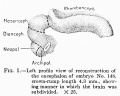
- Harvard No. 714, 4-mm embryo. Described in detail by Bremer (1906). Probably belongs to stage 12 rather than stage 11 or stage 13. It shows some unusual features, such as arrested or delayed closure of the rostral neuropore.
- No. 102 and No. 126, Department of Anatomy, Tohoku University, Sendai. These two embryos were assigned to stage 12 but the somitic count is not given. Distribution of alkaline phosphatase was studied by Mori (1965). PMID 14291749
References
- ↑ Girgis A. Description of a Human Embryo of Twenty-two paired Somites J. Anat.: 1926, 60(Pt 4);382-410. PMID 17104111 | PMC1249865
- ↑ C M West A Human Embryo of Twenty-five Somites. J. Anat.: 1937, 71(Pt 2);169-200.1 PubMed 17104635 PMC1252340
- ↑ D Waterston A Human Embryo of Twenty-seven Pairs of Somites, Embedded in Decidua. J Anat Physiol: 1914, 49(Pt 1);90-118 PMID 17233016 | PMC1288995
Subcategories
This category has the following 26 subcategories, out of 26 total.
C
- Carnegie Embryo 1062
- Carnegie Embryo 209
- Carnegie Embryo 2197
- Carnegie Embryo 250
- Carnegie Embryo 384
- Carnegie Embryo 4479
- Carnegie Embryo 4736
- Carnegie Embryo 4759
- Carnegie Embryo 4784
- Carnegie Embryo 5035
- Carnegie Embryo 5048
- Carnegie Embryo 5056
- Carnegie Embryo 5206
- Carnegie Embryo 5300
- Carnegie Embryo 5923
- Carnegie Embryo 6097
- Carnegie Embryo 6144
- Carnegie Embryo 6937
- Carnegie Embryo 7724
- Carnegie Embryo 7999
- Carnegie Embryo 8505a
- Carnegie Embryo 8505b
- Carnegie Embryo 8941
- Carnegie Embryo 8942
- Carnegie Embryo 9154
Pages in category 'Carnegie Stage 12'
The following 68 pages are in this category, out of 68 total.
B
C
- Carnegie stage 12
- Template:Carnegie stage 12 links
- Template:CE1062
- Template:CE209
- Template:CE2197
- Template:CE250
- Template:CE384
- Template:CE4479
- Template:CE4736
- Template:CE4759
- Template:CE4784
- Template:CE486
- Template:CE5035
- Template:CE5048
- Template:CE5056
- Template:CE5206
- Template:CE5300
- Template:CE5923
- Template:CE6144
- Template:CE6488
- Template:CE6937
- Template:CE7724
- Template:CE7852
- Template:CE7999
- Template:CE8505a
- Template:CE8505b
- Template:CE8941
- Template:CE8942
- Template:CE8943
- Template:CE8944
- Template:CE8963
- Template:CE8964
- Template:CE9154
- Template:CS12
P
- Paper - A Human Embryo of Twenty-five Somites
- Paper - A Human Embryo of Twenty-seven Pairs of Somites, Embedded in Decidua
- History:Paper - A Human Embryo of Twenty-seven Pairs of Somites, Embedded in Decidua
- Paper - A very Young Human Embryo found embedded in a "Decidual Cast" of the Uterus
- Paper - Description of a 4 mm human embryo (1906)
- Special:Badtitle/NS501:Paper - Description of a Human Embryo of 13-14 Mesodermic Somites
- Paper - Description of a Human Embryo of Twenty-three Paired Somites
- History:Paper - Description of a Human Embryo of Twenty-two paired Somites
- Paper - Normal development of early human embryos: Observation of 90 specimens at Carnegie stages 7 to 13
- Paper - The Anatomy of Human Embryos with Seventeen to Twenty-three Pairs of Somites
- Paper - The development of the human brain stage 12
- Paper - The Disappearance of the Precervical Sinus
R
Media in category 'Carnegie Stage 12'
The following 200 files are in this category, out of 210 total.
(previous page) (next page) B220849-01.jpg 1,311 × 849; 325 KB
 B220849-02.jpg 1,311 × 849; 358 KB
 B220849-03.jpg 1,388 × 1,040; 346 KB
 Bremer1906 fig01.jpg 1,884 × 1,426; 338 KB
 Bremer1906 fig02-5.jpg 1,200 × 1,434; 256 KB
 Bremer1906 fig06.jpg 1,300 × 927; 126 KB
 Bremer1906 fig07.jpg 875 × 1,000; 54 KB
 Bremer1906 fig08.jpg 1,154 × 576; 124 KB
 Bremer1906 fig09.jpg 915 × 1,200; 208 KB
 Carnegie stage 12 OPT.jpg 800 × 801; 46 KB
 Congdon1922-30.jpg 1,133 × 1,000; 176 KB
 Frazer1926 fig01.jpg 1,200 × 804; 137 KB
 Frazer1926 plate01.jpg 1,914 × 2,681; 469 KB
 Girgis01.jpg 988 × 618; 57 KB
 Girgis02.jpg 1,103 × 1,119; 108 KB
 Girgis03.jpg 1,008 × 969; 168 KB
 Girgis04.jpg 387 × 1,082; 48 KB
 Girgis05.jpg 1,007 × 673; 90 KB
 Girgis06.jpg 552 × 800; 47 KB
 Girgis07.jpg 313 × 937; 29 KB
 Girgis08.jpg 270 × 1,018; 37 KB
 Girgis09.jpg 342 × 1,033; 54 KB
 Girgis10.jpg 466 × 1,000; 106 KB
 Gray0472.jpg 550 × 653; 57 KB
 HillH4 Stage 12 bf01.jpg 841 × 1,121; 75 KB
 HillH4 Stage 12 bf02.jpg 841 × 1,121; 75 KB
 Human neural crest cell migration-in vitro.jpg 1,280 × 959; 163 KB
 Jenkins001.jpg 754 × 602; 98 KB
 Johnson1917 fig01.jpg 853 × 1,000; 165 KB
 Johnson1917 plate01.jpg 1,280 × 1,000; 148 KB
 Johnson1917 plate01fig01.jpg 516 × 800; 56 KB
 Johnson1917 plate01fig02.jpg 495 × 800; 52 KB
 Johnson1917 plate02.jpg 827 × 1,000; 114 KB
 Johnson1917 plate02fig01.jpg 800 × 460; 46 KB
 Johnson1917 plate02fig02.jpg 800 × 542; 55 KB
 Johnson1917 plate02fig03.jpg 800 × 447; 42 KB
 Johnson1917 plate02fig04.jpg 644 × 800; 67 KB
 Johnson1917 plate03.jpg 826 × 1,000; 107 KB
 Johnson1917 plate03fig01.jpg 800 × 613; 57 KB
 Johnson1917 plate03fig02.jpg 800 × 620; 42 KB
 Johnson1917 plate03fig03.jpg 599 × 800; 60 KB
 Johnson1917 plate03fig04.jpg 435 × 800; 45 KB
 Johnson1917 plate04.jpg 869 × 1,000; 149 KB
 Johnson1917 plate05.jpg 1,214 × 1,000; 170 KB
 Johnson1917 plate06.jpg 828 × 1,000; 117 KB
 Johnson1917 plate07.jpg 1,200 × 871; 158 KB
 Johnson1917 plate08.jpg 830 × 1,000; 146 KB
 Keibel Mall 2 414.jpg 1,000 × 1,162; 132 KB
 Keibel Mall 2 415.jpg 1,280 × 986; 201 KB
 Keibel Mall 2 416.jpg 1,000 × 738; 77 KB
 Keibel Mall 2 417.jpg 1,280 × 1,449; 179 KB
 Keibel Mall 2 418.jpg 1,280 × 1,510; 165 KB
 Keibel Mall 2 542.jpg 1,280 × 1,670; 276 KB
 Keibel Mall 2 543.jpg 1,280 × 1,193; 241 KB
 Keibel Mall 2 546.jpg 1,000 × 777; 67 KB
 Kollmann345.jpg 557 × 467; 27 KB
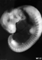 ME22 001.jpg 1,055 × 1,500; 214 KB
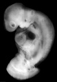 ME50 001.jpg 1,392 × 2,000; 184 KB
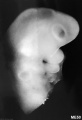 ME50 002.jpg 1,028 × 1,500; 245 KB
 Model Carnegie Embryo 588-1.jpg 750 × 1,000; 134 KB
 Model Carnegie Embryo 588.jpg 800 × 1,107; 191 KB
 Sgalitzer1941 fig01.jpg 577 × 1,200; 55 KB
 Sgalitzer1941 fig06.jpg 469 × 600; 35 KB
 Stage12 bf1.jpg 482 × 1,000; 20 KB
 Stage12 bf10.jpg 338 × 500; 11 KB
 Stage12 bf1a.jpg 386 × 800; 15 KB
 Stage12 bf1b.jpg 290 × 600; 11 KB
 Stage12 bf1c.jpg 193 × 400; 6 KB
 Stage12 bf2.jpg 471 × 1,000; 17 KB
 Stage12 bf2a.jpg 377 × 800; 12 KB
 Stage12 bf2b.jpg 283 × 600; 9 KB
 Stage12 bf2c.jpg 189 × 400; 5 KB
 Stage12 bf3.jpg 466 × 1,000; 43 KB
 Stage12 bf3a.jpg 373 × 800; 31 KB
 Stage12 bf3b.jpg 280 × 600; 21 KB
 Stage12 bf3c.jpg 187 × 400; 11 KB
 Stage12 bf4.jpg 933 × 1,000; 97 KB
 Stage12 bf4a.jpg 746 × 800; 67 KB
 Stage12 bf4b.jpg 560 × 600; 41 KB
 Stage12 bf4c.jpg 373 × 400; 10 KB
 Stage12 bf5.jpg 1,000 × 750; 59 KB
 Stage12 bf5a.jpg 800 × 600; 39 KB
 Stage12 bf5b.jpg 600 × 450; 23 KB
 Stage12 bf5c.jpg 400 × 300; 6 KB
 Stage12 bf6.jpg 350 × 500; 20 KB
 Stage12 bf7.jpg 350 × 500; 18 KB
 Stage12 bf8.jpg 733 × 500; 42 KB
 Stage12 bf9.jpg 338 × 500; 11 KB
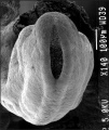
 Stage12 sem1.jpg 472 × 1,000; 69 KB
 Stage12 sem10.jpg 1,740 × 3,832; 454 KB
 Stage12 sem10a.jpg 454 × 1,000; 67 KB
 Stage12 sem10b.jpg 363 × 800; 48 KB
 Stage12 sem10c.jpg 272 × 600; 30 KB
 Stage12 sem1a.jpg 378 × 800; 24 KB
 Stage12 sem1b.jpg 284 × 600; 15 KB
 Stage12 sem1c.jpg 189 × 400; 8 KB
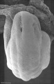
 Stage12 sem2.jpg 1,787 × 2,303; 313 KB
 Stage12 sem2a.jpg 776 × 1,000; 100 KB
 Stage12 sem2b.jpg 621 × 800; 73 KB
 Stage12 sem2c.jpg 466 × 600; 47 KB
 Stage12 SEM3.jpg 507 × 600; 68 KB
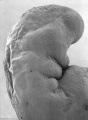
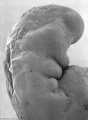
 Stage12 sem3.jpg 1,462 × 2,253; 297 KB
 Stage12 sem3a.jpg 649 × 1,000; 95 KB
 Stage12 sem3b.jpg 519 × 800; 67 KB
 Stage12 sem3c.jpg 389 × 600; 43 KB
 Stage12 sem4.jpg 1,245 × 1,693; 242 KB
 Stage12 sem4a.jpg 735 × 1,000; 107 KB
 Stage12 sem4b.jpg 588 × 800; 76 KB
 Stage12 sem4c.jpg 441 × 600; 47 KB
 Stage12 sem5.jpg 1,245 × 1,695; 217 KB
 Stage12 sem5a.jpg 735 × 1,000; 103 KB
 Stage12 sem5b.jpg 588 × 800; 74 KB
 Stage12 sem5c.jpg 441 × 600; 47 KB
 Stage12 sem6.jpg 1,620 × 1,612; 190 KB
 Stage12 sem6a.jpg 1,000 × 995; 100 KB
 Stage12 sem6b.jpg 800 × 796; 74 KB
 Stage12 sem6c.jpg 600 × 597; 49 KB
 Stage12 sem7.jpg 2,520 × 2,715; 479 KB
 Stage12 sem7a.jpg 1,000 × 1,077; 139 KB
 Stage12 sem7b.jpg 800 × 862; 102 KB
 Stage12 sem7c.jpg 600 × 647; 68 KB
 Stage12 sem8.jpg 1,376 × 2,123; 299 KB
 Stage12 sem8a.jpg 648 × 1,000; 107 KB
 Stage12 sem8b.jpg 518 × 800; 78 KB
 Stage12 sem8c.jpg 389 × 600; 51 KB
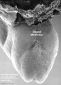
 Stage12 sem9 cloacal membrane.jpg 1,220 × 1,696; 239 KB
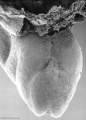
 Stage12 sem9.jpg 1,220 × 1,696; 229 KB
 Stage12 sem9a cloacal membrane.jpg 719 × 1,000; 115 KB
 Stage12 sem9a.jpg 719 × 1,000; 110 KB
 Stage12 sem9b.jpg 575 × 800; 81 KB
 Stage12 sem9c.jpg 431 × 600; 53 KB
 Streeter1957 fig01.jpg 1,292 × 1,500; 218 KB
 Thompson plate03.jpg 679 × 1,112; 77 KB
 Waterston01.jpg 1,094 × 618; 184 KB
 Waterston02.jpg 661 × 729; 130 KB
 Waterston03.jpg 800 × 547; 100 KB
 Waterston04.jpg 500 × 763; 75 KB
 Waterston05.jpg 414 × 678; 81 KB
 Waterston06.jpg 720 × 538; 92 KB
 Waterston07.jpg 562 × 655; 79 KB
 Waterston08.jpg 600 × 672; 89 KB
 Waterston09.jpg 600 × 774; 105 KB
 Waterston10.jpg 600 × 717; 83 KB
 Waterston11.jpg 500 × 728; 75 KB
 Waterston12.jpg 300 × 420; 18 KB
 Waterston13.jpg 429 × 681; 63 KB
 Waterston14.jpg 438 × 680; 66 KB
 Waterston15.jpg 500 × 674; 65 KB
 Waterston16.jpg 593 × 675; 73 KB
 Waterston17.jpg 500 × 710; 74 KB
 Waterston18.jpg 500 × 662; 71 KB
 Waterston19.jpg 500 × 642; 66 KB
 Waterston1914 fig001.jpg 1,422 × 1,082; 345 KB
 Waterston1914 fig01.jpg 1,200 × 671; 195 KB
 Waterston1914 fig02.jpg 1,485 × 1,017; 290 KB
 Waterston1914 fig03.jpg 1,418 × 982; 210 KB
 Waterston1914 fig04.jpg 1,945 × 1,043; 428 KB
 Waterston1914 figure animation.gif 489 × 752; 325 KB
 Waterston20.jpg 500 × 720; 75 KB
 Wen1928-Fig01.jpg 1,280 × 783; 80 KB
 Wen1928-Fig01a.jpg 375 × 763; 25 KB
 Wen1928-Fig01b.jpg 471 × 759; 31 KB
 Wen1928-Fig01c.jpg 398 × 641; 28 KB
 Wen1928-Fig02.jpg 900 × 915; 127 KB
 Wen1928-Fig03.jpg 809 × 900; 116 KB
 Wen1928-Fig04.jpg 1,200 × 1,392; 323 KB
 Wen1928-Fig05.jpg 1,200 × 1,404; 288 KB
 Wen1928-Fig06.jpg 1,280 × 917; 280 KB
 Wen1928-Fig07.jpg 1,265 × 648; 166 KB
 Wen1928-Fig08.jpg 1,000 × 619; 76 KB
 Wen1928-Fig09.jpg 755 × 1,000; 91 KB
 Wen1928-Fig10.jpg 495 × 1,200; 108 KB
 Wen1928-Fig11.jpg 1,000 × 543; 102 KB
 Wen1928-Fig12.jpg 1,000 × 921; 140 KB
 Wen1928-Fig13.jpg 851 × 800; 148 KB
 Wen1928-Fig14.jpg 1,111 × 1,200; 216 KB
 Wen1928-Fig15.jpg 773 × 1,200; 100 KB
 Wen1928-Fig16.jpg 702 × 1,500; 195 KB
 Wen1928-Fig17.jpg 877 × 1,000; 186 KB
 Wen1928-Fig18.jpg 1,000 × 962; 331 KB
 Wen1928-Fig19.jpg 1,200 × 1,259; 238 KB
 Wen1928-Fig20.jpg 824 × 1,012; 171 KB
 Wen1928-Fig21.jpg 787 × 1,012; 175 KB
 Wen1928-Fig22.jpg 758 × 1,103; 133 KB
 Wen1928-Fig23.jpg 806 × 1,103; 218 KB
 Wen1928-Fig24.jpg 730 × 1,000; 142 KB
 Wen1928-Fig25.jpg 660 × 1,000; 169 KB
 Wen1928-Fig26.jpg 850 × 800; 173 KB
 Wen1928-Fig27.jpg 668 × 800; 139 KB
 Wen1928-Fig28.jpg 1,200 × 899; 200 KB
 Wen1928-Fig29.jpg 1,200 × 884; 133 KB
 Wen1928-Plate02.jpg 1,280 × 788; 170 KB
 West01.jpg 868 × 1,008; 103 KB
 West02.jpg 619 × 549; 34 KB
 West03.jpg 237 × 1,025; 25 KB
 West04.jpg 946 × 631; 61 KB
 West05.jpg 457 × 759; 30 KB
 West06.jpg 308 × 802; 39 KB
 West07.jpg 984 × 153; 15 KB
 West08.jpg 808 × 376; 36 KB


