Carnegie stage 20: Difference between revisions
| Line 58: | Line 58: | ||
<gallery> | <gallery> | ||
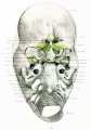
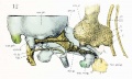
File:Lewis1920 fig01.jpg|Skull - Dorsal aspect of base with the basioccipital in horizontal plane. | |||
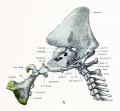
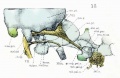
File:Lewis1920 fig02.jpg|Skull - Right half base of cartilaginous skull. | |||
File:Lewis1920 fig03.jpg|Skull - Dorsal aspect of cartilaginous and membranous skull. | |||
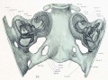
File:Lewis1920 fig05.jpg|Skull - Median sagittal aspect. | |||
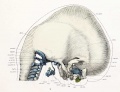
File:Lewis1920 fig06.jpg|Skull - Ventral aspect of base. | |||
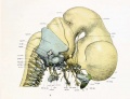
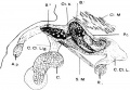
File:Lewis1920 fig07.jpg|Skull - Lateral aspect and cervical vertebrae with brain and cervical cord and hypophysis. | |||
File:Lewis1920 fig08.jpg|Skull - Lateral aspect and cervical vertebra with brain, cervical cord, and nerves. | |||
File:Lewis1920 fig09.jpg|Skull - Lateral view and cervical vertebrae with overlying membranous skull and dorsal membrane. | |||
File:Lewis1920 fig10.jpg|Skull - Dorsal aspect of sphenoid cartilage. | |||
File:Lewis1920 fig11.jpg|Skull - Dorsal aspect of sphenoid cartilage. | |||
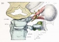
File:Lewis1920 fig12.jpg|Skull - Lateral view of right otic region. | |||
File:Lewis1920 fig13.jpg|Skull - Lateral view of right otic region showing relations of facial nerve. | |||
File:Lewis1920 fig14.jpg|Skull - Lateral view of base with deeper muscles of occipital region, mouth and pharynx. | |||
File:Lewis1920 fig15.jpg|Skull - Lateral view of part of cartilaginous and membranous skull. | |||
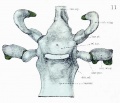
File:Lewis1920 fig16.jpg|Skull - Dorsal view of temporal and occipital cartilages. | |||
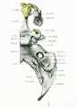
File:Human embryonic shoulder girdle 02.jpg|Human embryonic shoulder girdle | File:Human embryonic shoulder girdle 02.jpg|Human embryonic shoulder girdle | ||
</gallery> | </gallery> | ||
Revision as of 08:42, 15 April 2012
Introduction

Facts
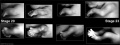
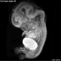
Week 8, 51 - 53 days, 18 - 22 mm
Events
Events Ectoderm: sensory placodes, lens pit, otocyst, nasal pits moved ventrally, fourth ventricle of brain
Mesoderm: heart prominence, ossification continues
Head: forebrain, eye, external acoustic meatus
Features
scalp vascular plexus, eylid, eye, nose, external acoustic meatus, auricle of external ear, arm, elbow, wrist, liver prominence, digital rays
Identify: straightening of trunk, pigmented eye, eyelid, nose, external acoustic meatus, scalp vascular plexus, digital rays, liver prominance, thigh, ankle, foot plate, umbilical cord
- Links: Week 8 | System Development | Lecture - Limb | Lecture - Head Development | Lecture - Sensory | Science Practical - Head | Science Practical - Sensory | Science Practical - Urogenital | Category:Carnegie Stage 20 | Stage 21
- Carnegie Stages: 1 | 2 | 3 | 4 | 5 | 6 | 7 | 8 | 9 | 10 | 11 | 12 | 13 | 14 | 15 | 16 | 17 | 18 | 19 | 20 | 21 | 22 | 23 | About Stages | Timeline
Bright Field


Kyoto Collection

View: This is a dorsolateral view of embryo. Amniotic membrane removed.
Image source: Embryology page Created: 19.03.1999
Image source: The Kyoto Collection images are reproduced with the permission of Prof. Kohei Shiota and Prof. Shigehito Yamada, Anatomy and Developmental Biology, Kyoto University Graduate School of Medicine, Kyoto, Japan for educational purposes only and cannot be reproduced electronically or in writing without permission.
Carnegie Collection


- Carnegie stage 20: 8517 Right | 8517 Anterior | 8517 Left | 7906 Right | 7906 Anterior | 7906 Left | 7274 Right | 7274 Anterior | 7274 Left
Additional Images
Stage 20-23 limbs
Stage 20 Optical Projection Tomography

Historic Images
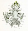
Skull - Dorsal aspect of base with the basioccipital in horizontal plane.
Skull - Right half base of cartilaginous skull.
Skull - Dorsal aspect of cartilaginous and membranous skull.
Skull - Median sagittal aspect.
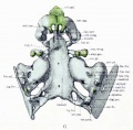
Skull - Ventral aspect of base.
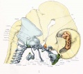
Skull - Lateral aspect and cervical vertebrae with brain and cervical cord and hypophysis.
Skull - Lateral aspect and cervical vertebra with brain, cervical cord, and nerves.
Skull - Lateral view and cervical vertebrae with overlying membranous skull and dorsal membrane.
Skull - Dorsal aspect of sphenoid cartilage.
Skull - Dorsal aspect of sphenoid cartilage.
Skull - Lateral view of right otic region.
Skull - Lateral view of right otic region showing relations of facial nerve.

Skull - Lateral view of base with deeper muscles of occipital region, mouth and pharynx.

Skull - Lateral view of part of cartilaginous and membranous skull.
Skull - Dorsal view of temporal and occipital cartilages.
Human embryonic shoulder girdle


- Carnegie Stages: 1 | 2 | 3 | 4 | 5 | 6 | 7 | 8 | 9 | 10 | 11 | 12 | 13 | 14 | 15 | 16 | 17 | 18 | 19 | 20 | 21 | 22 | 23 | About Stages | Timeline
Cite this page: Hill, M.A. (2024, June 27) Embryology Carnegie stage 20. Retrieved from https://embryology.med.unsw.edu.au/embryology/index.php/Carnegie_stage_20
- © Dr Mark Hill 2024, UNSW Embryology ISBN: 978 0 7334 2609 4 - UNSW CRICOS Provider Code No. 00098G