Histology Stains: Difference between revisions
(→Eosin) |
|||
| Line 348: | Line 348: | ||
---- | ---- | ||
Some above text modified from: Theory and practice of histological techniques. 3rd edt. By Bancroft JD and Stevens A. Churchill Livingstone, 1990. | Some above text modified from: | ||
# Theory and practice of histological techniques. 3rd edt. By Bancroft JD and Stevens A. Churchill Livingstone, 1990. | |||
# Glossary of Histological and Micro-Anatomical terms (including historical origins and eponyms) compiled by Dr Brian Freeman, Department of Anatomy, School of Medical Sciences, revised 2000. | |||
== External Links == | == External Links == | ||
Revision as of 06:08, 28 October 2011
Introduction
This page gives a general overview of some histological stains used to identify structures in cells and tissues. This stains information should also be considered in relation to Histology Fixatives. To see related histology images use the Category:Histology link.
Medicine Foundations students do not need to know stain information in this detail.
Common Stains and Their Reactions
|
Common Stains and Their Reactions | |||||
| Haematoxylin | mucins - light blue | ||||
| Eosin | colloid - pinkmuscle - red | ||||
| Iron Haematoxylin | |||||
| Van Gieson | muscle: yellow/browncartilage - pink | ||||
| Verhoeff's Elastin | elastic fibres - black | ||||
| Tartrazine | |||||
| Silver Impregnation | reticular fibres - black | ||||
| Methyl Green | |||||
| Nuclear Fast Red | |||||
| Gomori's Trichrome | keratin - redmuscle - purple/red | ||||
| Heidenhain's Azan | muscle - red | ||||
| Osmium tetroxide | myelin, lipids - black | ||||
| Alcian Blue | mucins, - blue | ||||
| Periodic acid-Schiff (PAS) | mucins, glycogen, glycocalyx - magenta | ||||
| Phosphotungstic Acid-Hematoxylin (PTAH) | muscle bands - blue | ||||
| Masson's Trichrome | cartilage, mucins - blue or green; muscle - red | ||||
| Luxol Fast Blue | myelin - blue~ | ||||
| Aldehyde Fuchsin | elastic fibres, mast cells - deep purple | ||||
| Light Green | |||||
| Gallocyanin | nucleic acids, Nissl granules - dark blue | ||||
| Romanowsky(e.g. Leishman's stain) | acidophils - redbasophils - blueazurophilic - purple | ||||
| Aldehyde Pararosanilin | elastic fibres - purple | ||||
Alcian Blue
- Stains mucopolysaccharides or glycosaminoglycans
- cationic dye (positively charged molecule) for the demonstration of glycosaminoglycans.
- binds anionic (negative) sites on the polysaccharide.
- can be combined with H&E and VG staining methods.
Alzarian Blue
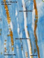
Cardiac muscle Alzarian Blue

Alizarine Brilliant Blue R
Alizarin Red
- Useful for identifying bone or other hight calcium structures.
- Stains insoluble calcium cations
- bright red stain
- Other metals such as barium, aluminium, mercury and magnesium (dark red)
Golgi Method
- (Golgi stain) A selective silver stain technique developed by Camillo Golgi (1843–1926) in 1873.
- This historic technique allowed Santiago Ramón y Cajal (1852–1934) to interpret the structure of the central nervous system.
- There are also a range of other silver staining techniques (see silver staining reticular fibres).
- Links: Neural System Development | Cahal
Gram Stain

A bacterial staining procedure using crystal violet and pink safranin counterstain that generally divides bacteria into either gram-positive or gram-negative and useful for considering associated pharmacology. The procedure was named after Hans Christian Gram (1853 - 1938).
Gram-positive bacteria
- Purple crystal violet stain is trapped by layer of peptidoglycan.
- peptidoglycan forms outer layer of the cell.
Gram-negative bacteria
- Outer membrane prevents stain from reaching peptidoglycan layer in the periplasm.
- outer membrane is composed of four major components: lipopolysaccharide, phospholipids, beta-barrel proteins, and lipoproteins.
- outer membrane then permeabilized.
- Pink safranin counterstain is trapped by peptidoglycan layer.
- Links: Histology Stains | Abnormal Development - Bacterial Infection | Medical Microbiology - Gram stain procedure
Haematoxylin and Eosin
- Acronym "H and E" stain. (H&E, HE)
- UK - Haematoxylin, USA - Hematoxylin
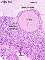
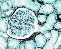
Ovary Histology

Ovary Histology
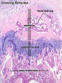
Endochondral ossification


Haematoxylin
- UK - Haematoxylin, USA - Hematoxylin
- Stains nuclei blue to dark-blue.
- Stains the matrix of hyaline cartilage, myxomatous, and mucoid material pale blue.
- Stains myelin weakly but is not noticeable if combined with eosin stain.
Eosin
- Stains cytoplasm pink to red; red blood cells are also bright red.
- Common counterstain to hematoxylin.
- Stain intensity varies with the formula as well as the fixative.
- Eosin - (Greek, eos = dawn, rose-coloured) an acidic dye staining the basic cytoplasmic proteins pink.
- eosinophil - (Greek, + philein = to love) a type of blood cell with distinct cytoplasmic granules which stain pink with eosin.
- eosinophilic - having an affinity for eosin dye.
Masson’s Trichrome Stain
- Stains nuclei deep blue, skeletal and smooth muscles red, collagen and mucin blue.
- Stains brain and spinal cord parenchymal tissue dusky pink to red.
- Used to evaluate fibrosis
- Striations in skeletal muscles also shows up much better in Masson’s trichrome than in hematoxylin and eosin stain.
- Although called a trichrome, four dyes (hematoxylin, Biebrich scarlet, acid fuchsin, and analine blue) are utilized.
Methenamine Silver
(Jone's Methenamine Silver)
- Stains the basement membrane of the glomerulus in the kidney.
- A routine stain on kidney biopsies.
- Periodic acid oxidizes the carbohydrate components of the basement membrane which produce aldehydes.
- Released aldehydes reduce the silver to a visible metallic silver (black).
Kidney - medullary ray
Kidney - glomerulus
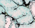
Kidney - proximal tubule


- Links: Renal System Histology
Papanicolaou stain
(Papanicolaou's stain, Pap stain) a multichromatic (five dyes) staining histological technique developed by George Papanikolaou, used to differentiate cells in smear preparations of various bodily secretions.
- Links: Papanicolaou stain
Periodic acid-Schiff (PAS)
- Stains glycogen, mucin, fungus, basement membrane and other substances.
- Stain used to detect fungal organisms and cytoplasmic accumulation of glycogen.
- Stains lysosomes granules red-purple, can be used in recognition of macrophages.
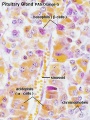
Pituitary histology
Pituitary histology


Phloxine
- derivatives of eosin
- used in the hematoxylin phloxine saffron (HPS) stain
PhosphoTungstic Acid Hematoxylin (PTAH)
- Stains nucleus and cytoplasm detail and connective tissue fibers.
- Stains collagen pink, fibrin blue, and striated muscle blue.
- Historic stain used to show CNS reactive astrocytes now used immunochemistry for glial fibrillary acidic protein (GFAP).
Tartrazine
- can be combined with Phloxine and Haem
- tartrazine acts as a yellow counterstain for tissues stained red with phloxine
- pancreatic islets - display pancreatic beta cells
Toluidine Blue
- Stains nucleus blue and cytoplasm light blue.
- A synthetic dye in the thiazins family.
Verhoeff-Van Gieson
- Verhoeff-Van Gieson or elastic-Van Gieson (EVG) stain.
- This is a combination of Verhoeff’s elastic stain which is a hematoxylin stain containing ferric chloride and Wright’s iodine solution and Van Gieson stain which contains acid fuchsin, picric acid, and hematoxylin.
- Stains elastic fibers blue-black to black, collagen pale red, other tissue elements yellow, and nuclei blue to black.
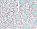
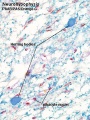
Colon Histology

Whipf's Polychrome

Some above text modified from:
- Theory and practice of histological techniques. 3rd edt. By Bancroft JD and Stevens A. Churchill Livingstone, 1990.
- Glossary of Histological and Micro-Anatomical terms (including historical origins and eponyms) compiled by Dr Brian Freeman, Department of Anatomy, School of Medical Sciences, revised 2000.
External Links
External Links Notice - The dynamic nature of the internet may mean that some of these listed links may no longer function. If the link no longer works search the web with the link text or name. Links to any external commercial sites are provided for information purposes only and should never be considered an endorsement. UNSW Embryology is provided as an educational resource with no clinical information or commercial affiliation.
- Mayo Medical Labs Mallory's Phosphotungstic Acid Hematoxylin (PTAH) Stain
Glossary Links
- Glossary: A | B | C | D | E | F | G | H | I | J | K | L | M | N | O | P | Q | R | S | T | U | V | W | X | Y | Z | Numbers | Symbols | Term Link
Cite this page: Hill, M.A. (2024, June 26) Embryology Histology Stains. Retrieved from https://embryology.med.unsw.edu.au/embryology/index.php/Histology_Stains
- © Dr Mark Hill 2024, UNSW Embryology ISBN: 978 0 7334 2609 4 - UNSW CRICOS Provider Code No. 00098G