Carnegie stage 23: Difference between revisions
| Line 78: | Line 78: | ||
{{Historic Disclaimer}} | {{Historic Disclaimer}} | ||
<gallery> | <gallery> | ||
File:Human_embryonic_shoulder_girdle_04.jpg| | File:Human_embryonic_shoulder_girdle_04.jpg|1913 Clavicle | ||
File:Fawcett1910 fig03.jpg|1910 Skull | File:Fawcett1910 fig03.jpg|1910 Skull | ||
</gallery> | </gallery> | ||
Revision as of 17:04, 23 March 2016
| Embryology - 5 Jun 2024 |
|---|
| Google Translate - select your language from the list shown below (this will open a new external page) |
|
العربية | català | 中文 | 中國傳統的 | français | Deutsche | עִברִית | हिंदी | bahasa Indonesia | italiano | 日本語 | 한국어 | မြန်မာ | Pilipino | Polskie | português | ਪੰਜਾਬੀ ਦੇ | Română | русский | Español | Swahili | Svensk | ไทย | Türkçe | اردو | ייִדיש | Tiếng Việt These external translations are automated and may not be accurate. (More? About Translations) |
Introduction

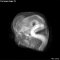
This is the final Carnegie stage of embryonic development in Week 8. After this development is considered fetal for the remainder of the pregnancy.
Facts
Week 8, 56 - 60 days, 27 - 31 mm
Gestational age GA week 10
Events
- Ectoderm:
- Mesoderm: ossification continues
- Head: eyelids, external ears, rounded head
- Body: straightening of trunk, intestines herniated at umbilicus
- Limbs: hands and feet turned inward
Features
- scalp vascular plexus, eylid, eye, nose, auricle of external ear, mouth, shoulder, arm, elbow, wrist, toes separated, sole of foot, umbilical cord
- Links: Week 8 | System Development | Lecture - Limb | Lecture - Head Development | Lecture - Sensory | Science Practical - Head | Science Practical - Sensory | Science Practical - Urogenital | Category:Carnegie Stage 23 | Fetal Development
| Week: | 1 | 2 | 3 | 4 | 5 | 6 | 7 | 8 |
| Carnegie stage: | 1 2 3 4 | 5 6 | 7 8 9 | 10 11 12 13 | 14 15 | 16 17 | 18 19 | 20 21 22 23 |
- Carnegie Stages: 1 | 2 | 3 | 4 | 5 | 6 | 7 | 8 | 9 | 10 | 11 | 12 | 13 | 14 | 15 | 16 | 17 | 18 | 19 | 20 | 21 | 22 | 23 | About Stages | Timeline
Kyoto Collection

View: This is a dorsolateral view of embryo. Amniotic membrane removed.
Image source: Embryology page Created: 19.03.1999
Image source: The Kyoto Collection images are reproduced with the permission of Prof. Kohei Shiota and Prof. Shigehito Yamada, Anatomy and Developmental Biology, Kyoto University Graduate School of Medicine, Kyoto, Japan for educational purposes only and cannot be reproduced electronically or in writing without permission.
Carnegie Collection


- Carnegie stage 23: 4570 right | 4570 anterior | 4570 left | 4570 posterior | 4570 large right | 4570 large left | Rotation | Carnegie Embryos
| iBook - Carnegie Embryos | |
|---|---|

|
|
Hill Collection
| Hill HH12 | |
|---|---|

|

|

|

|
- Links: Hill Collection
Additional Images
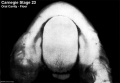
Oral cavity floor
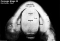
Oral cavity floor (labeled)

Oral cavity roof
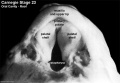
Oral cavity roof (labeled)

External ear Stages 14-23 and adult
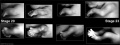
Stage 20-23 limbs
Stage 23 Optical Projection Tomography


Historic Images
| Historic Disclaimer - information about historic embryology pages |
|---|
 |
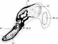
1913 Clavicle
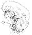
1910 Skull


- Carnegie Stages: 1 | 2 | 3 | 4 | 5 | 6 | 7 | 8 | 9 | 10 | 11 | 12 | 13 | 14 | 15 | 16 | 17 | 18 | 19 | 20 | 21 | 22 | 23 | About Stages | Timeline
Cite this page: Hill, M.A. (2024, June 5) Embryology Carnegie stage 23. Retrieved from https://embryology.med.unsw.edu.au/embryology/index.php/Carnegie_stage_23
- © Dr Mark Hill 2024, UNSW Embryology ISBN: 978 0 7334 2609 4 - UNSW CRICOS Provider Code No. 00098G