Neural - Cranial Nerve Development: Difference between revisions
mNo edit summary |
mNo edit summary |
||
| Line 79: | Line 79: | ||
|} | |} | ||
'''Cranial Nerve Development:''' [[Neural_Exam_-_3_month_cranial_nerves|3 months]] | [[Neural Exam - 12 month Cranial Nerves|12 months]] | '''Cranial Nerve Development:''' [[Neural_Exam_-_3_month_cranial_nerves|3 months]] | [[Neural Exam - 12 month Cranial Nerves|12 months]] | [[Neural Exam - 18 Cranial Nerves|18 months]] | ||
Revision as of 08:33, 16 February 2016
| Embryology - 26 Jun 2024 |
|---|
| Google Translate - select your language from the list shown below (this will open a new external page) |
|
العربية | català | 中文 | 中國傳統的 | français | Deutsche | עִברִית | हिंदी | bahasa Indonesia | italiano | 日本語 | 한국어 | မြန်မာ | Pilipino | Polskie | português | ਪੰਜਾਬੀ ਦੇ | Română | русский | Español | Swahili | Svensk | ไทย | Türkçe | اردو | ייִדיש | Tiếng Việt These external translations are automated and may not be accurate. (More? About Translations) |
Introduction

Neural development is one of the earliest systems to begin and the last to be completed after birth. This development generates the most complex structure within the embryo and the long time period of development means in utero insult during pregnancy may have consequences to development of the nervous system.
Differences between birds and mammals:
- both - have retinal axons projecting topographically to targets in the brain.
- birds - the visual fibers from the entire retina decussate at the optic chiasm.
- mammals - some axons from the temporal retina diverge at the midline to project ipsilaterally.
Neural development beginnings quite early, therefore also look at notes covering Week 3- neural tube and Week 4-early nervous system. Development of the neural crest and sensory systems (hearing/vision/smell) are only introduced in these notes and are covered in other notes sections.
| Cranial Nerves | |
|---|---|
| CN I | Olfactory |
| CN II | Optic |
| CN III | Oculomotor |
| CN IV | Trochlear |
| CN V | Trigeminal |
| CN VI | Abducent |
| CN VII | Facial |
| CN VIII | Acoustic |
| CN IX | Glossopharyngeal |
| CN X | Vagus |
| CN XI | Accessory |
| CN XII | Hypoglossal |
| Cranial Nerves | ||||
|---|---|---|---|---|
| Nerve Number | Name | Type | Origin | Function |
| CN I | Olfactory | sensory | telencephalon | smell placode |
| CN II | Optic | sensory | retinal ganglial cells | vision |
| CN III | Oculomotor | motor | anterior midbrain | extraocular muscles eye movements and pupil dilation (motor) |
| CN IV | Trochlear | motor | dorsal midbrain | extraocular muscles (superior oblique muscle) |
| CN V | Trigeminal | motor/sensory | pons | touch, mastication |
| CN VI | Abducent | motor | extraocular muscles | control eye movements (lateral rectus muscle) |
| CN VII | Facial | motor/sensory | pons | facial expression, taste (tongue anterior and central regions) regulate salivary production. |
| CN VIII | Acoustic | sensory | vestibular and cochlear nuclei | hearing, placode |
| CN IX | Glossopharyngeal | motor/sensory | medulla | swallowing and speech, taste (tongue posterior region) |
| CN X | Vagus | motor/sensory | medulla | larynx and pharynx muscles (speech and swallowing), regulates heartbeat, sweating, and peristalsis |
| CN XI | Accessory | motor | motor neurons | sternocleidomastoid and trapezius muscles |
| CN XII | Hypoglossal | motor | motor neurons | tongue muscles (speech, eating and other oral functions) |
| Cranial Nerve Links: Neural | Neural Crest | CN I | CN II | CN III| CN IV | CN V | CN VI | CN VII | CN VIII | CN IX | CN X | CN XI | CN XII | placodes | Category:Cranial Nerve |
Some Recent Findings
|
| More recent papers |
|---|
 This table allows an automated computer search of the external PubMed database using the listed "Search term" text link.
More? References | Discussion Page | Journal Searches | 2019 References | 2020 References Search term: Cranial Nerve Development <pubmed limit=5>Cranial Nerve Development</pubmed> |
Development Overview
Neuralation begins at the trilaminar embryo with formation of the notochord and somites, both of which underly the ectoderm and do not contribute to the nervous system, but are involved with patterning its initial formation. The central portion of the ectoderm then forms the neural plate that folds to form the neural tube, that will eventually form the entire central nervous system.
- Early developmental sequence: Epiblast - Ectoderm - Neural Plate - Neural groove and Neural Crest - Neural Tube and Neural Crest
| Neural Tube | Primary Vesicles | Secondary Vesicles | Adult Structures |
|---|---|---|---|
| week 3 | week 4 | week 5 | adult |
| prosencephalon (forebrain) | telencephalon | Rhinencephalon, Amygdala, hippocampus, cerebrum (cortex), hypothalamus, pituitary | Basal Ganglia, lateral ventricles | |
| diencephalon | epithalamus, thalamus, Subthalamus, pineal, posterior commissure, pretectum, third ventricle | ||
| mesencephalon (midbrain) | mesencephalon | tectum, Cerebral peduncle, cerebral aqueduct, pons | |
| rhombencephalon (hindbrain) | metencephalon | cerebellum | |
| myelencephalon | medulla oblongata, isthmus | ||
| spinal cord, pyramidal decussation, central canal | |||
Motor and Sensory

|

|
| Cranial motor nerves brainstem nuclei of origin | Primary Terminal Nuclei of the Afferent (sensory) Cranial Nerves |
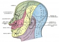
Cranial Nerve VII
(N. Facialis; Seventh Nerve; CN VII)
The facial nerve (Figs. 788, 790) consists of a motor and a sensory part, the latter being frequently described under the name of the nervus intermedius (pars intermedii of Wrisberg) (Fig. 788). The two parts emerge at the lower border of the pons in the recess between the olive and the inferior peduncle, the motor part being the more medial, immediately to the lateral side of the sensory part is the acoustic nerve.

Gray Fig. 788. Plan of the Facial and Intermediate Nerves and their Communication with Other Nerves
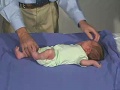
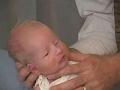
Neonatal - Clinical
Examination of the baby’s cranial nerve function is often accomplished by observing spontaneous activity.
| Newborn - Cranial Nerves | |||||||
|---|---|---|---|---|---|---|---|
|
| ||||||
| Normal | Abnormal |
Cranial Nerve Development: 3 months | 12 months | 18 months
- Links: Neural Exam Movies
Additional Images
Historic Images
| Historic Disclaimer - information about historic embryology pages |
|---|
|
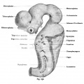
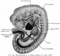
Kollman Fig. 627
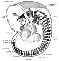
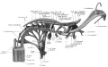
Kollman Fig. 636 cranial nerves human embryo of 10.2 mm CRL
Bailey Fig. 164
Keibel Mall Fig.32
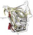
Gray Fig. 778 Maxillary Nerve
Gray Fig. 781 Mandibular division of the Trigeminal Nerve
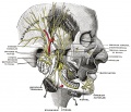
Gray Fig. 784 CN V
Lewis Fig. 8 (1920)


References
- ↑ <pubmed>20558153</pubmed>
Reviews
<pubmed>19206138</pubmed> <pubmed>11882306</pubmed>
Articles
<pubmed>18230116</pubmed>
Search PubMed
Search Pubmed: Tectum Embryology | Tectum Development
Glossary Links
- Glossary: A | B | C | D | E | F | G | H | I | J | K | L | M | N | O | P | Q | R | S | T | U | V | W | X | Y | Z | Numbers | Symbols | Term Link
Cite this page: Hill, M.A. (2024, June 26) Embryology Neural - Cranial Nerve Development. Retrieved from https://embryology.med.unsw.edu.au/embryology/index.php/Neural_-_Cranial_Nerve_Development
- © Dr Mark Hill 2024, UNSW Embryology ISBN: 978 0 7334 2609 4 - UNSW CRICOS Provider Code No. 00098G